Articoli

Tutto sul nostro settore


Febbraio 23, 2015 Newsletter

Era il 2000 quando i primi risultati sul sequenziale di tutto il DNA umano fu concluso. Furono svelati i segreti di come siamo fatti, a quali malattie siamo predisposti è come possiamo curarci. Ma leggere la sequenza di basi del nostro materiale genetico racconta solo una parte di chi siamo e a cosa siamo predisposti ad avere nel tempo.

È allo stesso modo importante anche sapere come vengono espressi (come funzionano i geni).Oggi sappiamo qualcosa di più al riguardo grazie al Roadmap Epigenomics Project, i cui risultati sono stati pubblicati su riviste del gruppo Nature.

Gli studi in questione descrivono un fattore molto importante che influenza l’attività dei geni, l’epigenomica, ovvero mostrano come, in diversi tipi di cellule umane, il DNA viene letto, il modo cioè in cui una stessa informazione genetica (tutte le cellule di uno stesso organismo hanno lo stesso dna) è usata (espressa) in modo diverso da cellula a cellula.

Ma cos’è l’epigenetica? È come funziona? A cosa serve?

Per epigenetica si intende l’insieme dei fenomeni che stanno al di fuori e al di sopra del DNA e quindi dei geni, che in qualche modo hanno una influenza sull’espressione dell’attività dei geni. Il come è codificato da tutte le modifiche che non alterano la sequenza genetica stessa ma stanno, per così dire, sopra (da qui la parola epigenetica).

Sono modifiche epigenetiche per esempio la metilazione del DNA (l’aggiunta di gruppi metilici alle basi di citosina) o il trasferimento di un gruppo acetile sugli istoni (proteine che legano il dna).
Per rendere meglio l’idea, ogni cellula può esprimere il proprio dnai n modo diverso a seconda del proprio lavoro (se deve essere una cellula cardiaca sarà diversa da un neurone), proprio come una stessa orchestra può suonare un pezzo in molti modi diversi.

I numerosi risultati pubblicati su Nature raccontano quindi di queste diverse sinfonie, suonate dalle cellule, scoperte analizzando campioni di tessuto embrionali (in fase di sviluppo e differenziazione) e adulto. Si scopre così, per esempio, che la trasformazione da staminali a cellule neuronali è orchestrata da specifici processi di metilazione, e più in generale la differenziazione delle cellule staminali è influenzata dalle modificazioni della cromatina (l’insieme del dna e delle proteine attaccate a esso), che influenza a sua volta il pattern di espressione genica.

Ma l’analisi epigenomica condotta dal gruppo di ricercatori, ha anche mostrato che l’epigenoma di una cellula cancerosa si porta dietro le impronte del tipo cellulare da cui ha avuto origine. O ancora: alterazioni epigenomiche nelle cellule del giro cingolato (un gruppo di cellule nervose che formano una regione specifica del cervello) potrebbero avere un ruolo nella sindrome da deficit di attenzione e iperattività.

Lo studio dell’epigenomica quindi potrebbe aprire le porte alla comprensione dell’origine delle malattie, e al tempo stesso alla comprensione di come gli stili di vita influenzino il nostro genoma.Le modifiche epigenetiche infatti sono in parte ereditarie, come i geni stessi, in parte dipendono dalle condizioni ambientali, quali la dieta delle mamme durante la gravidanza.

Concludendo, da queste osservazioni e conferme da parte dei ricercatori, possiamo affermare che siamo di fronte ad un punto di svolta per quanto riguarda la nutrizione e come questa sia in grado di influenzare l’attività dei geni, che a Loro volta si metteranno al lavoro, influenzando la salute o la possibile Malattia.

La nutrigenomica, da anni è al sevizio della medicina il cui scopo è quello di attivare programmi nutrizionali personalizzati che possono influenzare la salute e preventive le malattie.


Agosto 21, 2014 Newsletter

L’anguria o cocomero (Citrullus lanatus) è una pianta appartenente alla famiglia delle Curcubitaceae, tipica dell’Africa meridionale e tropicale. Il frutto ha un interno rosso o più raramente giallo; è ricco di semi, che possono essere neri, bianchi o gialli e hanno un effetto lievemente lassativo (utili quindi per riattivare un intestino pigro). La percentuale di acqua contenuta (92-93 %) risalta notevolmente rispetto al 7-8 % di zuccheri e allo 0,2-0,4 % di proteine e fibre, e ciò rende l’anguria molto dissetante.

Questo frutto si può considerare quindi depurativo e diuretico poiché, stimolando la diuresi, favorisce l’eliminazione di scorie in eccesso. Elevata è anche la presenza di sali minerali, in particolare potassio, fosforo e magnesio, vitamine A, C e quelle del gruppo B; per questo motivo l’anguria risulta senza dubbi un rimedio naturale contro la stanchezza e lo stress, prevenendo anche la ritenzione idrica. Inoltre sostanze antiossidanti, quali licopene e carotenoidi, garantiscono un benessere generale all’organismo con effetto preventivo su molte patologie e benefici anche sulla pelle.

Di recente è stata constatata la capacità di questo frutto contro le malattie cardiache e di ridurre i livelli del colesterolo cattivo. A risultare benefica è la citrulina, sostanza che rende l’anguria adatta a prevenire l’ipertensione. Tale amminoacido, presente però soprattutto nella parte bianca tra polpa e buccia, favorisce la vasodilatazione (allargamento dei vasi e quindi aumento del flusso sanguigno), conferendo quindi al frutto proprietà afrodisiache (utile soprattutto per i maschi, in quanto sembra produrre effetti simili al viagra).

Secondo alcuni studi basterebbe una fetta di anguria al giorno per aiutare il nostro organismo a ridurre il colesterolo nel sangue. Numerosi quindi sono i pregi di tale frutto ma attenzione per chi soffre di allergie e/o intolleranze.

L’anguria va evitata se si soffre di colite o gastrite: è possibile che la quantità di acido salicilico contenuta nella polpa del frutto possa causare reazioni avverse a livello intestinale. Per di più, le persone allergiche alle graminacee durante il periodo di pollinazione devono fare attenzione all’assunzione di alcuni alimenti tra cui proprio l’anguria in quanto questo frutto contiene e libera istamina. L’istamina è una sostanza che il corpo produce in risposta al contatto con una sostanza allergizzante: l’istamina, se presente in grandi quantità nel nostro organismo, determina una serie di reazioni quali arrossamenti eritemi, ponfi, produzione di muco nelle vie aeree, asma e diarrea. L’assunzione di alimenti che la contengono, come ad esempio proprio l’anguria, può amplificare la risposta allergica, incrementando l’infiammazione e i sintomi elencati in precedenza.

Una ricetta rinfrescante con l’anguria la potete trovare sul sito di Nonna Paperina: Sorbetto di anguria


Marzo 30, 2014 Newsletter

Cos’è la terapia chelante?

La terapia chelante rappresenta la prima scelta terapeutica nel trattamento di rimozioni dei metalli pesanti dall’organismo, e la principale indicazione nella prevenzione dei danni generati dai radicali liberi.Tra tutte le sostanze inquinanti, i metalli pesanti sono da considerare un elemento pericoloso con cui possiamo venire, volenti o nolenti, in contatto nella nostra quotidianità. Essi, infatti, possono trovarsi a nostra insaputa nei cibi, tessuti per abiti e vestiti, rivestimenti ambientali, vernici, farmaci, per citare alcuni dei principali veicoli.

I metalli sono tutti tossici?

Non tutti i metalli sono tossici, e soprattutto alcuni come il ferro, rame, selenio, zinco, a determinate concentrazioni sono assolutamente indispensabili per lo svolgimento delle normali funzioni metaboliche dell’organismo, il problema è che a dosi elevate risultano essere tossici e dannosi.

Quali sono i metalli che possono risultare tossici?

Alcuni metalli tossici, come il piombo, mercurio, alluminio, cadmio, esercitano effetti dannosi sull’organismo anche a bassissime dosi o concentrazioni.

Dove si accumulano i metalli?

I metalli si accumulano lentamente e progressivamente nel nostro corpo ad esempio a livello del tessuto adiposo, nelle ossa, nel fegato, nel cervello, nella tiroide.

Dove si trovano i principali metalli pesanti e tossici e come li introduciamo nell’organismo?

Le principali fonti maggiori di mercurio sono:

  • amalgame per otturazioni dentarie

  • thimerosal ( vaccini )

  • sostanze detergenti per pavimenti

  • pesci di grossa taglia ( tonno, spada )

  • cosmetici

  • talco

  • coloranti

  • diuretici

  • lassativi

  • tatuaggi

  • supposte anti infiammatorie e per emorroidi

  • pomate anti psoriasi

Le fonti di tossicità da alluminio:

  • lattine di birra

  • lattine in generale

  • lievito in polvere

  • aspirine tamponate

  • pentole e stoviglie di alluminio

  • carta di alluminio per o cibi

  • usato come conservante in alcuni condimenti per i primi, sott’aceto, cosmetici,

  • antiacidi

Le fonti di tossicità per il piombo:

  • vernici

  • inquinanti ambientali come gli scarichi dei motori delle auto

  • saldature e fonderie

  • sigarette

  • insetticidi

  • conserve

  • acqua di rubinetto

  • ortaggi coltivati in zone ad alto rischio  di inquinamento

Cosa crea il metallo tossico nell’organismo?

Il metallo blocca il lavoro di particolari enzimi all’interno delle cellule, progressivamente si riduce la produzione di energia fino ad arrivare al blocco cellulare.

Quali sono i sintomi da intossicazione da metalli pesanti?

La sintomatologia da metalli tossici è molto varia e può coinvolgere numerosi organi e apparati. In base al tessuto intaccato, avremo dei sintomi tipici e specifici correlati, ad esempio problemi neurologici se i metalli si depositano nel sistema nervoso centrale, disfunzioni ormonali tiroidee se si depositano nella tiroide, disturbi intestinali, polmonari. etc…

Siamo tutti a rischio?

Potenzialmente sì. Le categorie più a rischio sono bambini e anziani, o malati cronici.

Quali indagini si possono eseguire per sapere se si è predisposti ad accumulare i metalli pesanti?

Il mineralogramma rappresenta l’indagine non invasiva di elezione per la ricerca di metalli pesanti nel nostro organismo; la procedura è semplice e non invasiva, si tagliano i capelli della zona nucale, circa un grammo (1 cucchiaio), in pratica si tratta di una biopsia tessutale vera e propria. Grazie al mineralogramma, possiamo  scoprire l’accumulo qualitativo e quantitativo dei vari tipi di metallo pesante e dove tende la loro localizzazione. Si sceglie il capello perché rappresenta un tessuto stabile nel tempo ed affidabile.

Un’altra analisi utile si può eseguire attraverso l’esame delle urine.L’analisi della presenza di metalli pesanti nell’urina è indice del loro accumulo nell’organismo. Si possono ricercare i principali metalli tossici.

 Possiamo curare questa intossicazione? Esiste una terapia efficace?

Si esiste una terapia efficace che ci chiama CHELAZIONE. Ci sono alcuni principi attivi molto efficaci che si utilizzano da diversi anni con successi terapeutici.

Cosa si usa per chelare i metalli pesanti dall’organismo?

La prima scelta cade sull’uso di EDTA ( acido Etilen Diammino Tetracetico ) endovena, la metodica richiede esperienza da parte dell’operatore e consiste in una seduta terapeutica della durata da 1 fino a 2 ore.

Altre sostanze utilizzate sempre per via endo vena, sono acido lipoico, glutatione ridotto, ATP, Vitamina C ad alti dosaggi, N-Acetil-Cisteina. Queste sono tutte sostanze ad azione chelante, che hanno cioè lo scopo di eliminare i radicali liberi che si formano dalle reazioni biochimiche degli alimenti e dei metalli tossici.

A volte è possibile associare una terapia per via orale, con prodotti gastroprotetti, in grado di catturare le molecole dannose dall’organismo ed eliminarle attraverso i reni, fegato e intestino.

Meccanismi d’azione e benefici della terapia chelante:

  • chelazione, rimozione dei metalli tossici

  • chelazione dei depositi di calcio presenti sulle pareti dei capillari del microcircolo, con attivazione e ossigenazione dei capillari periferici

  • ha effetto anti radicali liberi ( corregge lo stress ossidativo )

  • riduce l’adesione delle piastrine

  • riduce il colesterolo e la sua adesione nelle arterie

  Indicazioni della terapia chelante ( dove la causa è un accumulo di metalli e   sostanze tossiche negli organi )

  • patologie della circolazione e del cuore ( ischemia, arterioscelrosi )

  • patologie della retina con elevato stress ossidativo

  • patologie renali

  • ipertensione arteriosa

  • patologie del fegato

  • invecchiamento ( aging )

  • intossicazione da agenti tossici

  • MCS ( sensibilità chimica multipla )

  • prevenzione e trattamento dei disturbi generati dall’arteriosclerosi

  • intossicazione da metalli pesanti

  • dolori osteo articolari causati da depositi di tossine nei tessuti

  • malattie del collagene ( collagenopatie )


Modulation of the anticancer immunity by natural agents: inhibition of T regulatory lymphocyte generation by arabinoxylan in patients with locally limited or metastatic solid tumors

lissoniResearch Article (download PDF version)

Paolo Lissoni1,*, Giusy Messina1, Fernando Brivio2, Luca Fumagalli2, Luigi Vigoré3, Franco Rovelli3, Luisa Maruelli4, Mauro Miceli4, Paolo Marchiori4, Giorgio Porro1, Michael Held5, Giuseppe di Fede6, Toshi Uchiyamada7

1 Division of Radiation Oncology, San Gerardo Hospital, Milan, Italy
2 Division of Surgery, San Gerardo Hospital, Milan, Italy
3 Laboratory of Immunomicrobiology, San Gerardo Hospital, Milan, Italy
4 Natur-Spiritual, Milan, Italy
5 Biological Medicine Center, Rome, Italy
6 Institute of Biological Medicine, Milan, Italy
7 Daiwa Pharmaceuticals, Tokyo, Japan

dr. Paolo Lissoni

*Correspondence: Dr. Paolo Lissoni, Divisione di Radioterapia Oncologica, Ospedale S.Gerardo, 20052 Monza, Milano, Italy; Fax: +390392332284, e-mail: p.lissoni@hsgerardo.org
Key words: Anticancer immunity, arabinoxylan, immunostimulation, T regulatory lymphocytes
Abbreviations: interleukin 10, (IL-10); interleukin 6, (IL-6); interleukin-2, (IL-2); interleukn 12, (IL-12); NK cells, (CD16+CD56+); T cytotoxic lymphocytes, (CD8+); T helper lymphocytes, (TH), (CD4+); T lymphocites, (CD3+); Transforming growth factor beta, (TGF-β) T-regulatory lymphocytes, (T-reg), (CD4+CD25+)

Received: 30 September 2008; Revised: 1 November 2008
Accepted: 17 November 2008; electronically published: December 2008

Summary

In the last years, several immunomodulating antitumor agents have demonstrated in the nature, particularly from Aloe plant and rice bran. However, the major problem concerning the natural antitumor agents is to define their immune mechanisms of action in relation to the more recent advances in tumor immunobiology. At present, the main cause responsible for the lack of an effective antitumor response in advanced cancer patients is belived to be represented by the generation of a subtype of T helper lymphocytes (CD4+) with suppressive activity on anticancer immunity, the so-called T regulatory lymphocytes (T reg), which may be clinically identified as CD4+CD25+ cells. On this basis, a study was planned to evaluate the effect of rice bran extract arabinoxylan on T reg cell count and percentage in solid tumor patients in relation to the various lymphocyte subpopulations. The study included 22 evaluable cancer patients, 16 of whom had an untreatable metastatic solid tumor. Arabinoxylan was given orally at a dose of 2000 mg/day for the first month, followed by a dose of 1000 mg/day for the next month. In each patient we evaluated by monoclonal antibodies the absolute number of lymphocytes, T lymphocytes (CD3+), T helper (TH) lymphocytes (CD4+), T cytotoxic lymphocytes (CD8+), NK cells (CD16+CD56+), T reg lymphocytes (CD4+CD25+) and TH/T reg ratio before and after 2 months of therapy. No substantial change occurred on therapy in the mean number of lymphocytes, CD3+, CD8+ and NK cells. On the other hand, the mean number of TH cells increased, whereas that of T reg cell decreased on treatment, even though none of these differences was statistically significant. On the contrary, TH/T reg mean ratio significantly enhanced after arabinoxylan therapy. In addition to its previously demonstrated stimulatory action on NK function, this study shows that arabinoxylan may inhibit the production of T reg cells, which are responsible for cancer-related immunosuppression, with a following improvement in the anticancer immunity. If further studies will confirm these results, arabinoxylan could be successfully associated with chemotherapy to induce not only a cytotoxic destruction of cancer cells, but also an improvement in the immune status.

I. Introduction
The recent advances in the definition of the mechanisms responsible for tumor progression have suggested the possibility to control cancer growth not only trough chemotherapy-induced cancer cell destruction, but also by stimulating the anticancer immunity. In addiction to the exisence of endogenous antitumor molecules, several agents capable of stimulating the anticancer immunity have alsso isolated from plants. However, the immunomodulatory effects of most natural immunomodulating agents need to be better investigated in an attempt to establish their mechanisms of action in relation to the most recent discoveries concerning the physiopathology of the anticancer immunity. At present, Aloe extracts (Lissoni et al, 1998) and arabinoxylan extract from rice bran (Ghoneum and Jewett, 2000) would represent some of the potential natural agents which could be utilized in the complementary therapy of human neoplasms. Today, it is known that the antitumor immune response is the end-result of several interactions involving cytokines and immune cells, provided by stimulatory or suppressive effects on the anticancer immunity (Atzpodien and Kirchner, 1990; Rosenberg, 1992). Therefore, the lack of an effective anticancer immune response in most cancer patients with advanced disease would simply depend on the prevalence of immunosuppressive mechansisms with respect to the immunostimulatory ones (Atzpodien and Kirchner, 1990). The anticancer immunity is mainly activated by T helper-type 1 lymphocytes by releasing IL-2 (Whittington and Faulds, 1993), and by dentritic cells, which act as antigen-presenting cells producing IL-12 (Banks et al, 1995), T cytotoxic lymphocytes and NK-LAK system, which are involved in the induction of the antigen-dependent and antigen-independent cytotoxicity, respectively (Atzpodien and Kirchner, 1990). Therefore, IL-2 and IL-12 would represent the main anticancer cytokines in humans. On the contrary, the suppression of the anticancer immune response is mediated by several cytokines, namely IL-10 (Moore et al, 1993), IL-6 (Matsuda and Hirano, 1990) and TGF-β (Shevach, 2002). Recently, however, it has been demonstrated that the various endogenous suppressive factors would exert their inhibitory immune effect through a common end-mechanism, consisting of the generation of a subtype of T helper lymphocytes (CD4+cells), provided by a fundamental suppressive activity on the anticancer immunity, the so-called T regulatory lymphocyte (T reg) (Dieckmann et al, 2001), which at present seems to constitute the main mechanism responsible for cancer-related immunosuppressive status. T reg cells may be identified by the simultaneous expression of the alpha-chain of IL-2 receptor (CD25) and CD4 antigen (Dieckmann et al, 2001). Then, T reg cells may be clinically recognized as CD4+CD25+ lymphocytes. Therefore, each eventual natural immunomodulating agent would have to be investigated in relation to its possible effect on T reg generation since, at least from a theoretical point of view, each natural agent capable of counteracting T reg activity could positively influence the prognosis of the neoplastic disease by improving the efficacy of the anticancer immune response. Moreover, our previous preliminary studies have suggested that the percentage of T reg cells with respect to the total number of T helper cells, as expressed as CD4/CD4CD25 ratio, may represent an optimal synthetic immune index to investigate the functional status of the anticancer immunity in the single cancer patient, by representing the synthesis of the actions of the great number of immunostimulating and immunosuppressive factors involved in the modulation of the anticancer immunity (Dieckmann et al, 2001). Within the great number of natural agents derived from plants and potentially usefull to be employed in the complementary therapy of cancer, arabinoxylan would seem to represent one of the potential natural agent, because of its efficacy in improving the clinical status of cancer patients (Ghoneum and Jewett, 2000; Ghoneum and Gollapudi, 2005; Markus et al, 2006; Ghoneum et al, 2007). The immunomodulating properties of this nautral substances extracted from plants have been confirmed by experimental studies, but unfortunately most experiments have been limited to the investigations of they effects on non-specific immune parameters for the anticancer immunity, such as NK cell cytotoxicity. In contrast, since reg cells play a fundamental role in suppressing the generation of the anticancer immunty, each potential antitumor immunomodulatory natural substances, would have to be investigated also in relation to their eventual influence on T reg cell system. On the basis of the recent discoveries in tumor immunobiology (Dieckmann et al, 2001; Shevach, 2002), a study was planned to investigate the possible influence of arabinoxylan on both absolute number of T reg cells and their ratio with respect to the total CD4+ T cells in a group of solid tumor patients, affected by locally limited or metastatic disease.

II. Materials and methods
The study included 24 consecutive patients, 18 of whom had a metastatic solid tumor, which did not respond to the conventional anticancer chemotherapies and for whom no other effective standard treatment was available, while the remaining 6 patients had been surgically treated for a locally limited neoplasm. Patients were followed at Biological Medical Institute of Milan and the protocol was approved by the Director of the Institute. Eligibility criteria were, as follows:histologically proven locally limited or metastatic solid tumor, no double tumor, no chronic therapy with corticosteroids because of their immunosuppressive effects and no concomitant treatment with other immunomodulating agents,such as interferons,interleukins and monoclonal antibodies. At the time of the start of arabinoxylan therapy, patients with untreatable metastatic cancer were under treatment with the only supportive care, consisting of anti-inflammatory agents for pain, anti-dopaminergic drugs for nausea and vomiting and with the pineal hormone melatonin for the therapy of the neoplastic cachexia (Banks et al, 1995). Patients were considered as fully evaluable when they had received arabinoxylan therapy for at least 2 consecutive months. Arabinoxylan was given orally at a dose of 1000 mg twice/day for the first month, followed by a dose of 1000 mg/day for the next month. Arabinoxylan was supplied by DAIWA Pharmaceutical (Tokyo, Japan). It was derived from rice bran treated enzymatically with an extract of the shiitake mushrooms. It is a polysaccharide containing β-1,4-xylopironase hemicellulose, commercially available and known as Biobran. For the immune investigation, venous blood samples were collected in the morning after an overnight fast before the onset of arabinoxylan therapy and after 2 consecutive months of treatment. In each blood sample, we evaluated the absolute number of total lymphocytes, T lymphocytes (CD3+), T helper (TH) lymphocytes (CD4+), T cytotoxic lymphocytes (CD8+), NK cells (CD16+ CD56+ and T regulatory (T reg) lymphocytes (CD4+ CD25+). The different lymphocyte subsets were measured with a flow cytometric assay by using specific monoclonal antibodies supplied by Becton-Dickinson (Milan, Italy). Moreover, because of the importance not only of their absolute number, but also of their percentage with respect to the other lymphocyte subsets, namely to that of CD4+ cells, CD4/CD4CD25 ratio, corresponding to TH/T reg ratio, was also determined before and after therapy. Normal values (95% confidence limits) of T reg number and TH/T reg ratio observed in our laboratory were below 240/mm3 and above 4.0, respectively. Data were reported as mean +/- SE and statistically analyzed by the Student’s t test, the analysis of variance and the chi-square test, as appropriate.

III. Results
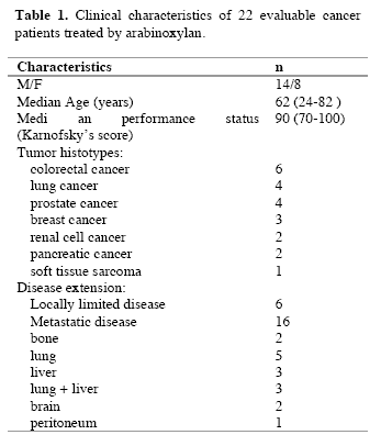
Evaluable patients were 22/24, while the remaining 2 patients, both affected by untreatable disseminated liver metastases due to colorectal cancer, rapidly died for disease progression before concluding the two planned months of arabinoxylan therapy. The clinical characteristics of the evaluable patients are reported in Table 1. Figure 1 illustrates changes in the mean number of total lymphocytes, T lymphocytes, T cytotoxic lymphocytes and NK cells occurring after 2 months of arabinoxylan therapy. No substantial variation was found in the mean number of lymphocytes, T lymphocytes, T cytotoxic lymphocytes and NK cells under arabinoxylan treatment. In contrast, as illustrated in Figure 2, TH and T reg mean numbers increased and decreased, respectively, after arabinoxylan therapy, without, however statistically significant differences with respect to the values seen prior to therapy. On the contrary, a statistically significant increase in TH/T reg mean ratio was achieved after arabinoxylan therapy (p<0.025). The increase in TH/T reg ratio under arabinoxylan therapy was more pronounced in patients with an abnormally low ratio prior to therapy with respect to that occurring in those with normal pre-treatment ratio, however without statistically significant differences ( 2.3 +/- 0.4 vs 1.7 +/- 0.5). In more detail,
090722_linfocitiT_table1
090722_linfocitiT_1

Figure 1. Changes in the number of lymphocytes, Tlymphocytes (CD3), T cytotoxic lymphocytes (CD8) and NK cells (CD16 CD56) after 2 months of arabinoxylan therapy.
090722_linfocitiT_2

Figure 2. Changes in the mean number of T helper (TH) lymphocytes (CD4) and T regulatory lymphocytes (cd4 cd24) and in TH/T reg mean ratio.

before arabinoxylan therapy, an abnormally low TH/T reg ratio was present in 12/22 (55%) evaluable patients. Arabinoxylan treatment induced a normalization of TH/T reg ratio in 5/12 (42%) patients with an abnormally low ratio prior to therapy. The percentage of arabinoxylan-induced TH/T reg normalization obtained in lymphocytopenic patients was not significantly different from that achieved in patients with normal pre-treatment lymphocyte count ( 3/7(43%) vs 2/5(40%) ). No toxicity was observed under arabinoxylan treatment, which was well tolerated in all patients. Asthenia was present in 8/22 (36%) evaluable patients. An evident relief of asthenia, as assessed by a specific patient report, was obtained under arabinoxylan therapy in 5/8 (63%) patients.

IV. Discussion
Previous experimental studies had already demonstrated some immunomodulating properties of arabinoxylan, in particular consisting of stimulation of NK cytotoxic function (Ghoneum, 1998), whereas NK cell number did not seem to be influenced by arabinoxylan administration. However, it has to be remarked that NK cells were belived to be fundamental in the antitumor immunity until some years ago, before the discovery of the essential role played by the antitumor cytokines, such as IL-2 and IL-12 (Whittington and Faulds, 1993) and dendritic cells, because of their function as antigen-presenting cells (Banks et al, 1995). In fact, it has to be considered that the cytotoxic activity of NK cells is effective only against artificial laboratory cancer cell lines, whose biological malignant properties are different from those presented by fresh human tumor cells (Whittington and Faulds, 1993). In addition, NK cells have been proven to be also able to destroy fresh human cancer cells only after the activation of their cytotoxic function by IL-2 (Atzpodien and Kirchner, 1990). From this point of view, arabinoxylan had been already proven to amplify the stimulatory effect of IL-2 on NK-mediated antitumor cytotoxicity (Ghoneum and Jewett, 2000). In contrast, no study has been performed up to now to evaluate the possible influence of arabinoxylan not only on the mechanisms responsible for the generation of an effective anticancer immune response, but also on those involved in the suppression of anticancer immunity. The results of this preliminary study, carried out to evaluate the influence of arabinoxylan on T reg cells, which represent the most important cells involved in the suppression of the antitumor cytotoxic immune response, demonstrates that arabinoxylan may counteract T reg cell generation by reducing their number and percentage with respect to the total amounts of CD4+ cells and circulating lymphocytes. Since NK cell function is inhibited by T reg activation (Shevach, 2002), the previously demonstrated arabinoxylan-induced stimulation of NK cell cytotoxic function might depend at least in part on its capacity of counteracting T reg generation (Dieckmann et al, 2001). Moreover, this study would suggest that the inhibitory action of arabinoxylan on T reg generation is more pronounced in patients with an abnormally high percentage of T reg cells prior to therapy, with a following pre-treatment abnormally low TH/T reg ratio before therapy, whereas its effect was less evident in patients with a pre-treatment value of TH/T reg ratio within the normal range. Therefore, the influence of arabinoxylan on T reg generation would consist of a modulatory action rather than an inhibitory activity. This finding could explain a potential favourable immunomodulatory effect of arabinoxylan also in patients with autoimmune diseases (Ghoneum, 1998), who in contrast to cancer patients would tend to present abnormally low amounts of T reg cells. In any case, the importance of the inhibition of T reg generation in the induction of an effective anticancer immune response has been recently confirmed by the evidence that the block of T reg activity by specific monoclonal antibodies may induce objective tumor regressions in humans (Yang et al, 2007). Obviously, the major problem is the exact identification of he T reg cell population. Even though T reg cells may express other immune markers, namely FOX-p2 cytoplasmatic antigen, most clinicians are in agreement to identify the CD4+CD25+ cells as T reg lymphocytes (12). In any case, further studies, by evaluating other immune markers, will be required to better identify T reg cells population, namely FOX-p3, even though recently some Authors have shown that FOX-p3 expression by T reg cells is associated with a lower suppressive activity (Dieckmann et al, 2001; Shevach, 2002). Moreover, it has to be remarked that several patients included in the present study were concomitantly under palliative therapy with the anti-cachectic pineal hormone melatonin (Brzezinski, 1997), which may also play immunomodulating effects (Maestroni, 1993). Therefore, further randomized studies with arabinoxylan alone versus arabinoxylan plus melatonin will be required to better define the immunomodulating action of arabinoxylan. If further clinical and experimental studies will confirm the inhibitory action of arabinoxylan on T reg cell system, it could be included in cytokine-based immunotherapies to enhance their efficacy by counteracting T reg cell generation.

References
Atzpodien J, Kirchner H (1990) Cancer, cytokines and cytotoxic cells:interleukin-2 in the immunotherapy of human neoplasms. Klin Wochenschr 14, 1-10.
Banks RE, Patel PM, Selby PJ (1995) Interleukin-12:a novel clinical player in cytokine therapy. Br J cancer 71, 655-659.
Brzezinski A (1997) Melatonin in humans. N Engl J Med 336, 185-195.
Dieckmann D, Plottner H, Berchtold S, Berger T, Schuler G (2001) Ex vivo isolation and characterization of CD4+CD25+ T cells with regulatory properties from human blood. J Exp Med 193, 1303-1310.
Ghoneum M (1998) Enhancement of human natural killer cell activity by modified arabinoxylane fro rice bran(MGN-3). Int J Immunother 14, 89-99.
Ghoneum M, Gollapudi S (2005) Synergistic of arabinoxilan rice bran (MGN-3/Biobran in S. Cerevisiae-induced apoptosis of monolayer breast cancer MFC-7 cells. Anticancer Res 25(6B), 4187-96.
Ghoneum M, Brown J, Gollapudi S (2007) Yeast therapy for the treatment of cancer and its enhancement by MGN-3/Biobran, an arabinoxylan rice bran. Cellular Signaling and Apoptosis Research (Ed. Alex R. Demasi) Cap IV: 185-200.
Ghoneum M, Jewett A (2000) Production of tumor necrosis factor-alpha and interferon-gamma from human peripheral blood lymphocytes by MGN-3, a modified arabinoxylan from rice bran, and its synergy with interleukin-2 in vitro. Cancer Detect Prevent 24, 314-324.
Lissoni P, Giani L, Zerbini S, Trabattoni P, Rovelli F (1998) Biotherapy with the pineal immunomodulating hormone melatonin versus melatonin plus Aloe vera in untreatable advanced solid neoplasms. Nat Immun 16, 27-33.
Maestroni JGM (1993) The immunoneuroendocrine role of melatonin. J Pineal Res 14, 1-10.
Markus J, Miller A, Smith M, Orengo I (2006) Metastatic hemangiopericytoma of the skin treated with wide local excision and MGN-3. Dermatol Surg 32, 145-147.
Matsuda T, Hirano T (1990) Interleukin-6 (IL-6). Biotherapy 2, 363-371.
Moore KW, O’Garra A, De Waal-Malefyt R (1993) Interleukin-10. Ann Rev Immunol 11, 165-174.
Rosenberg SA (1992) The immunotherapy and gene therapy of cancer. J Clin Oncol 10, 181-191.
Shevach EM (2002) CD4+CD25+ suppressor T cells:more questions than answers. Nat Rev Immunol 2, 389-400.
Whittington R, Faulds D (1993) Interleukin-2. Drugs 46, 446-514.
Yang JC, Hughes M, Kammula U, Royal R, Sherry RM, Topalian SL, Suri KB, Levy C, Allen T, Mavroukakis, Lowy I, White DE, Rosenberg SA (2007) Ipilimubab (anti-CTLA4 antibody)causes regression of metastatic renal cell cancer associated with enteritis and hypophysitis. J Immunother 30, 825-830.

(download PDF version)


A clinical study of T-regulatory lymphocyte function in cancer patients in relation to tumor histotype, disease extention, lymphocyte subtypes and cortisol secretion

Research Article (download PDF version)

Luigi Vigorè1, Fernando Brivio2, Luca Fumagalli2, Roberto Vezzo1, Giusy Messina6, Franco Rovelli6, Massimo Colciago3, Giovanna D’Amico4, Giuseppe Di Fede5, Paolo Lissoni6

1 Laboratory of Immunomicrobiology, St.Gerardo Hospital, Monza, Milan, Italy
2 Division of Surgery, St.Gerardo Hospital, Monza, Milan, Italy
3 I.N.R.C.A Laboratory of Analyses, Casatenovo, Lecco, Italy
4 Research Center “Fondazione Tettamanti” Clinica Pediatrica Università Milano-Bicocca, Italy
5 Institute of Biological Medicine, Milan, Italy
6 Division of Radiation Oncology, St.Gerardo Hospital, Monza, Milan, Italy

*Correspondence: Dr. Paolo Lissoni, Divisione di Radioterapia Oncologica, Ospedale S.Gerardo, 20052 Monza, Milano, Italy; Fax: +390392332284, E-mail: p.lissoni@hsgerardo.org
Key words: Anticancer immunity, immunosuppression, T regulatory lymphocytes
Abbreviations: cytotoxic T lymphocyte-associated antigen-4, (CTLA-4); glucocorticoid-induced TNF-α receptor, (GITR); myeloidderived suppressor cells, (MDSC); NK cells, (CD16CD56); T cytotoxic lymphocytes, (CD8); T helper lymphocytes, (CD4); Tregulatory lymphocytes, (T-reg)

Received: 24 July 2008; Revised: 11 September 2008
Accepted: 12 September 2008; electronically published: October 2008

Summary

Several clinical investigations showed that the immune status is a prognostic variable in cancer patients, even tough the evaluation of the anticancer immunity is not generally considered in the medical oncology. Several immune parameters, including lymphocyte subsets and cytokine blood concentration, had been proposed to quantify the functional status of the anticancer immunity, but recent discoveries would suggest that the end-result of the various immune interactions is represented by a subtype of CD4 lymphocytes capable of suppressing the antitumor immune reaction, the so called T-regulatory lymphocytes (T-reg). This study was performed to detect T-reg count and percentage in solid tumor patients, in relation to tumor histotype, disease extension, lymphocyte sub-populations and cortisol circadian secretion. The study included 114 consecutive cancer patients affected by the most frequent tumor histotypes, 69 of whom showed a metastatic disease. In each patient we evaluated T-reg cells, identified as CD4+CD25+, in relation to T helper (CD4), T cytotoxic (CD8) and NK (CD16CD56) cells. Abnormally high values of T-reg cells were seen in 52/114 (46%) patients, and the percentage of high values of T-reg was significantly higher in metastatic patients than in non-metastatic ones. In contrast, no significant difference was seen in relation to tumor histotype. Patients with increased T-reg count had a significantly lower NK cell number. Finally no significant difference in T-reg number was seen between patients with altered or normal rithm of cortisol. The study confirmed that, irrespectively of tumor histotype the metastatic disease is associated with a progressive and increased T-reg generation, with a following suppression of anticancer immunity.

I. Introduction
At present, there is no doubt about the existence of a sub-type of T lymphocytes, the so-called T regulatory lymphocytes (T-reg), capable of suppressing the cellular immune responses,including the anticancer immunity (Thomton and Shevach, 2000; Shevach, 2002; von Herrath and Harrison, 2003; Schwartz, 2005; von Boehmer, 2005; Ziegler, 2006; Zou, 2006). However, the exact definition
of T-reg cells in terms of cell surface marker expression still remains controversial, particularly from a clinical point of view. All authors are in agreement to consider Treg lymphocytes as CD4+CD25+ cells, but at present it is still unknown whether the expression of CD4 and CD25 antigens may be sufficient to identify T-reg cells (Thomton and Shevach, 2000; Shevach, 2002; von Herrath and Harrison, 2003; Schwartz, 2005; von Boehmer, 2005; Ziegler, 2006; Zou, 2006), since several authors retain that the intracytoplasmatic expression of the FOX p3 protein is essential for the differentiation into T-reg cells (Ziegler, 2006; Zou, 2006).Recently, however, some preliminary observations would suggest that the cytoplasmatic expression of FOX p3 by CD4+CD25+ cells may be associated at least in some experimental conditions with a diminished, rather than with an enhanced immunosuppressive activity of T-reg cells (Siddiqui et al, 2007). In contrast, all authors agree that the expression of CD152 antigen, also called cytotoxic T lymphocyteassociated antigen-4 (CTLA-4) (Vasu et al, 2004), is fundamental for the immunosuppressive activity of T-reg cells (Takahashi et al, 2000), since the block of its expression by using anti-CTLA-4 monoclonal antibodies may abolish the suppressive activity of T-reg cells, with a following stimulation of the anticancer immunity in cancer patients (Knutson and Disis, 2007) and an enhanced incidence of autoimmune diseases in the healthy subjects (Lan et al, 2005). Therefore, the addition of a third marker, such as CD152 antigen, may allow to define a more homogeneous cell population provided by a regulatory activity with respect to the simple CD4+CD25+ expression (Dieckmann and Plottner, 2001). In fact, the suppressive regulatory action of CD4+CD25+CD152+ has appeared to be clearly higher than that played by the simple CD4+CD25+ T lymphocytes (Leong et al, 2006).This finding is not surprising, since the simple expression of CD25 marker, corresponding to the !-chain of IL-2 receptor, is not an exclusive characteristic of T-reg lymphocytes, but it is a non-specific property of the overall activated T lymphocytes (Thomton and Shevach, 2000; Shevach, 2002; von Herrath and Harrison, 2003; Schwartz, 2005; von Boehmer, 2005; Ziegler, 2006; Zou, 2006). At present, preliminary clinical studies would show that the percent of circulating CD4+CD25+ cells may be about 10% of the all CD4+ lymphocytes, and that of CD4+CD25+CD152+ cells may be about 40% of the total CD4+CD25+ cells, then the expected percent of CD4+CD25+CD152+ in the healthy subjects would be less than 5% of the total circulating CD4+ lymphocytes (Jago et al, 2004). Finally, the expression of glucocorticoidinduced TNF-α receptor (GITR) is also associated with an evident suppressive activity by T-reg lymphocytes (Kanamaru et al, 2004), which in fact are stimulated by
cortisol (Sthephens et al, 2004), that in contrast may inhibit the activity of the most other T lymphocytes, namely that of T helper lymphocytes, with a following diminished production of IL-2 (Claman, 1998). As far as the mechanisms responsible for T-reg-induced suppression of the anticancer immunity are concerned, several experimental observations have shown that T-reg cells may suppress the antitumor immune response through the release of immunosuppressive cytokines, namely IL-10 and TGF-β (Dieckmann et al, 2002), even though other authors would suggest that the suppressive activity of Treg cells on CD4+ and CD8+ lymphocyte activation may be relatively independent from the action of cytokines, by mainly requiring cell surface contact (Birebent et al, 2004). IL-2 has been proven to be essential for T-reg generation and some authors consider IL-2 as the main growth factor of T-reg lymphocytes (Antony and Restito, 2005), but more adequate studies have demonstrated that IL-2 may induce both stimulation and inhibition of T-reg generation and activation (Malek and Bayer, 2004). In fact, IL-2 has appeared to induce and promote T-reg differentiation only in the presence of TGF-β (Chen et al, 2003). Therefore, IL-2 would constitute the main human cytokine in influencing the characteristics of the anticancer immunity, since it may be responsible for both activation and suppression of an effective immune response against cancer cell proliferation and dissemination (Wang et al, 2001), namely depending on the whole status of the cytokine network, in particular on the presence or in the absence of adequate concentrations of TGF-β. In the absence of TGF-β, IL-2 stimulates the anticancer immunity, whereas it counteracts the generation of an effective antitumor immunity in the presence of TGF-β. In other words, IL-2 would physiologically control both tolerance and immunity, depending on the presence of TGF-β and other less known factors (Annunziato et al, 2002). In fact, under cancer immunotherapy with IL-2 the percent of T-reg cells has been shown to decrease in responding patients and to enhance in those with disease progression (Cesana et al, 2006). However, the regulation of T-reg functions does not depend only on immune factors, since it is also under a neuroendocrine control (Ji et al, 2004). In particular, cortisol has appeared to stimulate T-reg cell generation (Ji et al, 2004), with a following enhanced release of IL-10, by representing the main mechanism responsible for cortisol-induced immunosuppression. From a clinical oncological point of view, preliminary observations showed an enhanced percent of circulating CD4+CD25+ lymphocytes in cancer patients, namely in those with advanced disease (Sasada et al, 2003). The present study was performed to better establish which is T-reg behaviour in cancer patients in relation to both tumor histotype and disease extension.

II. Materials and methods
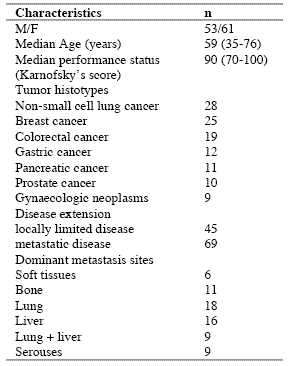
The study included 114 consecutive solid tumor patients with locally limited or metastatic disease, whose clinical characteristics are shown in Table 1. Lung cancer and gastrointestinal tumors were the most frequent neoplasms in our patients. For the immune detections, venous blood samples were collected in the morning after an overnight fast. Operable patients and metastatic patients were investigated before the surgical operation and before the onset of chemotherapy, respectively, in an attempt to exclude the possible influence of the various anticancer therapies on the immune status of patients.

In each sample, we have evaluated total lymphocyte count and the various lymphocyte subpopulations by a flow cytometric assay and monoclonal antibodies, including T helper lymphocytes (CD4), T cytotoxic lymphocytes (CD8), NK cells (CD16CD56), and T regulatory (T-reg) lymphocytes (CD4CD25). Normal values (95% confidence limits) of T-reg observed in our laboratory were below 240/mm3. Moreover, because of its importance in regulating lymphocyte functions and proliferation (Claman, 1998; Sthephens et al, 2004), the circadian rhythm of cortisol was also investigated by collecting blood samples at 8.00 A.M. and at 4.00 P.M., and cortisol serum concentrations were measured in duplicate by using the ECLA method. Data were reported as mean +/- SE, and statistically analyzed by the Student’s t test, the analysis of variance and the chi-square test, as appropriate.

III. Results
As reported in Table 2, an abnormally high number of T-reg was seen in 52/114 (46%) patients. Moreover, the percentage of cases with elevated number of T-reg observed in metastatic patients was significantly higher with respect to that found in non-metastatic patients (44/69 (64%) vs 8/45(18%), p < 0.01). Table 3 shows the mean number of T-reg and the mean percentages of T-reg with respect to both total lymphocytes and T helper (CD4+) lymphocytes observed in cancer patients in relation to their disease extension. The mean number of T-reg observed in metastatic patients was higher with respect to that found in patients with locally limited disease, without, however statistically significant differences. In contrast, the mean percentages of T-reg with respect to that of both lymphocytes and CD4 cells were significantly higher in metastatic patients than in the non-metastatic ones (p< 0.05 and p< 0.001,respectively). Moreover, within the metastatic group, patients with a normal lymphocyte count greater than 1500/mm3 showed a significantly higher mean number of T-reg with respect to the non-metastatic patients, whereas no difference was seen between nonmetastatic patients and metastatic patients with lymphocytopenia, consisting of lymphocyte count lower than 1500/mm3. In contrast, the mean percentages of T-reg with respect to total lymphocytes and CD4+ cells observed in both groups of metastatic patients with normal or low total lymphocyte count were significantly higher than in non-metastatic patients (lymphocytes: p< 0.025, CD4+ cells: p< 0.001).

Table 1. Clinical characteristics of 114 solid tumor patients
090722_clinicalstud1

Table 2. Percentages of abnormally high values of CD4+CD25+ lymphocytes

090722_clinicalstud2

* P < 0.01 vs non-metastatic patients

Table 3. Mean number of CD4+CD25+ lymphocytes and their mean percentages with respect to total lymphocytes and CD4+ lymphocytes in metastatic and non-metastatic patients

090722_clinicalstud3

* p<0.05 vs non-metastatic patients; ** p<0.025 vs non-metastatic patients; *** p<0.01 vs non-metastatic patients; **** p<0.001 vs nonmetastatic patients

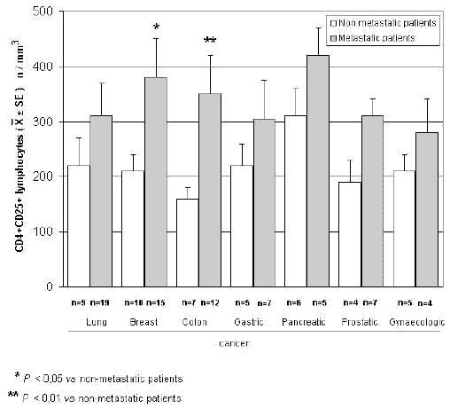
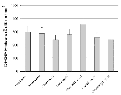
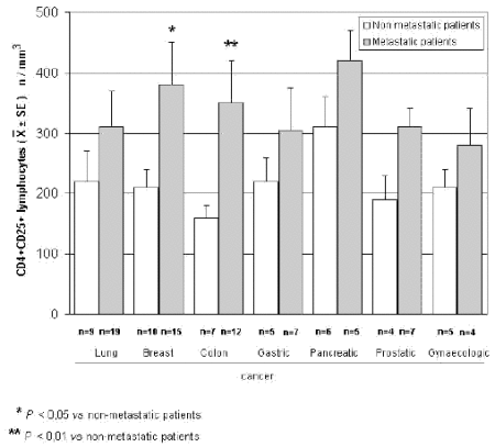
The mean counts of NK and CD8 cells in relation to that of T-reg are reported in Table 4. As shown, no significant difference in the mean number of CD8 lymphocytes was found between patients with normal or abnormally elevated number of T-reg. On the contrary, patients with elevated number of T-reg showed a significantly lower number of NK cells with respect to that found in those with normal T-reg count. Finally, Table 5 shows the circadian rhythm of cortisol in relation to total lymphocytes, CD4+ cells and T-reg mean number. A normal cortisol rhythm, with morning values greater at least than 50% with respect to the values occurring during the afternoon, was found in 85/114 (75%). Total lymphocyte and CD4+ cell mean numbers observed in patients with altered cortisol rhythm were significantly lower than those found in patients with normal cortisol circadianicity (p<0.01), whereas no significant difference was seen in the mean number of T reg. Figure 1 and Figure 2 illustrate T-reg mean numbers in relation to tumor histoptypes in the overall patients and with respect to their disease extension, respectively. No significant difference was seen in relation to tumor histotype. The highest values of T-reg were observed in pancreatic cancer patients, without however significant differences with respect to the overall other histotypes. The metastatic disease was associated with a higher number of T-reg with respect to the non-metastatic group in all tumor histotypes, even though a statistically significant differences occurred for the only breast cancer (p<0.05) and colorectal cancer (p< 0.01).

Table 4. Mean values of NK cells and CD8+ lymphocytes in cancer patients with normal or abnormally high values of CD4+CD25+ lymphocytes

090722_clinicalstud4

* p<0.05 vs normal values of CD4+CD25+ lymphocytes

Table 5. Mean numbers of total lymphocytes, T helper (CD4+) lymphocytes and T regulator lymphocytes (CD4+CD25+) in relation to cortisol circadian secretion in cancer patients
090722_clinicalstud5

* P<0.01 vs patients with altered cortisol rhythm
090722_clinicalstudfig1

Figure 1. CD4+CD25+ lymphocyte mean number in relation to tumor histotype
090722_clinicalstudfig2

Figure 2. CD4+CD25+ lymphocytes in relation to tumor histotype in metastatic and non-metastatic cancer patients

IV. Discussion
According to previous preliminary clinical investigations (Sasada et al, 2003; Cesana et al, 2006), this study confirms in a greater number of cancer patients that the metastatic disease is characterized by the evidence of an abnormally increased percentage of T-reg lymphocytes with respect to both total circulating lymphocytes and CD4+ lymphocytes. This finding does not seem to represent a specific characteristic of some tumor histotypes, then it could constitute a general alteration occurring during the progression of the neoplastic disease, by representing a fundamental immune parameter of cancer-related immunosuppression.

Several immune molecules have appeared to suppress the anticancer immunity, namely IL-6, IL-10, IL-1, TNF-α and TGF-β, but it seems that the common end result of their mechanisms of action may be represented by the stimulation of T-reg generation, with a consequent inhibition of the activation of an effective anticancer immune reaction. On the same way, several immune cells are able to suppress the anticancer immunity, including macrophages, T helper-2 lymphocytes and some myeloidderived suppressor cells, but also in this case they would act in a suppressive way by promoting the generation of Treg.

Then, the detection of T-reg amounts in terms of both absolute number and percentages with respect to total lymphocytes and CD4+ cells could constitute a simple and adequate clinical immune parameter to quantify the whole status of the anticancer immunity in the single cancer patient. Moreover, future clinical studies will be required to establish the possible prognostic significance of changes in T-reg percentage and number in relation to the anticancer efficacy of the various standard antitumor therapies. Moreover, it has to be remarked that T-reg lymphocytes would not represent the only immune cells involved in the suppression of the anticancer immunity. In fact, there is at least another fundamental immunosuppressive system, consisting of the monocytemacrophage cell lineage (Sica and Bronte, 2007). In more detail, it has been observed that the bone marrow may release myeloid precursors provided by suppressive activity on the antitumor immune response and defined as myeloid-derived suppressor cells (MDSC) (Kusmartsev and Gabrilovich, 2005). These cells have appeared to be characterized by the cell surface expression of GR-1, CD11b and CD80 antigens (Anderson et al, 2002; van Ginderachter et al, 2006). The myeloid suppressor cells would promote the generation and activation of T-reg lymphocytes, which at the other side would stimulate MDSC release from the bone marrow and M2 macrophage differentiation (Terabe et al, 2003; Wie et al, 2006).

Moreover, the myeloid suppressive cells would inhibit the anticancer immunity by promoting macrophage differentiation into the M2 sub-type (Ikemoto et al, 2003), which plays a clear inhibitory effect on the anticancer immunity, namely through the release of IL-6 (Ueno et al, 2000), whereas the M1 macrophage sub-type may either stimulate or suppress the antitumor immunity (Mantovani et al, 2004). M1 and M2 macrophage sub-types have appeared to be characterized by a high production of IL-12 or IL-10, respectively (Ueno et al, 2000).

Then, further studies by concomitantly evaluating T reg and MDSC count, will contribute to better define the immune mechanism responsible for the suppression of the anticancer immunity.

References
Anderson CF, Gerber JS, Mosser DM (2002) Modulating macrophage function with IgG immune complexes. J Endotoxin Res 8, 477-81.

Annunziato F, Cosmi L, Liotta F, Lazzeri E, Manetti R, Vanini V, Romagnani P, Maggi E, Romagnani S (2002) Phenotype,localization and mechanism of suppression of CD4+CD25+ human thymocytes. J Exp Med 196, 379-87.

Antony PA, Restito NP (2005) CD4+CD25+ T regulatory cells,immunotherapy of cancer,and interleukin-2. J Immunother 28, 120-8.

Birebent B, Lorho R, Lechartier H, de Guibert S, Alizadeh M,
Vu N, Beauplet A, Robillard N, Semana G (2004) Suppressive properties of human CD4+CD25+regulatory T cells are dependent on CTLA-4 expression. Eur J Immunol 34, 3485-96.

Cesana GC, DeRaffele G, Cohen S, Moroziewicz D, Mitcham J, Stoutenburg J, Cheung K, Hesdorffer C, Kim-Schulze S, Kaufman HL (2006) Characterization of CD4+CD25+ regulatory T cells in patients treated with high-dose interkleukin-2 for metastatic melanoma or renal cell carcinoma. J Clin Oncol 24, 1169-77.

Chen W, Jin W, Hardegen N, Lei KJ, Li L, Marinos N, McGrady G, Wahl SM (2003) Conversion of peripheral CD4+CD25- naïve T cells to CD4+CD25+ regulatory T cells by TGF-β induction of transcription factor Foxp 3. J Exp Med 198, 1875-86.

Claman HN (1998) Corticosteroids and the immune system. Adv Exp Med Biol 245, 203-10.

Dieckmann D, Bruett H, Ploettner H, Lutz MB, Schuler G (2002) Human CD4+CD25+ regulatory contact-dependent T cell induce IL-10 producing,contact-independent type-1-regulatory T cells. J Exp Med 196, 247-53.

Dieckmann D, Plottner H (2001) Ex vivo isolation and characterizationof CD4+CD25+ T cells with regulatory properties from human blood. J Exp Med 193, 1303-10.

Ehrke MJ, Mihich E, Berd D, Mastrangelo MJ (1989) Effects of anticancer drugs on the immune system. Semin Oncol 16, 230-9.

Ghiringhelli F, Larmonier N, Schmitt E, Parcellier A, Cathelin D, Garrido C, Chauffert B, Solary E, Bonnotte B, Martin F (2004) CD4+ CD25+ regulatory T cells suppress tumor immunity but are sensitive to cyclophosphamide which allows Immunotherapy of established tumors to be curative. Eur J Immunol 34, 336-44.

Ikemoto S, Yoshida N, Narita K, Wada S, Kishimoto T, Sugimura K, Nakatani T (2003) Role of tumor-associated macrophages in renal cell carcinoma. Oncol Rep 10, 1843-9.

Jago CB, Yates J, Camara NOS, Lechler RI, Lombardi AG (2004) Differential expression of CTLA-4 among T cell subsets. Clin Exp Immunol 136, 463-71.

Ji HB, Liao G, Faubion WA, Abadía-Molina AC, Cozzo C, Laroux FS, Caton A, Terhorst C (2004) Cutting edge, the natural ligand for glucocorticoid-induced TNF receptorrelated protein abrogates regulatory T cell suppression. J Immunol 172, 5823-7.

Kanamaru F, Youngnak P, Hashiguchi M, Nishioka T, Takahashi T, Sakaguchi S, Ishikawa I, Azuma M (2004) Costimulation via glucocorticoid-induced TNF receptor in both conventional and CD25+ regulatory CD4+ T cells. Immunol 172, 7306-14.

Knutson KL, Disis M, Salazar L (2007) CD4 regulatory T cells in human cancer pathogenesis. Cancer Immunol Immunother 556, 271-85.

Kusmartsev S, Gabrilovich DI (2005) STAT1 signaling regulates tumor-assolciated macrophage-mediate T cell deletion. J Immunol 174, 4880-91.

Lan RY, Ansari AA, Lian ZX, Gershwin ME (2005) Regulatory T cells, development,function,and role in autoimmunity. Autoimmun Rev 4, 351-63.

Leong PP, Mohammad R, Ibrahim N, Ithnin H, Abdullah M, Davis WC, Seow HF (2006) Phenotyping of lymphocytes expressing regulatory and effector markers in infiltrating ductal carcinoma of the breast. Immunol Lett 102, 229-36.

Malek TR, Bayer AL (2004) Tolerance not immunity crucially depends on IL-2. Nat Rev Immunol 4, 665-74.

Mantovani A, Sica A, Sozzani S, Allavena P, Vecchi A, Locati M (2004) The chemokine system in diverse forms of macrophage activation and polarization. Trends Immunol 25, 677-86.

Sasada T, Kimura M, Yoshida Y, Kanai M, Takabayashi A (2003) CD4+CD25+ regulatory T cells in patients with gastrointestinal malignancies, possible involvement of regulatory T cells in disease progression. Cancer 98, 1089-93.

Schwartz RH (2005) Natural regulatory T cells and selftolerance. Nat Immunol 6, 327-30.

Shevach EM (2002) CD4+CD25+ suppressor T cells, more questions than answers. Nat Rev Immunol 2, 389-400.

Sica A, Bronte V (2007) Altered macrophage differentiation and immune dysfunction in tumor development. J Clin Invest 117, 1155-66.

Siddiqui SA, Frigola X, Bonne-Annee S, et al (2007) Tumorinfiltrating Foxp3 CD4+CD25+ T cells predict poor survuival in renal cell carcinoma. Clin Cancer Res 13, 2075-81.

Stephens GL, McHugh RS, Whitters MJ, Young DA, Luxenberg D, Carreno BM, Collins M, Shevach EM (2004) Engagement of glucocorticoid-induced TNFR family-related receptor on effector T cells by its ligand mediates resistance to suppression by CD4+CD25+ T cells. J Immunol 173, 5008-20.

Takahashi T, Tagami T, Yamazaki S, Uede T, Shimizu J, Sakaguchi N, Mak TW, Sakaguchi S (2000) Immunologic and tolerance maintained by CD25+CD4+ regulatory T cells constitutively expressing cytotoxic T lymphocyte-associated antigen. J Exp Med 192, 1285-94.

Terabe M, Matsui S, Park JM, Mamura M, Noben-Trauth N, Donaldson DD, Chen W, Wahl SM, Ledbetter S, Pratt B, Letterio JJ, Paul WE, Berzofsky JA (2003) Transforming growth factor-β production and myeloid cells are an eggector mechanism trhough which CD1d-restricted T cells block cytotoxic T lymphocyte-mediated tumor immunosurveillance, abrogation prevents tumor recurrence. J Exp Med 198, 1741-52.

Thomton AM, Shevach EM (2000) Suppressor effector function of CD4+CD25+ immunoregulatory T cells is antigen nonspecific. J Immunol 164, 183-90.

Ueno T, Toi M, Saji H, Muta M, Bando H, Kuroi K, Koike M, Inadera H, Matsushima K (2000) Significance of macrophage chemo-attractant protein-1 in macrophage recruitment,angiogenesis and survival in human breast cancer. Clin Cancer Res 6, 3282-9.

Van Ginderachter JA, Movahedi K, Hassanzadeh Ghassabeh G, Meerschaut S, Beschin A, Raes G, De Baetselier P (2006) Classical and alternative activation of mononuclear phagocytes, picking the best of both worlds for tumor promotion. Immunobiology 211, 487-501.

Vasu C, Prabhakar BS, Holterman MJ (2004) Targeted CTLA-4 engagement induces CD4+CD25+CTLA-4 high T regulatory cells with target alloantigens specificity. J Immunol 173, 2866-76.

von Boehmer H (2005) Mechanisms of suppression by suppressor T cells. Nat Immunol 6, 338-44.

von Herrath MG, Harrison LC (2003) Regulatory lymphocytes, antigen-induced regulatory T cells in autoimmunity. Nat Rev Immunol 3, 223-32.

Wang XB, Zheng CY, Giscombe R, Lefvert AK (2001) Regulation of surface and intracellularexpression of CTLA-4 on human peripheral T cells. Scand J Immunol 54, 453-8.

Wie S, Kryczeck I, Zou W (2006) Regulatory T-cell compartmentalization and trafficking. Blood 108, 426-31.

Yang R, Cai Z, Zhang Y, Yutzy WH 4th, Roby KF, Roden RB (2006) CD80 in immune suppression by mouse ovarian carcinoma-associated Gr-1+CD11b+ myeloid cells. Cancer Res 66, 6807-15.

Ziegler SF (2006) FOXP3 of mice and men. Annu Rev Immunol 24, 209-26.

Zou W (2006) Regulatory T cells, tumour immunity and immunotherapy. Nat Rev Immunol 6, 295-307.

(download PDF version)



A phase II study of anastrozole plus the pineal anticancer hormone melatonin in the metastatic breast cancer women with poor clinical status

lissoniResearch Article (download PDF version)

Paolo Lissoni1*, Giuseppe Di Fede1, Antonio Battista2, Giusy Messina1, Remo Egardi1, Fernando Brivio3, Franco Rovelli1, Massimo Colciago4, Giuseppe Brera5

1 Institute of Biological Medicine, Milan
2 Azienda Sanitaria locale 2, Avellino;
3 Surgery Division, Bassini Hospital, Cinisello,Milan
4 I.N.R.C.A, Casatenovo, Lecco, Italy
5 Ambrosian University, Milan, Italy

dr. Paolo Lissoni

*Correspondence: Dr. Paolo Lissoni, Divisione di Radioterapia Oncologica, Ospedale S.Gerardo, 20052 Monza, Milano, Italy; Fax: +390392332284, E-mail: p.lissoni@hsgerardo.org
Key words: Anastrozole, breast cancer, melatonin, pineal gland
Abbreviations: melatonin, MLT; estrogen receptor, ER;

Received: 9 March 2009; Revised 1 April 2009;
Accepted: 13 April 2009; electronically published: 28 May 2009

Summary

The recent advances in the psychoneuroendocrinology have suggested the possibility to modulate tumor hormone dependency through a neuroendocrine approach. In particular, it has been proven that the pineal neurohormone melatonin (MLT) may stimulate estrogen receptor (ER) expression in breast cancer cells and inhibit the aromatase activity. On this basis, a study was planned to evacuate the efficacy of a concomitant treatment with the aromatase inhibitor anastrozole plus MLT in metastatic breast cancer. The study included 14 metastatic breast cancer women of poor clinical conditions with ER positive or unknown. Both anastrozole and MLT were given orally at a dose of 1 mg at noon and of 20 mg in the evening, respectively. The clinical response consisted of complete response in 2 and partial response in 6 patients. Then, an objective tumor regression was achieved in 8/14 (57%) patients, with a median duration of 26 months. No neoplastic cachexia occurred on treatment. This preliminary study shows that a neuroendocrine strategy with anastrozole plus the pineal hormone MLT may represent a new effective and well tolerated regimen in the treatment of metastatic breast cancer women, including those with poor clinical status, with therapeutic results apparently superior to those reported in the literature with the only aromatase inhibitor. Then, these results would justify further randomized studies of aromatase inhibitors with or without a concomitant administration of MLT, in an attempt to establish whether the pineal hormone may enhance the efficacy of the aromatase inibibitors in the treatment of human advanced breast cancer.

I. Introduction
Recent experimental studies have demonstrated that the hormone dependency is at least in part under a psychoneuroendocrine regulation (Cos et al, 2008;Grant et al, 2009). In particular, it has been shown that the pineal hormone melatonin (MLT), whose anticancer properties have been well demonstrated (Bartsch et al, 1981; Maestroni, 1993; Reiter et al, 2002), may in vitro stimulate estrogen receptor (ER) expression on breast cancer cell lines (Molis et al, 1995). Therefore, the hormone dependency of breast cancer cells would not depend only on intrinsic characteristics of cancer cells themselves, but also on host neuroendocrine regulation of tumor cell proliferation and differentiation (Bartsch et al, 2000). Moreover, cancer progression has been proven to be associated with pineal alterations, consisting of a progressive decline in MLT nocturnal production. (Maestroni, 1993). Therefore the advanced cancer would require a substitutive endocrine therapy with MLT (Bartsch et al, 1981; Maestroni, 1993). Previous preliminary clinical studies had already suggested that the concomitant administration of the pineal hormone MLT may apparently increase the efficacy of tamoxifen therapy in the treatment of metastatic breast cancer (Lissoni et al, 1995). Moreover, experimental studies have shown that the activity of aromatase enzyme, which is responsible for the peripheral production of estrogens from testosterone (Bagatell et al, 1994), is under a light/dark circadian rhythm (Bhatnagar et al, 1992). Because of the fundamental role of the pineal hormone MLT in the regulation of the daily photoperiod (Bartsch et al, 1981), it is possible to hypothesize that MLT may be involved in the control of the aromatase activity. In fact, recent studies have demonstrated an inhibitory action of MLT on the aromatase activity (Cos et al, 2005). This finding could reserve a prosiming application in the treatment of both early and advanced breast cancer. This statement is justified by the fact that the aromatase inhibitors represent a new class of agents in the endocrine treatment of breast cancer Plourde et al, 1994), with a potential efficacy superior to that achieved by the previous hormonal therapies with anti-estrogens, such as tamoxifene, even though tumor response rate obtained by the aromatase inhibitors are generally not greater than 40%. On this basis, a phase II study was planned in an attempt to evaluate the efficacy of a neuroendocrinotherapeutic regimen consisting of a concomitant administration of the aromatase inhibitor anastrozole and the pineal hormone MLT in metastatic breast cancer women with poor clinical conditions.

II. Materials and methods
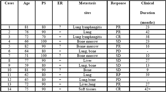
The study included 14 consecutive metastatic breast cancer women (median age: 72 years, range 51-82), who were followed at Biological Medicine Institute in Milan, or at Health Local Unit 2 of Avellino, from Feb. 2002 to Sept. 2003. Eligibility criteria were, as follows: histologically proven metastatic breast cancer, measurable lesions, ER positive or unknown, no ability to tolerate chemotherapy because of age, low performance status (PS), important clinical illnesses other than cancer and/or heavy chemotherapeutic pre-treatments, no previous endocrine therapies for the metastatic disease, no double tumor and life expectancy less than 1 year. Previous heavy chemotherapeutic treatment consisting of at least 3 chemotherapeutic lines was made in 11/14 (79 %) patients. Dominant metastasis sites were, as follows: soft tissues:1; bone:1; lung:7 (neoplastic lymphangitis:2); liver:1; lung + liver:1; bone marrow:3. Time-span since first diagnosis of the primary tumor was 44 months (31-66 months). All patients had an acceptable social conditions. The minimum and median follow-up periods were 60 months and 72 months respectively. In all patients, in the case of disease progression, at least to other endocrine therapeutic lines with other aromatase-inhibitors were planned. The experimental protocol, wich was approved by the Health Direction of Biological Medicine Institute of Milan, was explained to each patient and informed consent was obtained. The treatment consisted of anastrozole at a dose of 1 mg/day orally at noon, plus MLT at 20 mg/day orally in the evening, generally half-hour before sleeping, to correct cancer progression-related decline in MLT night secretion (10). Patients were considered to be evaluable when they were treated for at least 3 consecutive months. The clinical response was evaluated according to WHO criteria. Complete response (CR) was the complete disappearance of all neoplastic lesions for at least 1 month. Partial response (PR) was a reduction greater than 50 % of the sum of all neoplastic lesions, for at least 1 month. Stable disesase (SD) was no increase or decrease greater tha 25 % of tumor volume. Progressive Disease (PD) was an increase in tumor volume greater than 25 % or the appearance of new neoplastic lesions. PS was assessed according to Karnofsky’s score, consisting of the evaluation of the quality of life in relation to patient activity and bed-rest period. ER was positive in 10 and unknown in the remaining 4 patients. The median PS was 80% (range 70-100). Data were statistically evaluated by the chi-square test and the Student’s t test, as appropriate.

090722_phase2study

Table 1: Clinical characteristics of metastatic breast cancer women and their clinical response (WHO criteria) to a neuroendocrine regimen consisting of anastrozole plus the pineal hormone melatonin.

III. Results
All patients were fully evaluable for the clinical response. The clinical characteristics of patients and their individual clinical response to the treatment are reported in Table 1. As reported, a complete response (CR) was achieved in 2/14 (14%) (soft tissues:1; lung lymphangitis:1). A partial response (PR) was obtained in other 6/14 (43%) (bone:1; lung:3; liver:1; bone marrow:1). Then, an objective tumor response (CR + PR) was reached in 8/14 (57%) patients. The median duration of response was 26 months (range 9-42 months). A stable disease (SD) was seen in other 4/14 (29%), with a median duration of 25 months (range 10-27). Therefore, a disease-control (DC:CR + PR + SD) was achieved in 12/14 (86%) patients, whereas the remaining 2/14 (14%) patients had a progressive disease (PD). No significant difference in tumor response rate was observed between patients with positive or unknown ER ( 6/10(60%) vs 2/4(50%) ). An overall survival at 1 year and at 3 year was achieved in 11/14 (79 %) and in 5/14 (36 %) patients, respectively. Moreover, 3/14 (21%) patients were still alive at 5 years. The treatment was well tolerated in all patients.

Moreover, most patients experienced a relief of asthenia under the treatment and in no patient the neoplastic cachexia occurred. Finally, an evident increase in PS mean values was achieved under treatment, even though it did not reach the statistical significance (86 ±5 vs 93 ± 4, mean ± SE).

IV. Discussion
The results of this preliminary phase II study, by showing a percentage of 1-year survival greater than 70% in patients with live expectancy less than 1 year, would suggest that a neuroendocrine regimen consisting of the aromatase inhibitor anastrozole plus the pineal neurohormone MLT may represent a new effective therapeutic strategy in the treatment of metastatic breast cancer women, also in patients with poor clinical conditions, who would not be able to tolerate the most aggressive therapies. The concomitant administration of the pineal hormone would seem to enhance the efficacy of the aromatase inhibitor in terms of objective tumor regressions with respect to the results commonly reported in the literature with the only aromatase inhibitor (Plourde et al, 1994), which are generally lower than 40%.

The time to progression would seem to be apparently increased by the concomitant treatment with MLT.This finding is not surprising, since MLT could enhance the therapeutic anticancer acitivity of the aromatase inhibitors by either exerting direct antiproliferative antitumor effects (Bartsch et al, 1981; Maestroni, 1993; Reiter et al, 2002), or further inhibiting the aromatase activity by acting on gene and oncogene expression (Molis et al,1995; Cos et al, 2005). In addiction, MLT appeared to stimulate ER expression of breast cancer lines, by transforming ER negative into ER positive breast cancer, as observed in experimental conditions (Danforth et al, 1983).

Since the prognosis of ER positive breast cancers is clearly better than that of ER negative ones, MLT could per se improve the clinical couse of mammary tumors. Finally, because of its interesting therapeutic efficacy as a supportive care (Reiter et al, 2002), MLT would be responsible for the evident improvement in the relief of asthenia and in preventing the occurrence of the neoplastic cachexia. On the other hand, because of the inhibitory effect of MLT (Grant et al, 2009; Reiter et al, 2002) on cancer cell proliferation, the anticancer activity of this polyendocrine regimen would be due not only to an indirect effect, depending on a diminished estrogen production following aromatase enzyme inhibition, but also on a direct inhibition of cancer cell growth, due to MLT itself. Therefore, the results of this preliminary study may justify further clinical randomized investigations with the only aromatase inhibitor versus the concomitant treatment with MLT, in an attempt to confirm the ability of the pineal hormone to enhance the antitumor properties of the aromatase inhibitors in the treatment of metastatic breast cancer women with poor clinical conditions.

References
Bagatell CJ, Dahl KD, Bremner WJ (1994). The direct pituitary effect of testosterone to inhibit gonadotrophin secretion in men is partially mediated by aromatization to estradiol. J Androl 15:15-21.

Bartsch C, Bartsch H (1981) effect of melatonin on experimental tumors under different photoperiods and times of administration. J Neural Transm 52:269-279.

Bartsch H, Buchberger A, Franz H,Bartsch C,Maidonis I,Mecke D, Bayer E (2000) Effect of melatonin and pineal extracts on human ovarian and mammary tumor cells in a chemosensitivity assay. Life Sci 67:2953-2960.

Bhatnagar AS, Muller P, Schenkel L, Trunet PF, Beh I, Schieweck K. (1992). Inhibition of oestrogen biosynthesis and its consequences on gonadotrophin secretion in the male. J of Steroid Biochemistry and Molecular Biology;41:1021-1027

Cos S, Martinez-Campa C,Mediavilla MD, Sanchez-Barcelo E (2005). Melatonin modulates aromatase activity in MCF-7 human breast cancer cells. J Pineal Res 7:136-142.

Cos S, Gonzales A, Martinez-Campa C, Mediavilla MD, Alonzo-Gonzales C, Sanchez-Barcelo EJ (2008): Melatonin as a selective estrogen enzyme modulator. Curr Cancer Drug Targets. Dec 8(8): 691-702.

Danforth DN, Tamarkin L, Lipmann LE: (1983) Melatonin increase oestrogen receptor binding activity of human breast cancer cells. Nature, 305:323-325.

Grant S.G, Melan MA, Latimer JJ, Witt-Enderby PA (2009) : Melatonin and Breast cancer : cellular mechanism, clinical studies and future perspective. Expert Rev Mol Med 11:e5

Lissoni P, Barni S, Meregalli S, Fossati V, Cazzaniga M, Esposti D, Tancini G (1995) Modulation of cancer endocrine therapy by melatonin:a phase II study of tamoxifen plus melatonin in metastatic breast cancer patients progressing under tamoxifen alone. Br J Cancer 71:854-856.

Maestroni GJM (1993) The immunoneuroendocrine role of melatonin. J Pineal Res 14:1-10.

Molis TM, Spriggs LL,Jupiter Y,Hill SM (1995) Melatonin modulation of estrogen-regulated proteins, growth factors,and protooncogenes in human breast cancer. J Pineal Res 18:93-103.

Plourde PV, Dyroff M, Dukes MD (1994) Arimidex: a potent and selective fourth generation aromatase inhibitor. Breast Cancer Res Treat 30:103-111.

Reiter RJ, Tan DX, Sainz RM, Mayo JC, Lopez-Burillo S. (2002) Melatonin:reducing the toxicity and increasing the efficacy of drugs. Pharm Pharmacol 54:1299-1321

(download PDF version)


Maggio 30, 2012 Newsletter

Nonostante quella che può essere un’opinione comune, capita molto spesso che l’analisi bioimpedenziometrica rilevi particolari stati di malnutrizione e disidratazione in soggetti apparentemente sani a prescindere da età, sesso e classe sociale.

Risulta pertanto fondamentale questo esame per poter procedere nella definizione di un programma alimentare e di allenamento sulla base di un’attenta interpretazione dei dati. E’ inutile per esempio proporre un allenamento volto al condizionamento muscolare (la cosiddetta tonificazione) quando non vengono introdotti sufficienti “mattoni” per sostenere l’anabolismo cellulare.

Si è notato inoltre che in casi di insufficiente apporto proteico si ha un aumento dei fluidi extracellulari che può essere erroneamente scambiato per ritenzione idrica. Questo aumento invece è dato dall’acqua che residua dalla distruzione dei tessuti muscolari non supportati da un adeguata alimentazione e pertanto non va trattato con dei comuni diuretici bensì con una regolazione degli apporti proteici quotidiani e del carico di lavoro fisico nelle attività di tutti i giorni.
E molto comune infatti osservare in soggetti sovrallenati e sottoalimentati la classica plica edematosa, ossia un gonfiore liquido sottocutaneo riconoscibile anche al tatto per la scarsa consistenza del muscolo sottostante e per la lentezza con la quale i tessuti tornano normali dopo una pressione (delle dita o delle parti elastiche degli abiti).

Per rilevare uno stato di malnutrizione (PEM = protein-energy malnutrition) e definire la massa cellulare minima teorica basterà applicare il seguente calcolo:

Massa Cellulare Minima=(Altezza in cm – 100) x 0,3.

Se il dato acquisito è superiore alla massa cellulare (BCM) rilevata dal bioimpedenziometro, allora abbiamo un caso di malnutrizione alla quale sarà probabilmente associata un elevata quantità di acqua extracellulare.

Dovrebbe bastare, ove non ci siano particolari ragioni di natura patologica, introdurre 0,8-1 gr.di proteine per kg di peso corporeo, secondo le tabelle più comuni, fino a 1,8-2,2 per coloro che sono coinvolti in attività lavorative o sportive di una certa intensità e regolarità.

Personalmente preferisco lavorare considerando la parte metabolicamente attiva, quindi escludendo la massa grassa (FM) e calcolando fino a 1,5-2 gr per kg di massa magra (quindi Massa Cellulare, BCM + Massa Extracellulare, ECM) a seconda dell’attività quotidiana.

La qualità delle proteine da introdurre sarà argomento dei prossimi articoli

di Aaron Ciancio – Certified Fitness Trainer (CFT1) International Sport & Science Association


Luglio 30, 2011 Newsletter

Recentemente, un team di esperti gastroenterologia riunitisi nella “First International Consensus Conference on Gluten Sensitivity“, tenutasi a Londra l’11 e 12 febbraio 2011, ha stabilito l’esistenza della sensibilità al glutine, l’intolleranza alle proteine presenti nel glutine, in assenza di malattia celiaca.

L’esistenza di una condizione d’intolleranza al glutine è stata sospettata da diverso tempo. I medici che collaborano con l’Istituto di Medicina Biologica di Milano – IMBIO, conoscono questa entità clinica, che fino ad ora non aveva ricevuto una vera e propria collocazione clinica.

Nella pratica clinica, da più di dieci anni, abbiamo osservato casi di reazione al glutine in cui non sono coinvolti né meccanismi auto immuni come nella celiachia né di tipo allergico, mediato dalle IgE. Abbiamo notato che, dopo una dieta priva o a ridotto contenuto di glutine, i pazienti riscontravano un miglioramento di tutti i sintomi e i disturbi. Di recente, quindi, la nuova correlazione tra glutine e disturbi a essi correlati è definita “Gluten Sensitivity” (GS). L’interesse verso questa condizione è in continuo aumento come testimoniato dalla “First International consensus conference on gluten sensitività” tenutasi a Londra nel febbraio del 2011.

Gli studi condotti finora, hanno evidenziato come nella GS non esistano dei parametri immunologici e biochimici ben precisi, ma si tratti di una condizione alla quale si arriva per esclusione. In pratica, una volta esclusa la malattia celiaca attraverso le indagini biochimiche, gastroscopia con biopsia, si arriva a definire la GS, dove sembra sia coinvolto principalmente un meccanismo immunitario innato. Il sistema immunitario innato è rappresentato da cellule sentinella, i Granulociti Neutrofili (GN), particolarmente attivi verso alcune sostanze di origine alimentare.

Uno dei metodi che utilizziamo da diversi anni per scoprire eventuali intolleranze agli alimenti è il test ALCAT, un test citotossico computerizzato, che si effettua su sangue venoso.

Chi soffre di sensibilità al glutine presenta i seguenti sintomi: disturbi d’intestino irritabile (diarrea, dolore addominale, gonfiore, stipsi) cui si associa a volte stanchezza cronica, cefalea, eczema, sonnolenza, difficoltà di concentrazione, formicolii agli arti, depressione, alterazioni dell’umore, ipereccitabilità e ipercinesi (presente specialmente nei bambini), anemia ferropriva. Tali condizioni cliniche migliorano e regrediscono seguendo una dieta a rotazione priva di glutine o a ridotto introito di prodotti contenenti glutine.
Considerando che la sintomatologia della sensibilità al glutine è sovrapponibile alla celiachia, il paziente dovrà sottoporsi ai test specifici per escludere tale patologia.

Uno degli esami richiesti per la ricerca della “Gluten Sensitivity” è l’esame genetico. La GS presenta una positività per HLA-DQ2 e/o DQ8 in più nel 50% dei casi, nella celiachia la percentuale arriva al 99% dei celiaci, e nel 30% della popolazione normale.

Il trattamento terapeutico per chi è sensibile al glutine dipende dal momento in cui si esegue la diagnosi e dall’età del paziente. Oltre alla dieta, si pone molta importanza allo studio e al ripristino della funzionalità intestinale e dell’integrità delle pareti (mucosa) intestinali, primo approccio operativo volto al miglioramento dei sintomi clinici.

Un ciclo di sedute settimanali di pulizia intestinale (Idrocolonterapia) sotto la supervisione del medico, abbinato a un trattamento mirato a base di probiotici e prebiotici, prescritto in seguito al disbiosi test, esame che rivela le condizioni di eubiosi o disbiosi batterica, costituisce senz’altro un valido aiuto per la ripresa di uno stato di funzionalità intestinale ottimale.


Settembre 30, 2010 Newsletter

È opinione comune ritenere che la cellulite sia eminentemente un inestetismo (peraltro molto diffuso, visto che fa soffrire circa l’80 per cento delle donne), quando in verità si tratta di una vera e propria patologia, nella fattispecie del tessuto adiposo, definita pannicolopatia edemato-fibrosclerotica.

Come ben sa chi mal la sopporta, essa si concentra prevalentemente all’esterno delle cosce (nella zona laterale del bacino), all’interno delle stesse e sui glutei, interessando il “pannicolo adiposo” che si trova fra il derma (lo strato di tessuto connettivo giusto sotto l’epidermide) e la parte muscolare. Il pannicolo ha una sua impalcatura di sostegno (il tessuto reticolare e il collagene) e una vascolarizzazione (denominata microcircolo) attraverso la quale il tessuto adiposo fornisce l’energia all’organismo o l’accumula sotto forma di grasso.

Per motivi diversi – stress, stipsi, fattori ereditari, fumo, sedentarietà, abuso di medicinali (pillola anticoncezionale), cause ormonali (per esempio un cattivo funzionamento della ghiandola tiroidea, o ipotiroidismo), disturbi del ciclo mestruale (come si verificano nella Sindrome dell’ovaio micropolicistico), ma soprattutto cattiva alimentazione –, la circolazione venosa e linfatica (vasi che trasportano scorie) può rallentare e dai capillari possono uscire sostanze che invece dovrebbero essere eliminate. Parte così il meccanismo iniziale di accumulo di liquidi (edema), che s’infiltra fra le cellule del pannicolo adiposo. Queste si allargano, peggiorando lo stato edematoso e nel contempo il tessuto di sostegno (collagene e fibre reticolari) reagisce cercando d’impedire che le cellule adipose si allontanino, ma causando così la produzione di capsule che impediscono a loro volta gli scambi ematici fra la “cellulite” e il resto dell’organismo.

I processi esposti possono essere distinti in 4 fasi che evolvono per grado di gravità. I primi due stadi possono essere rallentati o addirittura bloccati con l’attività fisica (e la corsa in particolare); nel terzo e quarto la consistente struttura fibrosa dei noduli rende tutto molto difficile. La corsa, quindi, è certamente da proporre come prevenzione e cura per i primi 2-3 stadi, insieme ad alcune semplice regole di alimentazione e qualche stratagemma nutri-cosmeceutico per sconfiggere o tenere sotto controllo la cellulite.


Gli stadi della cellulite
La cellulite viene suddivisa in quattro stadi, o gradi, in base alla gravità dei sintomi
con i quali si manifesta e alla loro difficoltà di trattamento.

1 Cellulite edematosa È caratterizzata dalla presenza di gonfiore, ma la cute è ancora tesa  ed elastica e, se compressa, non resta segno né si avverte dolore.

2 Cellulite fibrosa La pelle, se compressa, ancora non duole ma, seppur ancora liscia, comincia ad assumere un colorito spento e si presenta “a buccia d’arancia” se viene stretta fra le dita. C’è formazione di tessuto fibrotico e la cattiva circolazione sanguigna può causare formicolii, pesantezza alle gambe, crampi.

3-4 Cellulite sclerotica Il tessuto connettivo s’indurisce ulteriormente e si formano i primi macronoduli che conferiscono alla pelle l’aspetto a buccia d’arancia “a vista”. Nel quarto stadio le zone cellulitiche si presentano isolate dal tessuto circostante. I macronoduli danno origine ad avvallamenti e protuberanze; la cute è dura e dolorante alla palpazione.
È la più difficile da trattare.


CORRI (FORTE) CHE TI PASSA
In verità il running non è, e non può essere, del tutto risolutivo. Un programma di attività deve prevedere almeno 3-4 sedute di allenamento divise in 3 sedute di corsa e una di tonificazione generale, ma soprattutto delle zone interessate alla cellulite. La corsa dev’essere affrontata secondo tabelle che prevedano la possibilità di migliorare le proprie caratteristiche fisico-organiche progressivamente e secondo le proprie caratteristiche e predisposizioni.

Vanno sfatati alcuni miti: camminare invece di correre (vale solo nei casi di situazioni gravi, dove il carico sulle articolazioni è importante), oppure allenarsi sotto la soglia lattacida: si dimentica che l’acido lattico è un potentissimo vasodilatatore e stimola la produzione di mitocondri nella supercompensazione fisiologica. A torto si potrebbe pensare che la vasodilatazione da acido lattico potrebbe peggiorare la perdita di materiale vasale, perché nella prevenzione – e anche negli stadi 1 e 2 della cellulite – l’azione tonico-trofica della corsa sul circolo ematico è di gran lunga maggiore.
Una limitazione nella produzione di acido lattico potrebbe valere nei primi momenti d’allenamento per soggetti con stadio 3 di cellulite.

Quindi: progressione nell’allenamento e, nelle uscite, lasciare almeno un allenamento con corsa variata. L’allenamento di tonificazione dovrà interessare in maggior misura le parti interessate dalla cellulite, quindi con esercizi di adduzione (per i muscoli interni della coscia: 10 minuti) e abduzione (per glutei e laterali della coscia: 10 minuti), per il bicipite il femorale (posteriore della coscia: 10 minuti), per il quadricipite (10 minuti) e alla pressa (15 minuti). Meglio sarebbe utilizzare il circuit training, intervallando gli esercizi a con 10 minuti di cyclette, corsa leggera, step.

100924_artcellulite

LA COSMETICA CHE AIUTA
Utilizzo d’integratori alimentari e interventi più prettamente cosmetici possono sovrapporsi e migliorare i risultati che si possono ottenere con la corsa e l’alimentazione (vedi Occhio a ciò che mangi nel box sotto). Certamente la possibilità d’intervenire in modo coordinato in-out consente di ottenere risultati apprezzabili nella lotta contro l’inestetismo in questione, ma non bisogna dimenticare che la cellulite è anche pelle a buccia d’arancia, colore dell’incarnato, pelle asfittica, condizioni che possono migliorare molto anche con interventi cosmetici. La maggior parte degli attivatori metabolici che stimolano la lipolisi si trovano in integratori alimentari: guaranà, arancio amaro, mate, caffeina e tè verde possono attivare preventivamente l’attività lipolitica rispetto alla corsa e quindi si possono ottenere risultati sinergici.

Un effetto simile si può ottenere anche con l’applicazione topica di una crema anticellulite prima di correre. Integratori ad uso sistemico possono contenere principi fitoterapici ad azione diuretica come l’orthosiphon stamineus, che per la presenza di sinenstesina determina un effetto drenante e andiedematoso, o altri estratti come la centella asiatica e l’ippocastano che producono benefici effetti sulla tonicità dei vasi sanguigni. Da evitare l’uso d’integratori a base di fucus vesciculosus, soprattutto in soggetti a rischio d’infiammazioni tiroidee: anche una tiroide “normale” può andare incontro, “caricandola” eccessivamente di iodio, a fenomeni di flogosi cronica.

In sostanza una corretta alimentazione, integrata quando ce ne sia la necessità da una ragionata supplementazione e unita a un corretto programma di corsa, magari anche questo integrato da qualche seduta con i pesi in palestra, potranno aiutare le donne, in maniera ragionevole e soprattutto a basso costo, a prevenire e, nei primi stadi, anche a curare il più diffuso inestetismo che le colpisce.
(Ha collaborato il dottor Carmine Orlandi)

da http://www.runnersworld.it – settembre 2010
di FULVIO MARZATICO


Gennaio 30, 2009 Newsletter

Potenziale utilità clinica di Deutrosulfazyme® CELLFOOD®.
Osservatorio Internazionale Stress Ossidativo, Radicali Liberi e Sistemi Antiossidanti
(Salerno)

090101_radicaliLo stress ossidativo, definito classicamente come l’effetto finale dello squilibrio fra produzione ed eliminazione di specie chimiche ossidanti, generalmente di natura radicalica e centrate sull’ossigeno (Reactive Oxygen Species, ROS), costituisce uno dei fattori di rischio emergenti per la salute. Ad esso, infatti, risultano associati non solo l’invecchiamento precoce, ma una serie di quadri morbosi, spesso di natura degenerativa e ad andamento cronico. In particolare, il Sistema Nervoso Centrale rappresenta, per varie ragioni (elevato consumo di ossigeno, alti livelli di ferro, concentrazione significativa di acidi grassi poliinsaturi, etc.) uno dei principali target dei ROS e, quindi, dello stress ossidativo che, a ragione, viene oggi considerato uno dei principali cofattori di malattie neurodegenerattive (malattia di Parkinson, malattia di Alzheimer, slerosi laterale amiotrofica, etc.).

Per questo motivo, negli ultimi anni il tradizionale approccio terapeutico a queste patologie si sta sempre più aprendo al contributo, talvolta determinante, degli integratori. In tale contesto, Deutrosulfazyme(R) [CELLFOOD®), Eurodream srl, La Spezia], una soluzione colloidale in fase disperdente acquosa a base di amminoacidi, minerali, enzimi e deuterio solfato in tracce, ha dimostrato di possedere potente azione antiossidante sia in vitro che in vitro, proponendosi come formulazione potenzialmente in grado di migliore il trofismo neuronale, più o meno gravemente compromesso dallo squilibrio del bilancio ossidativo nel corso di malattie croniche e degenerative.

Antiossidanti – I radicali liberi

I radicali liberi sono prodotti di “scarto” che si formano naturalmente all’interno delle cellule del corpo quando l’ossigeno viene utilizzato nei processi metabolici per produrre energia (ossidazione). Se sono in quantità minima aiutano il sistema immunitario nell’eliminazione dei germi e nella difesa dai batteri.
Dal punto di vista biochimico, i radicali liberi sono frammenti di molecola dotati di un elettrone spaiato (numero dispari di elettroni nell’ultimo strato, quando normalmente gli elettroni sono accoppiati), che si formano nelle cellule sia in seguito alle normali reazioni metaboliche sia in seguito a stimoli esterni: radiazioni ionizzanti, elevata tensione di ossigeno, sostanze chimiche di varia natura.
Tra i radicali liberi più pericolosi ci sono i composti dell’ossigeno, il cui effetto é simile a quello delle radiazioni: anione superossido, acqua ossigenata e idrossilico. Il più diffuso è l’anione superossido che è estremamente pericoloso perché distrugge l’ossido nitrico determinando ipertensione .
I radicali liberi sono fortemente ossidanti, molto reattivi e vanno alla ricerca disperata di ciò che hanno perduto: un elettrone per completare la doppietta.

Questo li porta a reagire con altre molecole, creando nuovi radicali instabili e dando inizio a reazioni a catena che finiscono per danneggiare irreversibilmente le strutture cellulari.
In condizioni fisiologiche vi è uno stato di equilibrio tra la produzione endogena di radicali liberi e la loro neutralizzazione da parte dei meccanismi anti-ossidanti di difesa. Quando prevale la produzione di radicali si viene a determinare un danno, definito stress ossidativo, che, a lungo andare, comporta una progressiva usura.

Oltre alle normali reazioni biochimiche di ossidazione cellulare, contribuiscono alla formazione dei radicali liberi:

  • alcune disfunzioni e stati patologici come le malattie cardiovascolari, l’artrite reumatoide, gli stati infiammatori in genere, i traumi al sistema nervoso, ecc.;

  • l’ischemia dei tessuti e conseguente riduzione dell’apporto di sangue;

  • le diete troppo ricche di proteine e di grassi animali saturi;

  • gli alimenti non tollerati;

  • la presenza di un eccesso di ferro che, nella prima fase della trasformazione, fa liberare dal perossido di idrogeno il radicale ossidrile, che è in grado di attivare reazioni chimiche ulteriormente dannose;

  • l’azione dei gas inquinanti e delle sostanze tossiche in genere (monossidi di carbonio e piombo prodotti dalla combustione dei motori; cadmio, piombo e mercurio prodotti dall’attività industriale; idrocarburi derivati dalle lavorazioni chimiche, ecc.);

  • il fumo di sigaretta, che è una vera e propria miniera di sostanze chimiche;

  • l’eccesso di alcool;

  • le radiazioni ionizzanti e quelle solari (ozono in eccesso e raggi UVA e UVB). Le radiazioni solari inducono sulla pelle processi di fotoossidazione che degradano gli acidi grassi polinsaturi delle membrane cellulari e conseguente formazione di radicali liberi;

  • i farmaci;

  • l’attività fisica intensa, sia di resistenza organica che di forza muscolare, causa un incremento notevole delle reazioni che utilizzano l’ossigeno (aumento della respirazione polmonare e dell’attività dei mitocondri delle cellule muscolari, ecc.) e conseguente surplus di formazione di perossido di idrogeno. Anche le reazioni biochimiche legate all’accumulo e rimozione dell’acido lattico dai muscoli affaticati, contribuiscono ad innalzare la soglia dei radicali liberi.

Secondo alcuni studiosi, la lisi della membrana cellulare da parte dei radicali liberi (perossili), è una delle cause del dolore muscolare. Lo stesso avviene per i globuli rossi, contribuendo a determinare o accentuare l’anemia negli atleti. L’atleta allenato è comunque in grado di fronteggiare la presenza di radicali liberi in maniera nettamente più efficace del sedentario o di chi pratica attività fisica saltuariamente.
Quando respiriamo, introduciamo ossigeno. Il 95% circa di questo ossigeno viene utilizzato dalla cellule per produrre energia; mentre la parte rimanente dà origine ai radicali liberi. Questo è un processo fisiologico, normale, e l’organismo di una persona sana è attrezzato per fare fronte alla presenza di questi radicali liberi difendendosi con un proprio sistema anti-radicali, che si chiama sistema antiossidante.
Questo sistema antiossidante comprende meccanismi enzimatici e meccanissmi non-enzimatici. Tra i primi vi è la superossidodismutasi, la catalasi e il glutatione ridotto. Tra le sostanze non enzimatiche ricordiamo la Vitamina E, la Vitamina C, i carotenoidi, i polifenoli, le antocianine, ecc.
Pertanto, alla formazione di radicali liberi il nostro organismo risponde mediante il suo sistema antiossidante.
Se però il quantitativo di radicali liberi prodotto è superiore a quello fisiologico, il nostro sistema antiossidante non è più in grado di neutralizzare questo eccesso, per cui i radicali liberi aggrediscono le cellule, provocando danni più o meno gravi (stress ossidativo).
L’azione distruttiva dei radicali liberi è indirizzata soprattutto sulle cellule, in particolare sui grassi che ne formano le membrane (liperossidazione), sugli zuccheri e sui fosfati, sulle proteine del loro nucleo centrale, specialmente sul DNA (acido desossiribonucleico) dove alterano le informazioni genetiche, sugli enzimi, ecc.
L’azione continua dei radicali liberi si evidenzia soprattutto nel precoce invecchiamento delle cellule e nell’insorgere di varie patologie gravi come il cancro, malattie dell’apparato cardiovascolare, diabete, sclerosi multipla, artrite reumatoide, enfisema polmonare, cataratta, morbo di Parkinson e Alzheimer, dermatiti, ecc.

Antiossidanti
Le sostanze antiossidanti riportano l’equilibrio chimico nei radicali liberi grazie alla possibilità di fornire loro gli elettroni di cui sono privi. L’organismo umano si difende naturalmente dai radicali liberi producendo degli antiossidanti endogeni come la superossido dismutasi, la catalasi e il glutatione.

Superata una certa soglia è necessario un apporto esterno di antiossidanti.
I principali sono:

  • Vitamine: vitamina C, multivitaminico, vitamina E, betacaroteni (provitamina A);

  • CELLFOOD® gocce

  • Pigmenti vegetali: polifenoli, bioflavonoidi;

  • Micronutrienti ed enzimi: selenio, rame, zinco, glutatione, coenzima Q10, ecc.).

Gli agenti antiossidanti possono agire singolarmente o interagire, proteggendosi a vicenda nel momento in cui vengono ossidati. Va tenuto presente che ciascun antiossidante ha un campo di azione limitato ad uno o due specifici radicali liberi. Pertanto solo un’alimentazione completa, con cibi che assicurano un ampio ed equilibrato spettro di nutrienti, può garantire un’efficace azione antiossidativa.

Le microalghe verdi-azzurre Klamath sono ricchissime di pigmenti antiossidanti, tra cui spiccano le potentissime ficocianine, la Klamath fornisce, oltre ai più importanti antiossidanti endogeni (SOD e glutatione), il più alto contenuto di betacarotene fra tutti i cibi conosciuti, perfettamente assimilabile perché presente nelle forme cis e trans in maniera bilanciata. Cosa ancora più importante, il betacarotene nella Klamath è parte dell’intero corredo dei carotenoidi (alpha, gamma, etc.). Gli studi CARET e PHS, promossi dallo statunitense National Cancer Institute e pubblicati dal New England Journal of Medicine nel 1996. hanno mostrato come il betacarotene sintetico, e lo stesso betacarotene naturale ma isolato, lungi dallo svolgere un’azione antiossidante, agiscono come fattori ossidativi e pro-tumorali. A partire da quel momento, è diventato chiaro che per ottenere una potente azione antiossidante e preventiva dei tumori è necessario assumere il betacarotene in entrambe le sue forme cis e trans, cioè come si trova nel suo stato naturale, e soprattutto nella sua naturale sinergia con altri carotenoidi (vedi Mayne S.T., et al., Beta-carotene, carotenoids and disease prevention in humans, in FASEB J., 10-7: 690-701 – 1996; Pryor W.A., et al., Beta-carotene: from biochemistry to clinical trials, in Nutr Rev., 58-2 Pt 1): 39-53 – 2000).

Elenco delle malattie sicuramente associate allo stress ossidativo

1. Aceruloplasminemia
3. Anemia falciforme
5. Apnea notturna
7. Artrite cronica giovanile
9. Asbestosi
11. Atassia di Friedreich
13. Aterosclerosi
15. Baropatie
17. Broncopneumopatie professionali
19. Cancro del polmone
21. Cataratta
23. Cirrosi biliare primitiva
25. Colite
27. Corea di Huntington
29. Degenerazione maculare
31. Diabete mellito
33. Dislipidemie
35. Eclampsia/preeclampsia
37. Epatopatia alcolica
39. Fibrosi cistica
41. Infarto del miocardio
43. Infezione ed infiammazione da
Helicobacter pylori
45. Insufficienza venosa
47. Ipertensione arteriosa
49. Ischemia/ictus cerebrale
51. Leucemia
53. Malattia di Alzheimer
55. Malattia di Parkinson
57. Malattia Reumatica
59. Miocardite
61. Obesità
63. Osteoporosi
65. Photoageing
67. Progeria
69. Retinopatia dei prematuri
71. Sclerodermia
73. Sclerosi multipla
75. Sepsi
77. Sindrome da distress respiratorio
79. Sindrome da Immunodeficienza
Acquisita (AIDS)81. Sindrome di Menière
83. Sindrome di Zellweger
85. Sinucleinopatie
87. Trapianto renale
89. Uremia
2. Amiloidosi sistemica
4. Angina instabile
6. Arteriopatia coronarica
8. Artrite psoriasica
10. Asma
12. Atassia teleangectasia
14. Atrite Reumatoide
16. Broncopneumopatia cronica ostruttiva
18. By-passo cardiopolmonare
20. Carcinoma a cellule renali
22. Cellulite
24. Cirrosi epatica
26. Compromissione cognitiva minima
28. Danno da ischemiariperfusione
30. Dermatite atopica
32. Disfunzione erettile
34. Displasia broncopolmonare grave
neonatale
36. Epatite cronica C
38. Fibroplasia retrolenticolare
40. Fibrosi polmonare idiopatica
42. Infertilità maschile
44. Insufficienza renale cronica/emodialisi
46. Iperomocisteinemia
48. Ipertensione polmonare
50. Leismaniosi cutanea
52. Lupus Eritematoso Sistemico
54. Malattia di Creutzfeldt–Jakob
56. Malattia parodontale
58. Meningite
60. Morbo di Crohn
62. Osteoartrosi
64. Pancreatite
66. Pneumopatie interstiziali
68. Psoriasi
70. Sarcoidosi
72. Sclerosi Laterale Amiotrofica
74. Scompenso cardiaco
76. Sferocitosi
78. Sindrome da fatica cronica
80. Sindrome di Down
82. Sindrome di Werner
84. Sindromi mielodisplastiche
86. Taupatie
88. Tubercolosi
90. Ustioni


Copyright by IMBIO 2017. All rights reserved.