Articoli

Tutto sul nostro settore


Ottobre 5, 2013 RASSEGNA STAMPA

Nutraceutica e nutrigenomica, rappresentano ormai la frontiera degli studi e delle terapie che riguardano la nutrizione, da anni uno dei temi più discussi nel mondo medico e farmaceutico. Per ribadirne l’importanza potremmo partire da un dato, che vuole il 37 per cento dei tumori generato da alimenti. Forse in molti avrebbero scommesso sulla relazione, piuttosto intuitiva, tra cibo e patologie oncologiche, ma ancora in pochi conoscono le innovazioni nel campo: studi e relative tecniche che hanno come obiettivo proprio individuare l’alimentazione migliore per scongiurare anche le malattie più gravi. A entrare nel dettaglio di queste nuove discipline è il professor Giuseppe Di Fede, direttore sanitario dell’Istituto di Medicina Biologica (Imbio) e dell’Istituto di Medicina Genetica Preventiva di Milano, entrambi fondati con la dottoressa Paola Carassai.

«Un terzo delle malattie oncologiche – insiste Di Fede – ha origine da un’alimentazione scorretta protratta negli anni. La prima soluzione sta nella prevenzione, che nasce a tavola e inizia da piccoli. La medicina, negli ultimi anni, ha fatto molti passi avanti fino alla definizione, per esempio, della nutraceutica, parola derivante dai termini nutrizione e farmaceutica. In questa prospettiva, il cibo non è più solo un alimento, ma è considerato alla stregua di un farmaco,che nutre le nostre cellule attraverso le sostanze in esso contenute.
Grazie alla nutraceutica, analizzando la targa genetica di un soggetto, si favorisce l’assunzione degli alimenti ai quali è ben predisposto, eliminando o riducendo quelli negativi».

Ma come identificare le patologie legate agli alimenti e quali opportunità di cura esistono?
«Sia le allergie sia le intolleranze – risponde Di Fede –, nonostante le notevoli differenze, sono rilevate e diagnosticate tramite esami effettuati sul sangue.
Tra questi il test più valido per metodica e attendibilità dei risultati è l’Alcat Test, utilizzato in tutto il mondo. L’Alcat Test è oggi lo strumento più utile perla diagnosi delle intolleranze alimentarie perla corretta impostazione di una terapia individuale che guidi verso il recupero della tolleranza immunologica.Il test consente, inoltre, di valutare la sensibilità agli additivi alimentari, ai conservanti, ai coloranti, ai contaminanti ambientali, agli antibiotici e agli anti infiammatori».

Per insegnare un nuovo modo di cucinare, seguendo le indicazioni emerse dal referto di un Alcat Test, Di Fede e Carassai hanno collaborato alla realizzazione del libro di Tiziana Colombo “Nichel. L’intolleranza? La Cuciniamo!”.

«Il libro – spiega il professore – contiene, oltre alle ricette, tutte le informazioni necessarie per affrontare i disturbi connessi all’intolleranza al nichel, passando in rassegna i singoli alimenti, i metodi di cottura, le pentole da utilizzare, e fornendo indicazioni anche in tema di cosmetici e di detersivi».

Ultimamente si è scoperto che alcuni alimenti sono capaci di stimolare in modo positivo l’espressione delle proteine prodotte dal Dna. «Come del resto – continua Di Fede – alcuni alimenti sono invece dannosi per la salute, nel senso che ci fanno invecchiare più velocemente. Nuovi test genomici, sono in grado di aiutarci in un percorso preventivo. In particolare lo studio delle malattie degenerative cerebrovascolari, artrosiche e nutrizionali, ha permesso di tracciare nuovi profili genomici di predisposizione e relative misure preventive personalizzate». In questo quadro Imbio si pone come un istituto che offre visite e terapie per diverse patologie e promuove la diffusione di strumenti diagnostici per un’azione di medicina predittiva e preventiva. «Il principio su cui si basa l’Istituto è la prevenzione e la cura creando per ogni singolo paziente un check up personalizzato. Imbio, inoltre, offre diverse terapie tra cui quelle sulle intolleranze alimentari, quelle di ipertermia preventiva e oncologica, l’immunoterapia biologica e la terapia antiossidante».

Rassegna stampa: Sanissimi- maggio 2013 – periodico di medicina, politiche sanitarie e ricerca, allegato mensile al quotidiano nazionale IL GIORNALE. di Renato Ferretti

GIUSEPPEDI FEDE PARLA DELLE NUOVE DISCIPLINE GRAZIE ALLE QUALI L’ALIMENTAZIONE ASSUME UNA NUOVA PROSPETTIVA «ANCHE CONTRO LE MALATTIE PIÙ GRAVI»

scarichi l’articolo (pdf 197 kb)


Modulation of the anticancer immunity by natural agents: inhibition of T regulatory lymphocyte generation by arabinoxylan in patients with locally limited or metastatic solid tumors

lissoniResearch Article (download PDF version)

Paolo Lissoni1,*, Giusy Messina1, Fernando Brivio2, Luca Fumagalli2, Luigi Vigoré3, Franco Rovelli3, Luisa Maruelli4, Mauro Miceli4, Paolo Marchiori4, Giorgio Porro1, Michael Held5, Giuseppe di Fede6, Toshi Uchiyamada7

1 Division of Radiation Oncology, San Gerardo Hospital, Milan, Italy
2 Division of Surgery, San Gerardo Hospital, Milan, Italy
3 Laboratory of Immunomicrobiology, San Gerardo Hospital, Milan, Italy
4 Natur-Spiritual, Milan, Italy
5 Biological Medicine Center, Rome, Italy
6 Institute of Biological Medicine, Milan, Italy
7 Daiwa Pharmaceuticals, Tokyo, Japan

dr. Paolo Lissoni

*Correspondence: Dr. Paolo Lissoni, Divisione di Radioterapia Oncologica, Ospedale S.Gerardo, 20052 Monza, Milano, Italy; Fax: +390392332284, e-mail: p.lissoni@hsgerardo.org
Key words: Anticancer immunity, arabinoxylan, immunostimulation, T regulatory lymphocytes
Abbreviations: interleukin 10, (IL-10); interleukin 6, (IL-6); interleukin-2, (IL-2); interleukn 12, (IL-12); NK cells, (CD16+CD56+); T cytotoxic lymphocytes, (CD8+); T helper lymphocytes, (TH), (CD4+); T lymphocites, (CD3+); Transforming growth factor beta, (TGF-β) T-regulatory lymphocytes, (T-reg), (CD4+CD25+)

Received: 30 September 2008; Revised: 1 November 2008
Accepted: 17 November 2008; electronically published: December 2008

Summary

In the last years, several immunomodulating antitumor agents have demonstrated in the nature, particularly from Aloe plant and rice bran. However, the major problem concerning the natural antitumor agents is to define their immune mechanisms of action in relation to the more recent advances in tumor immunobiology. At present, the main cause responsible for the lack of an effective antitumor response in advanced cancer patients is belived to be represented by the generation of a subtype of T helper lymphocytes (CD4+) with suppressive activity on anticancer immunity, the so-called T regulatory lymphocytes (T reg), which may be clinically identified as CD4+CD25+ cells. On this basis, a study was planned to evaluate the effect of rice bran extract arabinoxylan on T reg cell count and percentage in solid tumor patients in relation to the various lymphocyte subpopulations. The study included 22 evaluable cancer patients, 16 of whom had an untreatable metastatic solid tumor. Arabinoxylan was given orally at a dose of 2000 mg/day for the first month, followed by a dose of 1000 mg/day for the next month. In each patient we evaluated by monoclonal antibodies the absolute number of lymphocytes, T lymphocytes (CD3+), T helper (TH) lymphocytes (CD4+), T cytotoxic lymphocytes (CD8+), NK cells (CD16+CD56+), T reg lymphocytes (CD4+CD25+) and TH/T reg ratio before and after 2 months of therapy. No substantial change occurred on therapy in the mean number of lymphocytes, CD3+, CD8+ and NK cells. On the other hand, the mean number of TH cells increased, whereas that of T reg cell decreased on treatment, even though none of these differences was statistically significant. On the contrary, TH/T reg mean ratio significantly enhanced after arabinoxylan therapy. In addition to its previously demonstrated stimulatory action on NK function, this study shows that arabinoxylan may inhibit the production of T reg cells, which are responsible for cancer-related immunosuppression, with a following improvement in the anticancer immunity. If further studies will confirm these results, arabinoxylan could be successfully associated with chemotherapy to induce not only a cytotoxic destruction of cancer cells, but also an improvement in the immune status.

I. Introduction
The recent advances in the definition of the mechanisms responsible for tumor progression have suggested the possibility to control cancer growth not only trough chemotherapy-induced cancer cell destruction, but also by stimulating the anticancer immunity. In addiction to the exisence of endogenous antitumor molecules, several agents capable of stimulating the anticancer immunity have alsso isolated from plants. However, the immunomodulatory effects of most natural immunomodulating agents need to be better investigated in an attempt to establish their mechanisms of action in relation to the most recent discoveries concerning the physiopathology of the anticancer immunity. At present, Aloe extracts (Lissoni et al, 1998) and arabinoxylan extract from rice bran (Ghoneum and Jewett, 2000) would represent some of the potential natural agents which could be utilized in the complementary therapy of human neoplasms. Today, it is known that the antitumor immune response is the end-result of several interactions involving cytokines and immune cells, provided by stimulatory or suppressive effects on the anticancer immunity (Atzpodien and Kirchner, 1990; Rosenberg, 1992). Therefore, the lack of an effective anticancer immune response in most cancer patients with advanced disease would simply depend on the prevalence of immunosuppressive mechansisms with respect to the immunostimulatory ones (Atzpodien and Kirchner, 1990). The anticancer immunity is mainly activated by T helper-type 1 lymphocytes by releasing IL-2 (Whittington and Faulds, 1993), and by dentritic cells, which act as antigen-presenting cells producing IL-12 (Banks et al, 1995), T cytotoxic lymphocytes and NK-LAK system, which are involved in the induction of the antigen-dependent and antigen-independent cytotoxicity, respectively (Atzpodien and Kirchner, 1990). Therefore, IL-2 and IL-12 would represent the main anticancer cytokines in humans. On the contrary, the suppression of the anticancer immune response is mediated by several cytokines, namely IL-10 (Moore et al, 1993), IL-6 (Matsuda and Hirano, 1990) and TGF-β (Shevach, 2002). Recently, however, it has been demonstrated that the various endogenous suppressive factors would exert their inhibitory immune effect through a common end-mechanism, consisting of the generation of a subtype of T helper lymphocytes (CD4+cells), provided by a fundamental suppressive activity on the anticancer immunity, the so-called T regulatory lymphocyte (T reg) (Dieckmann et al, 2001), which at present seems to constitute the main mechanism responsible for cancer-related immunosuppressive status. T reg cells may be identified by the simultaneous expression of the alpha-chain of IL-2 receptor (CD25) and CD4 antigen (Dieckmann et al, 2001). Then, T reg cells may be clinically recognized as CD4+CD25+ lymphocytes. Therefore, each eventual natural immunomodulating agent would have to be investigated in relation to its possible effect on T reg generation since, at least from a theoretical point of view, each natural agent capable of counteracting T reg activity could positively influence the prognosis of the neoplastic disease by improving the efficacy of the anticancer immune response. Moreover, our previous preliminary studies have suggested that the percentage of T reg cells with respect to the total number of T helper cells, as expressed as CD4/CD4CD25 ratio, may represent an optimal synthetic immune index to investigate the functional status of the anticancer immunity in the single cancer patient, by representing the synthesis of the actions of the great number of immunostimulating and immunosuppressive factors involved in the modulation of the anticancer immunity (Dieckmann et al, 2001). Within the great number of natural agents derived from plants and potentially usefull to be employed in the complementary therapy of cancer, arabinoxylan would seem to represent one of the potential natural agent, because of its efficacy in improving the clinical status of cancer patients (Ghoneum and Jewett, 2000; Ghoneum and Gollapudi, 2005; Markus et al, 2006; Ghoneum et al, 2007). The immunomodulating properties of this nautral substances extracted from plants have been confirmed by experimental studies, but unfortunately most experiments have been limited to the investigations of they effects on non-specific immune parameters for the anticancer immunity, such as NK cell cytotoxicity. In contrast, since reg cells play a fundamental role in suppressing the generation of the anticancer immunty, each potential antitumor immunomodulatory natural substances, would have to be investigated also in relation to their eventual influence on T reg cell system. On the basis of the recent discoveries in tumor immunobiology (Dieckmann et al, 2001; Shevach, 2002), a study was planned to investigate the possible influence of arabinoxylan on both absolute number of T reg cells and their ratio with respect to the total CD4+ T cells in a group of solid tumor patients, affected by locally limited or metastatic disease.

II. Materials and methods
The study included 24 consecutive patients, 18 of whom had a metastatic solid tumor, which did not respond to the conventional anticancer chemotherapies and for whom no other effective standard treatment was available, while the remaining 6 patients had been surgically treated for a locally limited neoplasm. Patients were followed at Biological Medical Institute of Milan and the protocol was approved by the Director of the Institute. Eligibility criteria were, as follows:histologically proven locally limited or metastatic solid tumor, no double tumor, no chronic therapy with corticosteroids because of their immunosuppressive effects and no concomitant treatment with other immunomodulating agents,such as interferons,interleukins and monoclonal antibodies. At the time of the start of arabinoxylan therapy, patients with untreatable metastatic cancer were under treatment with the only supportive care, consisting of anti-inflammatory agents for pain, anti-dopaminergic drugs for nausea and vomiting and with the pineal hormone melatonin for the therapy of the neoplastic cachexia (Banks et al, 1995). Patients were considered as fully evaluable when they had received arabinoxylan therapy for at least 2 consecutive months. Arabinoxylan was given orally at a dose of 1000 mg twice/day for the first month, followed by a dose of 1000 mg/day for the next month. Arabinoxylan was supplied by DAIWA Pharmaceutical (Tokyo, Japan). It was derived from rice bran treated enzymatically with an extract of the shiitake mushrooms. It is a polysaccharide containing β-1,4-xylopironase hemicellulose, commercially available and known as Biobran. For the immune investigation, venous blood samples were collected in the morning after an overnight fast before the onset of arabinoxylan therapy and after 2 consecutive months of treatment. In each blood sample, we evaluated the absolute number of total lymphocytes, T lymphocytes (CD3+), T helper (TH) lymphocytes (CD4+), T cytotoxic lymphocytes (CD8+), NK cells (CD16+ CD56+ and T regulatory (T reg) lymphocytes (CD4+ CD25+). The different lymphocyte subsets were measured with a flow cytometric assay by using specific monoclonal antibodies supplied by Becton-Dickinson (Milan, Italy). Moreover, because of the importance not only of their absolute number, but also of their percentage with respect to the other lymphocyte subsets, namely to that of CD4+ cells, CD4/CD4CD25 ratio, corresponding to TH/T reg ratio, was also determined before and after therapy. Normal values (95% confidence limits) of T reg number and TH/T reg ratio observed in our laboratory were below 240/mm3 and above 4.0, respectively. Data were reported as mean +/- SE and statistically analyzed by the Student’s t test, the analysis of variance and the chi-square test, as appropriate.

III. Results
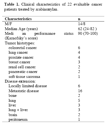
Evaluable patients were 22/24, while the remaining 2 patients, both affected by untreatable disseminated liver metastases due to colorectal cancer, rapidly died for disease progression before concluding the two planned months of arabinoxylan therapy. The clinical characteristics of the evaluable patients are reported in Table 1. Figure 1 illustrates changes in the mean number of total lymphocytes, T lymphocytes, T cytotoxic lymphocytes and NK cells occurring after 2 months of arabinoxylan therapy. No substantial variation was found in the mean number of lymphocytes, T lymphocytes, T cytotoxic lymphocytes and NK cells under arabinoxylan treatment. In contrast, as illustrated in Figure 2, TH and T reg mean numbers increased and decreased, respectively, after arabinoxylan therapy, without, however statistically significant differences with respect to the values seen prior to therapy. On the contrary, a statistically significant increase in TH/T reg mean ratio was achieved after arabinoxylan therapy (p<0.025). The increase in TH/T reg ratio under arabinoxylan therapy was more pronounced in patients with an abnormally low ratio prior to therapy with respect to that occurring in those with normal pre-treatment ratio, however without statistically significant differences ( 2.3 +/- 0.4 vs 1.7 +/- 0.5). In more detail,
090722_linfocitiT_table1
090722_linfocitiT_1

Figure 1. Changes in the number of lymphocytes, Tlymphocytes (CD3), T cytotoxic lymphocytes (CD8) and NK cells (CD16 CD56) after 2 months of arabinoxylan therapy.
090722_linfocitiT_2

Figure 2. Changes in the mean number of T helper (TH) lymphocytes (CD4) and T regulatory lymphocytes (cd4 cd24) and in TH/T reg mean ratio.

before arabinoxylan therapy, an abnormally low TH/T reg ratio was present in 12/22 (55%) evaluable patients. Arabinoxylan treatment induced a normalization of TH/T reg ratio in 5/12 (42%) patients with an abnormally low ratio prior to therapy. The percentage of arabinoxylan-induced TH/T reg normalization obtained in lymphocytopenic patients was not significantly different from that achieved in patients with normal pre-treatment lymphocyte count ( 3/7(43%) vs 2/5(40%) ). No toxicity was observed under arabinoxylan treatment, which was well tolerated in all patients. Asthenia was present in 8/22 (36%) evaluable patients. An evident relief of asthenia, as assessed by a specific patient report, was obtained under arabinoxylan therapy in 5/8 (63%) patients.

IV. Discussion
Previous experimental studies had already demonstrated some immunomodulating properties of arabinoxylan, in particular consisting of stimulation of NK cytotoxic function (Ghoneum, 1998), whereas NK cell number did not seem to be influenced by arabinoxylan administration. However, it has to be remarked that NK cells were belived to be fundamental in the antitumor immunity until some years ago, before the discovery of the essential role played by the antitumor cytokines, such as IL-2 and IL-12 (Whittington and Faulds, 1993) and dendritic cells, because of their function as antigen-presenting cells (Banks et al, 1995). In fact, it has to be considered that the cytotoxic activity of NK cells is effective only against artificial laboratory cancer cell lines, whose biological malignant properties are different from those presented by fresh human tumor cells (Whittington and Faulds, 1993). In addition, NK cells have been proven to be also able to destroy fresh human cancer cells only after the activation of their cytotoxic function by IL-2 (Atzpodien and Kirchner, 1990). From this point of view, arabinoxylan had been already proven to amplify the stimulatory effect of IL-2 on NK-mediated antitumor cytotoxicity (Ghoneum and Jewett, 2000). In contrast, no study has been performed up to now to evaluate the possible influence of arabinoxylan not only on the mechanisms responsible for the generation of an effective anticancer immune response, but also on those involved in the suppression of anticancer immunity. The results of this preliminary study, carried out to evaluate the influence of arabinoxylan on T reg cells, which represent the most important cells involved in the suppression of the antitumor cytotoxic immune response, demonstrates that arabinoxylan may counteract T reg cell generation by reducing their number and percentage with respect to the total amounts of CD4+ cells and circulating lymphocytes. Since NK cell function is inhibited by T reg activation (Shevach, 2002), the previously demonstrated arabinoxylan-induced stimulation of NK cell cytotoxic function might depend at least in part on its capacity of counteracting T reg generation (Dieckmann et al, 2001). Moreover, this study would suggest that the inhibitory action of arabinoxylan on T reg generation is more pronounced in patients with an abnormally high percentage of T reg cells prior to therapy, with a following pre-treatment abnormally low TH/T reg ratio before therapy, whereas its effect was less evident in patients with a pre-treatment value of TH/T reg ratio within the normal range. Therefore, the influence of arabinoxylan on T reg generation would consist of a modulatory action rather than an inhibitory activity. This finding could explain a potential favourable immunomodulatory effect of arabinoxylan also in patients with autoimmune diseases (Ghoneum, 1998), who in contrast to cancer patients would tend to present abnormally low amounts of T reg cells. In any case, the importance of the inhibition of T reg generation in the induction of an effective anticancer immune response has been recently confirmed by the evidence that the block of T reg activity by specific monoclonal antibodies may induce objective tumor regressions in humans (Yang et al, 2007). Obviously, the major problem is the exact identification of he T reg cell population. Even though T reg cells may express other immune markers, namely FOX-p2 cytoplasmatic antigen, most clinicians are in agreement to identify the CD4+CD25+ cells as T reg lymphocytes (12). In any case, further studies, by evaluating other immune markers, will be required to better identify T reg cells population, namely FOX-p3, even though recently some Authors have shown that FOX-p3 expression by T reg cells is associated with a lower suppressive activity (Dieckmann et al, 2001; Shevach, 2002). Moreover, it has to be remarked that several patients included in the present study were concomitantly under palliative therapy with the anti-cachectic pineal hormone melatonin (Brzezinski, 1997), which may also play immunomodulating effects (Maestroni, 1993). Therefore, further randomized studies with arabinoxylan alone versus arabinoxylan plus melatonin will be required to better define the immunomodulating action of arabinoxylan. If further clinical and experimental studies will confirm the inhibitory action of arabinoxylan on T reg cell system, it could be included in cytokine-based immunotherapies to enhance their efficacy by counteracting T reg cell generation.

References
Atzpodien J, Kirchner H (1990) Cancer, cytokines and cytotoxic cells:interleukin-2 in the immunotherapy of human neoplasms. Klin Wochenschr 14, 1-10.
Banks RE, Patel PM, Selby PJ (1995) Interleukin-12:a novel clinical player in cytokine therapy. Br J cancer 71, 655-659.
Brzezinski A (1997) Melatonin in humans. N Engl J Med 336, 185-195.
Dieckmann D, Plottner H, Berchtold S, Berger T, Schuler G (2001) Ex vivo isolation and characterization of CD4+CD25+ T cells with regulatory properties from human blood. J Exp Med 193, 1303-1310.
Ghoneum M (1998) Enhancement of human natural killer cell activity by modified arabinoxylane fro rice bran(MGN-3). Int J Immunother 14, 89-99.
Ghoneum M, Gollapudi S (2005) Synergistic of arabinoxilan rice bran (MGN-3/Biobran in S. Cerevisiae-induced apoptosis of monolayer breast cancer MFC-7 cells. Anticancer Res 25(6B), 4187-96.
Ghoneum M, Brown J, Gollapudi S (2007) Yeast therapy for the treatment of cancer and its enhancement by MGN-3/Biobran, an arabinoxylan rice bran. Cellular Signaling and Apoptosis Research (Ed. Alex R. Demasi) Cap IV: 185-200.
Ghoneum M, Jewett A (2000) Production of tumor necrosis factor-alpha and interferon-gamma from human peripheral blood lymphocytes by MGN-3, a modified arabinoxylan from rice bran, and its synergy with interleukin-2 in vitro. Cancer Detect Prevent 24, 314-324.
Lissoni P, Giani L, Zerbini S, Trabattoni P, Rovelli F (1998) Biotherapy with the pineal immunomodulating hormone melatonin versus melatonin plus Aloe vera in untreatable advanced solid neoplasms. Nat Immun 16, 27-33.
Maestroni JGM (1993) The immunoneuroendocrine role of melatonin. J Pineal Res 14, 1-10.
Markus J, Miller A, Smith M, Orengo I (2006) Metastatic hemangiopericytoma of the skin treated with wide local excision and MGN-3. Dermatol Surg 32, 145-147.
Matsuda T, Hirano T (1990) Interleukin-6 (IL-6). Biotherapy 2, 363-371.
Moore KW, O’Garra A, De Waal-Malefyt R (1993) Interleukin-10. Ann Rev Immunol 11, 165-174.
Rosenberg SA (1992) The immunotherapy and gene therapy of cancer. J Clin Oncol 10, 181-191.
Shevach EM (2002) CD4+CD25+ suppressor T cells:more questions than answers. Nat Rev Immunol 2, 389-400.
Whittington R, Faulds D (1993) Interleukin-2. Drugs 46, 446-514.
Yang JC, Hughes M, Kammula U, Royal R, Sherry RM, Topalian SL, Suri KB, Levy C, Allen T, Mavroukakis, Lowy I, White DE, Rosenberg SA (2007) Ipilimubab (anti-CTLA4 antibody)causes regression of metastatic renal cell cancer associated with enteritis and hypophysitis. J Immunother 30, 825-830.

(download PDF version)


A clinical study of T-regulatory lymphocyte function in cancer patients in relation to tumor histotype, disease extention, lymphocyte subtypes and cortisol secretion

Research Article (download PDF version)

Luigi Vigorè1, Fernando Brivio2, Luca Fumagalli2, Roberto Vezzo1, Giusy Messina6, Franco Rovelli6, Massimo Colciago3, Giovanna D’Amico4, Giuseppe Di Fede5, Paolo Lissoni6

1 Laboratory of Immunomicrobiology, St.Gerardo Hospital, Monza, Milan, Italy
2 Division of Surgery, St.Gerardo Hospital, Monza, Milan, Italy
3 I.N.R.C.A Laboratory of Analyses, Casatenovo, Lecco, Italy
4 Research Center “Fondazione Tettamanti” Clinica Pediatrica Università Milano-Bicocca, Italy
5 Institute of Biological Medicine, Milan, Italy
6 Division of Radiation Oncology, St.Gerardo Hospital, Monza, Milan, Italy

*Correspondence: Dr. Paolo Lissoni, Divisione di Radioterapia Oncologica, Ospedale S.Gerardo, 20052 Monza, Milano, Italy; Fax: +390392332284, E-mail: p.lissoni@hsgerardo.org
Key words: Anticancer immunity, immunosuppression, T regulatory lymphocytes
Abbreviations: cytotoxic T lymphocyte-associated antigen-4, (CTLA-4); glucocorticoid-induced TNF-α receptor, (GITR); myeloidderived suppressor cells, (MDSC); NK cells, (CD16CD56); T cytotoxic lymphocytes, (CD8); T helper lymphocytes, (CD4); Tregulatory lymphocytes, (T-reg)

Received: 24 July 2008; Revised: 11 September 2008
Accepted: 12 September 2008; electronically published: October 2008

Summary

Several clinical investigations showed that the immune status is a prognostic variable in cancer patients, even tough the evaluation of the anticancer immunity is not generally considered in the medical oncology. Several immune parameters, including lymphocyte subsets and cytokine blood concentration, had been proposed to quantify the functional status of the anticancer immunity, but recent discoveries would suggest that the end-result of the various immune interactions is represented by a subtype of CD4 lymphocytes capable of suppressing the antitumor immune reaction, the so called T-regulatory lymphocytes (T-reg). This study was performed to detect T-reg count and percentage in solid tumor patients, in relation to tumor histotype, disease extension, lymphocyte sub-populations and cortisol circadian secretion. The study included 114 consecutive cancer patients affected by the most frequent tumor histotypes, 69 of whom showed a metastatic disease. In each patient we evaluated T-reg cells, identified as CD4+CD25+, in relation to T helper (CD4), T cytotoxic (CD8) and NK (CD16CD56) cells. Abnormally high values of T-reg cells were seen in 52/114 (46%) patients, and the percentage of high values of T-reg was significantly higher in metastatic patients than in non-metastatic ones. In contrast, no significant difference was seen in relation to tumor histotype. Patients with increased T-reg count had a significantly lower NK cell number. Finally no significant difference in T-reg number was seen between patients with altered or normal rithm of cortisol. The study confirmed that, irrespectively of tumor histotype the metastatic disease is associated with a progressive and increased T-reg generation, with a following suppression of anticancer immunity.

I. Introduction
At present, there is no doubt about the existence of a sub-type of T lymphocytes, the so-called T regulatory lymphocytes (T-reg), capable of suppressing the cellular immune responses,including the anticancer immunity (Thomton and Shevach, 2000; Shevach, 2002; von Herrath and Harrison, 2003; Schwartz, 2005; von Boehmer, 2005; Ziegler, 2006; Zou, 2006). However, the exact definition
of T-reg cells in terms of cell surface marker expression still remains controversial, particularly from a clinical point of view. All authors are in agreement to consider Treg lymphocytes as CD4+CD25+ cells, but at present it is still unknown whether the expression of CD4 and CD25 antigens may be sufficient to identify T-reg cells (Thomton and Shevach, 2000; Shevach, 2002; von Herrath and Harrison, 2003; Schwartz, 2005; von Boehmer, 2005; Ziegler, 2006; Zou, 2006), since several authors retain that the intracytoplasmatic expression of the FOX p3 protein is essential for the differentiation into T-reg cells (Ziegler, 2006; Zou, 2006).Recently, however, some preliminary observations would suggest that the cytoplasmatic expression of FOX p3 by CD4+CD25+ cells may be associated at least in some experimental conditions with a diminished, rather than with an enhanced immunosuppressive activity of T-reg cells (Siddiqui et al, 2007). In contrast, all authors agree that the expression of CD152 antigen, also called cytotoxic T lymphocyteassociated antigen-4 (CTLA-4) (Vasu et al, 2004), is fundamental for the immunosuppressive activity of T-reg cells (Takahashi et al, 2000), since the block of its expression by using anti-CTLA-4 monoclonal antibodies may abolish the suppressive activity of T-reg cells, with a following stimulation of the anticancer immunity in cancer patients (Knutson and Disis, 2007) and an enhanced incidence of autoimmune diseases in the healthy subjects (Lan et al, 2005). Therefore, the addition of a third marker, such as CD152 antigen, may allow to define a more homogeneous cell population provided by a regulatory activity with respect to the simple CD4+CD25+ expression (Dieckmann and Plottner, 2001). In fact, the suppressive regulatory action of CD4+CD25+CD152+ has appeared to be clearly higher than that played by the simple CD4+CD25+ T lymphocytes (Leong et al, 2006).This finding is not surprising, since the simple expression of CD25 marker, corresponding to the !-chain of IL-2 receptor, is not an exclusive characteristic of T-reg lymphocytes, but it is a non-specific property of the overall activated T lymphocytes (Thomton and Shevach, 2000; Shevach, 2002; von Herrath and Harrison, 2003; Schwartz, 2005; von Boehmer, 2005; Ziegler, 2006; Zou, 2006). At present, preliminary clinical studies would show that the percent of circulating CD4+CD25+ cells may be about 10% of the all CD4+ lymphocytes, and that of CD4+CD25+CD152+ cells may be about 40% of the total CD4+CD25+ cells, then the expected percent of CD4+CD25+CD152+ in the healthy subjects would be less than 5% of the total circulating CD4+ lymphocytes (Jago et al, 2004). Finally, the expression of glucocorticoidinduced TNF-α receptor (GITR) is also associated with an evident suppressive activity by T-reg lymphocytes (Kanamaru et al, 2004), which in fact are stimulated by
cortisol (Sthephens et al, 2004), that in contrast may inhibit the activity of the most other T lymphocytes, namely that of T helper lymphocytes, with a following diminished production of IL-2 (Claman, 1998). As far as the mechanisms responsible for T-reg-induced suppression of the anticancer immunity are concerned, several experimental observations have shown that T-reg cells may suppress the antitumor immune response through the release of immunosuppressive cytokines, namely IL-10 and TGF-β (Dieckmann et al, 2002), even though other authors would suggest that the suppressive activity of Treg cells on CD4+ and CD8+ lymphocyte activation may be relatively independent from the action of cytokines, by mainly requiring cell surface contact (Birebent et al, 2004). IL-2 has been proven to be essential for T-reg generation and some authors consider IL-2 as the main growth factor of T-reg lymphocytes (Antony and Restito, 2005), but more adequate studies have demonstrated that IL-2 may induce both stimulation and inhibition of T-reg generation and activation (Malek and Bayer, 2004). In fact, IL-2 has appeared to induce and promote T-reg differentiation only in the presence of TGF-β (Chen et al, 2003). Therefore, IL-2 would constitute the main human cytokine in influencing the characteristics of the anticancer immunity, since it may be responsible for both activation and suppression of an effective immune response against cancer cell proliferation and dissemination (Wang et al, 2001), namely depending on the whole status of the cytokine network, in particular on the presence or in the absence of adequate concentrations of TGF-β. In the absence of TGF-β, IL-2 stimulates the anticancer immunity, whereas it counteracts the generation of an effective antitumor immunity in the presence of TGF-β. In other words, IL-2 would physiologically control both tolerance and immunity, depending on the presence of TGF-β and other less known factors (Annunziato et al, 2002). In fact, under cancer immunotherapy with IL-2 the percent of T-reg cells has been shown to decrease in responding patients and to enhance in those with disease progression (Cesana et al, 2006). However, the regulation of T-reg functions does not depend only on immune factors, since it is also under a neuroendocrine control (Ji et al, 2004). In particular, cortisol has appeared to stimulate T-reg cell generation (Ji et al, 2004), with a following enhanced release of IL-10, by representing the main mechanism responsible for cortisol-induced immunosuppression. From a clinical oncological point of view, preliminary observations showed an enhanced percent of circulating CD4+CD25+ lymphocytes in cancer patients, namely in those with advanced disease (Sasada et al, 2003). The present study was performed to better establish which is T-reg behaviour in cancer patients in relation to both tumor histotype and disease extension.

II. Materials and methods
The study included 114 consecutive solid tumor patients with locally limited or metastatic disease, whose clinical characteristics are shown in Table 1. Lung cancer and gastrointestinal tumors were the most frequent neoplasms in our patients. For the immune detections, venous blood samples were collected in the morning after an overnight fast. Operable patients and metastatic patients were investigated before the surgical operation and before the onset of chemotherapy, respectively, in an attempt to exclude the possible influence of the various anticancer therapies on the immune status of patients.

In each sample, we have evaluated total lymphocyte count and the various lymphocyte subpopulations by a flow cytometric assay and monoclonal antibodies, including T helper lymphocytes (CD4), T cytotoxic lymphocytes (CD8), NK cells (CD16CD56), and T regulatory (T-reg) lymphocytes (CD4CD25). Normal values (95% confidence limits) of T-reg observed in our laboratory were below 240/mm3. Moreover, because of its importance in regulating lymphocyte functions and proliferation (Claman, 1998; Sthephens et al, 2004), the circadian rhythm of cortisol was also investigated by collecting blood samples at 8.00 A.M. and at 4.00 P.M., and cortisol serum concentrations were measured in duplicate by using the ECLA method. Data were reported as mean +/- SE, and statistically analyzed by the Student’s t test, the analysis of variance and the chi-square test, as appropriate.

III. Results
As reported in Table 2, an abnormally high number of T-reg was seen in 52/114 (46%) patients. Moreover, the percentage of cases with elevated number of T-reg observed in metastatic patients was significantly higher with respect to that found in non-metastatic patients (44/69 (64%) vs 8/45(18%), p < 0.01). Table 3 shows the mean number of T-reg and the mean percentages of T-reg with respect to both total lymphocytes and T helper (CD4+) lymphocytes observed in cancer patients in relation to their disease extension. The mean number of T-reg observed in metastatic patients was higher with respect to that found in patients with locally limited disease, without, however statistically significant differences. In contrast, the mean percentages of T-reg with respect to that of both lymphocytes and CD4 cells were significantly higher in metastatic patients than in the non-metastatic ones (p< 0.05 and p< 0.001,respectively). Moreover, within the metastatic group, patients with a normal lymphocyte count greater than 1500/mm3 showed a significantly higher mean number of T-reg with respect to the non-metastatic patients, whereas no difference was seen between nonmetastatic patients and metastatic patients with lymphocytopenia, consisting of lymphocyte count lower than 1500/mm3. In contrast, the mean percentages of T-reg with respect to total lymphocytes and CD4+ cells observed in both groups of metastatic patients with normal or low total lymphocyte count were significantly higher than in non-metastatic patients (lymphocytes: p< 0.025, CD4+ cells: p< 0.001).

Table 1. Clinical characteristics of 114 solid tumor patients
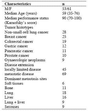
090722_clinicalstud1

Table 2. Percentages of abnormally high values of CD4+CD25+ lymphocytes

090722_clinicalstud2

* P < 0.01 vs non-metastatic patients

Table 3. Mean number of CD4+CD25+ lymphocytes and their mean percentages with respect to total lymphocytes and CD4+ lymphocytes in metastatic and non-metastatic patients

090722_clinicalstud3

* p<0.05 vs non-metastatic patients; ** p<0.025 vs non-metastatic patients; *** p<0.01 vs non-metastatic patients; **** p<0.001 vs nonmetastatic patients

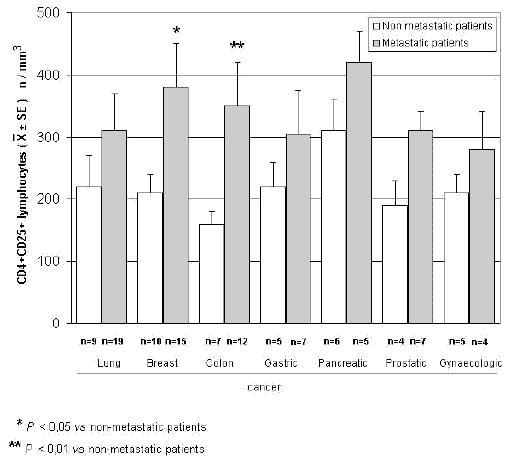
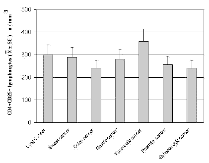
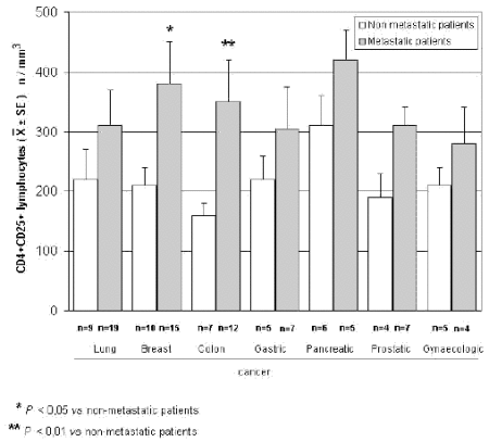
The mean counts of NK and CD8 cells in relation to that of T-reg are reported in Table 4. As shown, no significant difference in the mean number of CD8 lymphocytes was found between patients with normal or abnormally elevated number of T-reg. On the contrary, patients with elevated number of T-reg showed a significantly lower number of NK cells with respect to that found in those with normal T-reg count. Finally, Table 5 shows the circadian rhythm of cortisol in relation to total lymphocytes, CD4+ cells and T-reg mean number. A normal cortisol rhythm, with morning values greater at least than 50% with respect to the values occurring during the afternoon, was found in 85/114 (75%). Total lymphocyte and CD4+ cell mean numbers observed in patients with altered cortisol rhythm were significantly lower than those found in patients with normal cortisol circadianicity (p<0.01), whereas no significant difference was seen in the mean number of T reg. Figure 1 and Figure 2 illustrate T-reg mean numbers in relation to tumor histoptypes in the overall patients and with respect to their disease extension, respectively. No significant difference was seen in relation to tumor histotype. The highest values of T-reg were observed in pancreatic cancer patients, without however significant differences with respect to the overall other histotypes. The metastatic disease was associated with a higher number of T-reg with respect to the non-metastatic group in all tumor histotypes, even though a statistically significant differences occurred for the only breast cancer (p<0.05) and colorectal cancer (p< 0.01).

Table 4. Mean values of NK cells and CD8+ lymphocytes in cancer patients with normal or abnormally high values of CD4+CD25+ lymphocytes

090722_clinicalstud4

* p<0.05 vs normal values of CD4+CD25+ lymphocytes

Table 5. Mean numbers of total lymphocytes, T helper (CD4+) lymphocytes and T regulator lymphocytes (CD4+CD25+) in relation to cortisol circadian secretion in cancer patients
090722_clinicalstud5

* P<0.01 vs patients with altered cortisol rhythm
090722_clinicalstudfig1

Figure 1. CD4+CD25+ lymphocyte mean number in relation to tumor histotype
090722_clinicalstudfig2

Figure 2. CD4+CD25+ lymphocytes in relation to tumor histotype in metastatic and non-metastatic cancer patients

IV. Discussion
According to previous preliminary clinical investigations (Sasada et al, 2003; Cesana et al, 2006), this study confirms in a greater number of cancer patients that the metastatic disease is characterized by the evidence of an abnormally increased percentage of T-reg lymphocytes with respect to both total circulating lymphocytes and CD4+ lymphocytes. This finding does not seem to represent a specific characteristic of some tumor histotypes, then it could constitute a general alteration occurring during the progression of the neoplastic disease, by representing a fundamental immune parameter of cancer-related immunosuppression.

Several immune molecules have appeared to suppress the anticancer immunity, namely IL-6, IL-10, IL-1, TNF-α and TGF-β, but it seems that the common end result of their mechanisms of action may be represented by the stimulation of T-reg generation, with a consequent inhibition of the activation of an effective anticancer immune reaction. On the same way, several immune cells are able to suppress the anticancer immunity, including macrophages, T helper-2 lymphocytes and some myeloidderived suppressor cells, but also in this case they would act in a suppressive way by promoting the generation of Treg.

Then, the detection of T-reg amounts in terms of both absolute number and percentages with respect to total lymphocytes and CD4+ cells could constitute a simple and adequate clinical immune parameter to quantify the whole status of the anticancer immunity in the single cancer patient. Moreover, future clinical studies will be required to establish the possible prognostic significance of changes in T-reg percentage and number in relation to the anticancer efficacy of the various standard antitumor therapies. Moreover, it has to be remarked that T-reg lymphocytes would not represent the only immune cells involved in the suppression of the anticancer immunity. In fact, there is at least another fundamental immunosuppressive system, consisting of the monocytemacrophage cell lineage (Sica and Bronte, 2007). In more detail, it has been observed that the bone marrow may release myeloid precursors provided by suppressive activity on the antitumor immune response and defined as myeloid-derived suppressor cells (MDSC) (Kusmartsev and Gabrilovich, 2005). These cells have appeared to be characterized by the cell surface expression of GR-1, CD11b and CD80 antigens (Anderson et al, 2002; van Ginderachter et al, 2006). The myeloid suppressor cells would promote the generation and activation of T-reg lymphocytes, which at the other side would stimulate MDSC release from the bone marrow and M2 macrophage differentiation (Terabe et al, 2003; Wie et al, 2006).

Moreover, the myeloid suppressive cells would inhibit the anticancer immunity by promoting macrophage differentiation into the M2 sub-type (Ikemoto et al, 2003), which plays a clear inhibitory effect on the anticancer immunity, namely through the release of IL-6 (Ueno et al, 2000), whereas the M1 macrophage sub-type may either stimulate or suppress the antitumor immunity (Mantovani et al, 2004). M1 and M2 macrophage sub-types have appeared to be characterized by a high production of IL-12 or IL-10, respectively (Ueno et al, 2000).

Then, further studies by concomitantly evaluating T reg and MDSC count, will contribute to better define the immune mechanism responsible for the suppression of the anticancer immunity.

References
Anderson CF, Gerber JS, Mosser DM (2002) Modulating macrophage function with IgG immune complexes. J Endotoxin Res 8, 477-81.

Annunziato F, Cosmi L, Liotta F, Lazzeri E, Manetti R, Vanini V, Romagnani P, Maggi E, Romagnani S (2002) Phenotype,localization and mechanism of suppression of CD4+CD25+ human thymocytes. J Exp Med 196, 379-87.

Antony PA, Restito NP (2005) CD4+CD25+ T regulatory cells,immunotherapy of cancer,and interleukin-2. J Immunother 28, 120-8.

Birebent B, Lorho R, Lechartier H, de Guibert S, Alizadeh M,
Vu N, Beauplet A, Robillard N, Semana G (2004) Suppressive properties of human CD4+CD25+regulatory T cells are dependent on CTLA-4 expression. Eur J Immunol 34, 3485-96.

Cesana GC, DeRaffele G, Cohen S, Moroziewicz D, Mitcham J, Stoutenburg J, Cheung K, Hesdorffer C, Kim-Schulze S, Kaufman HL (2006) Characterization of CD4+CD25+ regulatory T cells in patients treated with high-dose interkleukin-2 for metastatic melanoma or renal cell carcinoma. J Clin Oncol 24, 1169-77.

Chen W, Jin W, Hardegen N, Lei KJ, Li L, Marinos N, McGrady G, Wahl SM (2003) Conversion of peripheral CD4+CD25- naïve T cells to CD4+CD25+ regulatory T cells by TGF-β induction of transcription factor Foxp 3. J Exp Med 198, 1875-86.

Claman HN (1998) Corticosteroids and the immune system. Adv Exp Med Biol 245, 203-10.

Dieckmann D, Bruett H, Ploettner H, Lutz MB, Schuler G (2002) Human CD4+CD25+ regulatory contact-dependent T cell induce IL-10 producing,contact-independent type-1-regulatory T cells. J Exp Med 196, 247-53.

Dieckmann D, Plottner H (2001) Ex vivo isolation and characterizationof CD4+CD25+ T cells with regulatory properties from human blood. J Exp Med 193, 1303-10.

Ehrke MJ, Mihich E, Berd D, Mastrangelo MJ (1989) Effects of anticancer drugs on the immune system. Semin Oncol 16, 230-9.

Ghiringhelli F, Larmonier N, Schmitt E, Parcellier A, Cathelin D, Garrido C, Chauffert B, Solary E, Bonnotte B, Martin F (2004) CD4+ CD25+ regulatory T cells suppress tumor immunity but are sensitive to cyclophosphamide which allows Immunotherapy of established tumors to be curative. Eur J Immunol 34, 336-44.

Ikemoto S, Yoshida N, Narita K, Wada S, Kishimoto T, Sugimura K, Nakatani T (2003) Role of tumor-associated macrophages in renal cell carcinoma. Oncol Rep 10, 1843-9.

Jago CB, Yates J, Camara NOS, Lechler RI, Lombardi AG (2004) Differential expression of CTLA-4 among T cell subsets. Clin Exp Immunol 136, 463-71.

Ji HB, Liao G, Faubion WA, Abadía-Molina AC, Cozzo C, Laroux FS, Caton A, Terhorst C (2004) Cutting edge, the natural ligand for glucocorticoid-induced TNF receptorrelated protein abrogates regulatory T cell suppression. J Immunol 172, 5823-7.

Kanamaru F, Youngnak P, Hashiguchi M, Nishioka T, Takahashi T, Sakaguchi S, Ishikawa I, Azuma M (2004) Costimulation via glucocorticoid-induced TNF receptor in both conventional and CD25+ regulatory CD4+ T cells. Immunol 172, 7306-14.

Knutson KL, Disis M, Salazar L (2007) CD4 regulatory T cells in human cancer pathogenesis. Cancer Immunol Immunother 556, 271-85.

Kusmartsev S, Gabrilovich DI (2005) STAT1 signaling regulates tumor-assolciated macrophage-mediate T cell deletion. J Immunol 174, 4880-91.

Lan RY, Ansari AA, Lian ZX, Gershwin ME (2005) Regulatory T cells, development,function,and role in autoimmunity. Autoimmun Rev 4, 351-63.

Leong PP, Mohammad R, Ibrahim N, Ithnin H, Abdullah M, Davis WC, Seow HF (2006) Phenotyping of lymphocytes expressing regulatory and effector markers in infiltrating ductal carcinoma of the breast. Immunol Lett 102, 229-36.

Malek TR, Bayer AL (2004) Tolerance not immunity crucially depends on IL-2. Nat Rev Immunol 4, 665-74.

Mantovani A, Sica A, Sozzani S, Allavena P, Vecchi A, Locati M (2004) The chemokine system in diverse forms of macrophage activation and polarization. Trends Immunol 25, 677-86.

Sasada T, Kimura M, Yoshida Y, Kanai M, Takabayashi A (2003) CD4+CD25+ regulatory T cells in patients with gastrointestinal malignancies, possible involvement of regulatory T cells in disease progression. Cancer 98, 1089-93.

Schwartz RH (2005) Natural regulatory T cells and selftolerance. Nat Immunol 6, 327-30.

Shevach EM (2002) CD4+CD25+ suppressor T cells, more questions than answers. Nat Rev Immunol 2, 389-400.

Sica A, Bronte V (2007) Altered macrophage differentiation and immune dysfunction in tumor development. J Clin Invest 117, 1155-66.

Siddiqui SA, Frigola X, Bonne-Annee S, et al (2007) Tumorinfiltrating Foxp3 CD4+CD25+ T cells predict poor survuival in renal cell carcinoma. Clin Cancer Res 13, 2075-81.

Stephens GL, McHugh RS, Whitters MJ, Young DA, Luxenberg D, Carreno BM, Collins M, Shevach EM (2004) Engagement of glucocorticoid-induced TNFR family-related receptor on effector T cells by its ligand mediates resistance to suppression by CD4+CD25+ T cells. J Immunol 173, 5008-20.

Takahashi T, Tagami T, Yamazaki S, Uede T, Shimizu J, Sakaguchi N, Mak TW, Sakaguchi S (2000) Immunologic and tolerance maintained by CD25+CD4+ regulatory T cells constitutively expressing cytotoxic T lymphocyte-associated antigen. J Exp Med 192, 1285-94.

Terabe M, Matsui S, Park JM, Mamura M, Noben-Trauth N, Donaldson DD, Chen W, Wahl SM, Ledbetter S, Pratt B, Letterio JJ, Paul WE, Berzofsky JA (2003) Transforming growth factor-β production and myeloid cells are an eggector mechanism trhough which CD1d-restricted T cells block cytotoxic T lymphocyte-mediated tumor immunosurveillance, abrogation prevents tumor recurrence. J Exp Med 198, 1741-52.

Thomton AM, Shevach EM (2000) Suppressor effector function of CD4+CD25+ immunoregulatory T cells is antigen nonspecific. J Immunol 164, 183-90.

Ueno T, Toi M, Saji H, Muta M, Bando H, Kuroi K, Koike M, Inadera H, Matsushima K (2000) Significance of macrophage chemo-attractant protein-1 in macrophage recruitment,angiogenesis and survival in human breast cancer. Clin Cancer Res 6, 3282-9.

Van Ginderachter JA, Movahedi K, Hassanzadeh Ghassabeh G, Meerschaut S, Beschin A, Raes G, De Baetselier P (2006) Classical and alternative activation of mononuclear phagocytes, picking the best of both worlds for tumor promotion. Immunobiology 211, 487-501.

Vasu C, Prabhakar BS, Holterman MJ (2004) Targeted CTLA-4 engagement induces CD4+CD25+CTLA-4 high T regulatory cells with target alloantigens specificity. J Immunol 173, 2866-76.

von Boehmer H (2005) Mechanisms of suppression by suppressor T cells. Nat Immunol 6, 338-44.

von Herrath MG, Harrison LC (2003) Regulatory lymphocytes, antigen-induced regulatory T cells in autoimmunity. Nat Rev Immunol 3, 223-32.

Wang XB, Zheng CY, Giscombe R, Lefvert AK (2001) Regulation of surface and intracellularexpression of CTLA-4 on human peripheral T cells. Scand J Immunol 54, 453-8.

Wie S, Kryczeck I, Zou W (2006) Regulatory T-cell compartmentalization and trafficking. Blood 108, 426-31.

Yang R, Cai Z, Zhang Y, Yutzy WH 4th, Roby KF, Roden RB (2006) CD80 in immune suppression by mouse ovarian carcinoma-associated Gr-1+CD11b+ myeloid cells. Cancer Res 66, 6807-15.

Ziegler SF (2006) FOXP3 of mice and men. Annu Rev Immunol 24, 209-26.

Zou W (2006) Regulatory T cells, tumour immunity and immunotherapy. Nat Rev Immunol 6, 295-307.

(download PDF version)



A phase II study of anastrozole plus the pineal anticancer hormone melatonin in the metastatic breast cancer women with poor clinical status

lissoniResearch Article (download PDF version)

Paolo Lissoni1*, Giuseppe Di Fede1, Antonio Battista2, Giusy Messina1, Remo Egardi1, Fernando Brivio3, Franco Rovelli1, Massimo Colciago4, Giuseppe Brera5

1 Institute of Biological Medicine, Milan
2 Azienda Sanitaria locale 2, Avellino;
3 Surgery Division, Bassini Hospital, Cinisello,Milan
4 I.N.R.C.A, Casatenovo, Lecco, Italy
5 Ambrosian University, Milan, Italy

dr. Paolo Lissoni

*Correspondence: Dr. Paolo Lissoni, Divisione di Radioterapia Oncologica, Ospedale S.Gerardo, 20052 Monza, Milano, Italy; Fax: +390392332284, E-mail: p.lissoni@hsgerardo.org
Key words: Anastrozole, breast cancer, melatonin, pineal gland
Abbreviations: melatonin, MLT; estrogen receptor, ER;

Received: 9 March 2009; Revised 1 April 2009;
Accepted: 13 April 2009; electronically published: 28 May 2009

Summary

The recent advances in the psychoneuroendocrinology have suggested the possibility to modulate tumor hormone dependency through a neuroendocrine approach. In particular, it has been proven that the pineal neurohormone melatonin (MLT) may stimulate estrogen receptor (ER) expression in breast cancer cells and inhibit the aromatase activity. On this basis, a study was planned to evacuate the efficacy of a concomitant treatment with the aromatase inhibitor anastrozole plus MLT in metastatic breast cancer. The study included 14 metastatic breast cancer women of poor clinical conditions with ER positive or unknown. Both anastrozole and MLT were given orally at a dose of 1 mg at noon and of 20 mg in the evening, respectively. The clinical response consisted of complete response in 2 and partial response in 6 patients. Then, an objective tumor regression was achieved in 8/14 (57%) patients, with a median duration of 26 months. No neoplastic cachexia occurred on treatment. This preliminary study shows that a neuroendocrine strategy with anastrozole plus the pineal hormone MLT may represent a new effective and well tolerated regimen in the treatment of metastatic breast cancer women, including those with poor clinical status, with therapeutic results apparently superior to those reported in the literature with the only aromatase inhibitor. Then, these results would justify further randomized studies of aromatase inhibitors with or without a concomitant administration of MLT, in an attempt to establish whether the pineal hormone may enhance the efficacy of the aromatase inibibitors in the treatment of human advanced breast cancer.

I. Introduction
Recent experimental studies have demonstrated that the hormone dependency is at least in part under a psychoneuroendocrine regulation (Cos et al, 2008;Grant et al, 2009). In particular, it has been shown that the pineal hormone melatonin (MLT), whose anticancer properties have been well demonstrated (Bartsch et al, 1981; Maestroni, 1993; Reiter et al, 2002), may in vitro stimulate estrogen receptor (ER) expression on breast cancer cell lines (Molis et al, 1995). Therefore, the hormone dependency of breast cancer cells would not depend only on intrinsic characteristics of cancer cells themselves, but also on host neuroendocrine regulation of tumor cell proliferation and differentiation (Bartsch et al, 2000). Moreover, cancer progression has been proven to be associated with pineal alterations, consisting of a progressive decline in MLT nocturnal production. (Maestroni, 1993). Therefore the advanced cancer would require a substitutive endocrine therapy with MLT (Bartsch et al, 1981; Maestroni, 1993). Previous preliminary clinical studies had already suggested that the concomitant administration of the pineal hormone MLT may apparently increase the efficacy of tamoxifen therapy in the treatment of metastatic breast cancer (Lissoni et al, 1995). Moreover, experimental studies have shown that the activity of aromatase enzyme, which is responsible for the peripheral production of estrogens from testosterone (Bagatell et al, 1994), is under a light/dark circadian rhythm (Bhatnagar et al, 1992). Because of the fundamental role of the pineal hormone MLT in the regulation of the daily photoperiod (Bartsch et al, 1981), it is possible to hypothesize that MLT may be involved in the control of the aromatase activity. In fact, recent studies have demonstrated an inhibitory action of MLT on the aromatase activity (Cos et al, 2005). This finding could reserve a prosiming application in the treatment of both early and advanced breast cancer. This statement is justified by the fact that the aromatase inhibitors represent a new class of agents in the endocrine treatment of breast cancer Plourde et al, 1994), with a potential efficacy superior to that achieved by the previous hormonal therapies with anti-estrogens, such as tamoxifene, even though tumor response rate obtained by the aromatase inhibitors are generally not greater than 40%. On this basis, a phase II study was planned in an attempt to evaluate the efficacy of a neuroendocrinotherapeutic regimen consisting of a concomitant administration of the aromatase inhibitor anastrozole and the pineal hormone MLT in metastatic breast cancer women with poor clinical conditions.

II. Materials and methods
The study included 14 consecutive metastatic breast cancer women (median age: 72 years, range 51-82), who were followed at Biological Medicine Institute in Milan, or at Health Local Unit 2 of Avellino, from Feb. 2002 to Sept. 2003. Eligibility criteria were, as follows: histologically proven metastatic breast cancer, measurable lesions, ER positive or unknown, no ability to tolerate chemotherapy because of age, low performance status (PS), important clinical illnesses other than cancer and/or heavy chemotherapeutic pre-treatments, no previous endocrine therapies for the metastatic disease, no double tumor and life expectancy less than 1 year. Previous heavy chemotherapeutic treatment consisting of at least 3 chemotherapeutic lines was made in 11/14 (79 %) patients. Dominant metastasis sites were, as follows: soft tissues:1; bone:1; lung:7 (neoplastic lymphangitis:2); liver:1; lung + liver:1; bone marrow:3. Time-span since first diagnosis of the primary tumor was 44 months (31-66 months). All patients had an acceptable social conditions. The minimum and median follow-up periods were 60 months and 72 months respectively. In all patients, in the case of disease progression, at least to other endocrine therapeutic lines with other aromatase-inhibitors were planned. The experimental protocol, wich was approved by the Health Direction of Biological Medicine Institute of Milan, was explained to each patient and informed consent was obtained. The treatment consisted of anastrozole at a dose of 1 mg/day orally at noon, plus MLT at 20 mg/day orally in the evening, generally half-hour before sleeping, to correct cancer progression-related decline in MLT night secretion (10). Patients were considered to be evaluable when they were treated for at least 3 consecutive months. The clinical response was evaluated according to WHO criteria. Complete response (CR) was the complete disappearance of all neoplastic lesions for at least 1 month. Partial response (PR) was a reduction greater than 50 % of the sum of all neoplastic lesions, for at least 1 month. Stable disesase (SD) was no increase or decrease greater tha 25 % of tumor volume. Progressive Disease (PD) was an increase in tumor volume greater than 25 % or the appearance of new neoplastic lesions. PS was assessed according to Karnofsky’s score, consisting of the evaluation of the quality of life in relation to patient activity and bed-rest period. ER was positive in 10 and unknown in the remaining 4 patients. The median PS was 80% (range 70-100). Data were statistically evaluated by the chi-square test and the Student’s t test, as appropriate.

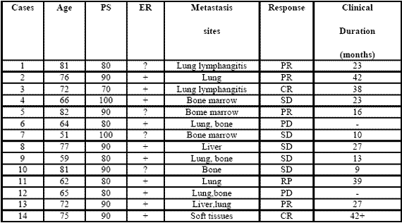
090722_phase2study

Table 1: Clinical characteristics of metastatic breast cancer women and their clinical response (WHO criteria) to a neuroendocrine regimen consisting of anastrozole plus the pineal hormone melatonin.

III. Results
All patients were fully evaluable for the clinical response. The clinical characteristics of patients and their individual clinical response to the treatment are reported in Table 1. As reported, a complete response (CR) was achieved in 2/14 (14%) (soft tissues:1; lung lymphangitis:1). A partial response (PR) was obtained in other 6/14 (43%) (bone:1; lung:3; liver:1; bone marrow:1). Then, an objective tumor response (CR + PR) was reached in 8/14 (57%) patients. The median duration of response was 26 months (range 9-42 months). A stable disease (SD) was seen in other 4/14 (29%), with a median duration of 25 months (range 10-27). Therefore, a disease-control (DC:CR + PR + SD) was achieved in 12/14 (86%) patients, whereas the remaining 2/14 (14%) patients had a progressive disease (PD). No significant difference in tumor response rate was observed between patients with positive or unknown ER ( 6/10(60%) vs 2/4(50%) ). An overall survival at 1 year and at 3 year was achieved in 11/14 (79 %) and in 5/14 (36 %) patients, respectively. Moreover, 3/14 (21%) patients were still alive at 5 years. The treatment was well tolerated in all patients.

Moreover, most patients experienced a relief of asthenia under the treatment and in no patient the neoplastic cachexia occurred. Finally, an evident increase in PS mean values was achieved under treatment, even though it did not reach the statistical significance (86 ±5 vs 93 ± 4, mean ± SE).

IV. Discussion
The results of this preliminary phase II study, by showing a percentage of 1-year survival greater than 70% in patients with live expectancy less than 1 year, would suggest that a neuroendocrine regimen consisting of the aromatase inhibitor anastrozole plus the pineal neurohormone MLT may represent a new effective therapeutic strategy in the treatment of metastatic breast cancer women, also in patients with poor clinical conditions, who would not be able to tolerate the most aggressive therapies. The concomitant administration of the pineal hormone would seem to enhance the efficacy of the aromatase inhibitor in terms of objective tumor regressions with respect to the results commonly reported in the literature with the only aromatase inhibitor (Plourde et al, 1994), which are generally lower than 40%.

The time to progression would seem to be apparently increased by the concomitant treatment with MLT.This finding is not surprising, since MLT could enhance the therapeutic anticancer acitivity of the aromatase inhibitors by either exerting direct antiproliferative antitumor effects (Bartsch et al, 1981; Maestroni, 1993; Reiter et al, 2002), or further inhibiting the aromatase activity by acting on gene and oncogene expression (Molis et al,1995; Cos et al, 2005). In addiction, MLT appeared to stimulate ER expression of breast cancer lines, by transforming ER negative into ER positive breast cancer, as observed in experimental conditions (Danforth et al, 1983).

Since the prognosis of ER positive breast cancers is clearly better than that of ER negative ones, MLT could per se improve the clinical couse of mammary tumors. Finally, because of its interesting therapeutic efficacy as a supportive care (Reiter et al, 2002), MLT would be responsible for the evident improvement in the relief of asthenia and in preventing the occurrence of the neoplastic cachexia. On the other hand, because of the inhibitory effect of MLT (Grant et al, 2009; Reiter et al, 2002) on cancer cell proliferation, the anticancer activity of this polyendocrine regimen would be due not only to an indirect effect, depending on a diminished estrogen production following aromatase enzyme inhibition, but also on a direct inhibition of cancer cell growth, due to MLT itself. Therefore, the results of this preliminary study may justify further clinical randomized investigations with the only aromatase inhibitor versus the concomitant treatment with MLT, in an attempt to confirm the ability of the pineal hormone to enhance the antitumor properties of the aromatase inhibitors in the treatment of metastatic breast cancer women with poor clinical conditions.

References
Bagatell CJ, Dahl KD, Bremner WJ (1994). The direct pituitary effect of testosterone to inhibit gonadotrophin secretion in men is partially mediated by aromatization to estradiol. J Androl 15:15-21.

Bartsch C, Bartsch H (1981) effect of melatonin on experimental tumors under different photoperiods and times of administration. J Neural Transm 52:269-279.

Bartsch H, Buchberger A, Franz H,Bartsch C,Maidonis I,Mecke D, Bayer E (2000) Effect of melatonin and pineal extracts on human ovarian and mammary tumor cells in a chemosensitivity assay. Life Sci 67:2953-2960.

Bhatnagar AS, Muller P, Schenkel L, Trunet PF, Beh I, Schieweck K. (1992). Inhibition of oestrogen biosynthesis and its consequences on gonadotrophin secretion in the male. J of Steroid Biochemistry and Molecular Biology;41:1021-1027

Cos S, Martinez-Campa C,Mediavilla MD, Sanchez-Barcelo E (2005). Melatonin modulates aromatase activity in MCF-7 human breast cancer cells. J Pineal Res 7:136-142.

Cos S, Gonzales A, Martinez-Campa C, Mediavilla MD, Alonzo-Gonzales C, Sanchez-Barcelo EJ (2008): Melatonin as a selective estrogen enzyme modulator. Curr Cancer Drug Targets. Dec 8(8): 691-702.

Danforth DN, Tamarkin L, Lipmann LE: (1983) Melatonin increase oestrogen receptor binding activity of human breast cancer cells. Nature, 305:323-325.

Grant S.G, Melan MA, Latimer JJ, Witt-Enderby PA (2009) : Melatonin and Breast cancer : cellular mechanism, clinical studies and future perspective. Expert Rev Mol Med 11:e5

Lissoni P, Barni S, Meregalli S, Fossati V, Cazzaniga M, Esposti D, Tancini G (1995) Modulation of cancer endocrine therapy by melatonin:a phase II study of tamoxifen plus melatonin in metastatic breast cancer patients progressing under tamoxifen alone. Br J Cancer 71:854-856.

Maestroni GJM (1993) The immunoneuroendocrine role of melatonin. J Pineal Res 14:1-10.

Molis TM, Spriggs LL,Jupiter Y,Hill SM (1995) Melatonin modulation of estrogen-regulated proteins, growth factors,and protooncogenes in human breast cancer. J Pineal Res 18:93-103.

Plourde PV, Dyroff M, Dukes MD (1994) Arimidex: a potent and selective fourth generation aromatase inhibitor. Breast Cancer Res Treat 30:103-111.

Reiter RJ, Tan DX, Sainz RM, Mayo JC, Lopez-Burillo S. (2002) Melatonin:reducing the toxicity and increasing the efficacy of drugs. Pharm Pharmacol 54:1299-1321

(download PDF version)


Evaluation of neopterin and ALCAT test in patients with food intolerance

d.ssa Berardi della IRCCS Policlinico San Matteo di Pavia reparto di dermatologia

Background:
Specific foods and food additives can induce adverse gastrointestinal (GI) and cutaneous reactions, including food allergies that involve an abnormal immunologic reaction to food proteins or food intolerance which is not pathogenetically immune-mediated.

Neopterin is synthesized by human monocyte-derived macrophages upon stimulation with interferon-gamma (IFN-gamma). Measurement of neopterin concentrations is useful for monitoring cell-mediated immune activation as there is a positive direct relationship between neopterin levels and severity of immune-mediated disorders. The aim of this study was to evaluate the neopterin serum levels in a group of patients with food intolerance.

Methods:
A group of forty-six patients (40 females, 6 males, median age 33 years) affected by cutaneous disease (27%) or GI symptoms (73%) consequent to food ingestion were tested by ALCAT and neopterin serum levels were measured.

The ALCAT test utilizes electronic haematology instrumentation and computerized data analysis to measure volumetric shifts in peripheral blood cells following incubation with food antigens.
Results are expressed in terms of percent change for cell volume and number. The degree of reactivity was determined by comparing a baseline distribution curve (of WBC) against the distribution curve generated by the analysis of each test agent/blood sample, and calculating the absolute differences between the curves and the standard deviation (SD). Any reactivity under 2 SD was considered non-reactive (negative) and these foods are allowed in the diet.

The Neopterin-MW EIA kit (DRG Instruments GmbH) employed an enzyme immunoassay technique to measure neopterin in a serum sample and was performed according to the Manufacturer’s Instructions and expressed as ng/mL.

Neopterin values were reported as medians with first and third quartiles (interquartile range). ALCAT test data were described as a percentage of non-reactive foodstuffs. Correlation between neopterin serum levels and percentage of non-reactive food was evaluated by means of the Spearman’s correlation coefficient (r).

Results:
The neopterin median value was 2 ng/mL (25°th-75°th: 1.50-2.25 ng/mL). There was a significant positive relationship between neopterin serum levels and percentage of non-reactive foodstuffs (p=0.05, r=0.302).

Conclusion:
This preliminary study confirms that food intolerance is not provoked by immunological mechanisms.

home page EAACI 2010 >>


Hsp10, Hsp70, and Hsp90 immunohistochemical levels change in ulcerative colitis after therapy

Research Article (download PDF version – complete 590 kb)

G. Tomasello,1 C. Sciumè,1 F. Rappa,2 V. Rodolico,3 M. Zerilli,3 A. Martorana,3 G. Cicero,4 R. De Luca,4 P. Damiani,5 F.M. Accardo,6 M. Romeo,7 F. Farina,2 G. Bonaventura,2 G. Modica,1 G. Zummo,2 E. Conway de Macario,8 A.J.L. Macario,8,9 F. Cappello2,9

1 Dipartimento di Discipline Chirurgiche ed Oncologiche, Università di Palermo;
2 Dipartimento di Biomedicina Sperimentale e Neuroscienze Cliniche, Università di Palermo, Italy;
3 Dipartimento di Patologia Umana, Università di Palermo, Italy;
4 Dipartimento di Oncologia, Università di Palermo, Italy;
5 Dipartimento di Medicina Interna e delle Malattie Emergenti, Università di Palermo, Italy;
6 Dipartimento di Contabilità Nazionale ed Analisi dei Processi Sociali, Università di Palermo, Italy;
7 Istituto di Medicina Biologica, Milano, Italy;
8 Department of Microbiology and Immunology, School of Medicine, University of Maryland
at Baltimore, and IMET, Baltimore, MD, USA;
9 Istituto Euro-Mediterraneo di Scienza e Tecnologia (IEMEST), Palermo, Italy

*Correspondence: Prof. Francesco Cappello, Dipartimento di Biomedicina Sperimentale e Neuroscienze Cliniche, Sezione di Anatomia Umana, via del Vespro 129, 90127, Palermo, Italy.
tel./Fax: +39.091.6553580. E-mail: francapp@hotmail.com

Abstract

Ulcerative colitis (UC) is a form of inflammatory bowel disease (IBD) characterized by damage of large bowel mucosa and frequent extra-intestinal autoimmune comorbidities.

The role played in IBD pathogenesis by molecular chaperones known to interact with components of the immune system involved in inflammation is unclear. We previously demonstrated that mucosal Hsp60 decreases in UC patients treated with conventional therapies (mesalazine, probiotics), suggesting that this chaperonin could be a reliable biomarker useful for monitoring response to treatment, and that it might play a role in pathogenesis.

In the present work we investigated three other heat shock protein/molecular chaperones: Hsp10, Hsp70, and Hsp90. We found that the levels of these proteins are increased in UC patients at the time of diagnosis and decrease after therapy, supporting the notion that these proteins deserve attention in the study of the mechanisms that promote the development and maintenance of IBD, and as biomarkers of this disease (e.g., to monitor response to treatment at the histological level).

(download PDF version – complete 590 kb)


Settembre 30, 2013 Newsletter

La Paleo Diet è una dieta che si basa su un principio molto semplice, ma altrettanto efficace: la comunanza dell’uomo moderno con le popolazioni del Paleolitico.

Dalle analisi del DNA è, infatti, emerso che gli essere umani rispetto ai loro antenati di 40.000 anni fa non sono cambiati quasi per nulla. Sulla base di questo principio si è osservata l’alimentazione dei nostri più antichi predecessori, che non avevano ancora scoperto l’agricoltura e la successiva elaborazione dei cibi.

In pratica i nostri antenati:

  • Non mangiavano latticini
  • Consumavano un solo zucchero raffinato: il miele
  • Si nutrivano di animali selvatici magri e quindi con un’alimentazione più proteica rispetto alla nostra
  • I carboidrati che assumevano venivano tutti da frutta e verdura con un’assunzione di fibre più alta (quindi non è vero che la PaleoDiet è una dieta esclusivamente proteica.)
  • I grassi che consumavano erano grassi “sani” (mono insaturi, polinsaturi e omega 3) e non quelli dannosi (saturi.)

Sebbene la Paleo Diet risulti essere il regime alimentare più idoneo sia per il dimagrimento (perdita di massa grassa e non magra) sia per una vita più lunga e salutare, anche con questo tipo dieta è possibile che si manifestino disturbi dovuti alle intolleranze alimentari.

Le intolleranze alimentari, definibili sinteticamente come reazioni negative del sistema immunitario nei confronti di alcuni alimenti o additivi presenti in essi, si manifestano con una sintomatologia molto varia:

  • ritenzione idrica
  • stanchezza e debolezza muscolare
  • alterazioni del metabolismo (sovrappeso e obesità)
  • problemi all’apparato gastroenterico
  • problemi all’apparato muscolo scheletrico
  • problemi all’apparato respiratorio.

Il metodo più affidabile per diagnosticarle è Alcat Test, un test di laboratorio standardizzato che si effettua tramite prelievo venoso e grazie al quale è possibile individuare gli alimenti da escludere e/o sostituire in un determinato piano alimentare.

Per citare un esempio concreto: se la mia dieta prevede come frutta le pere e come verdura gli spinaci, dovrei sicuramente sostituire questi alimenti ad alto contenuto di Nickel nel caso in cui, in seguito ai risultati ottenuti con ALCAT TEST, scoprissi di essere intollerante al Nickel.

Per allenarsi con un Personal trainer è importante impostare un piano alimentare efficace, come quello previsto dalla Paleo Diet (prescritto da un dietologo), e tenere in considerazione che, se la massa grassa rimane invariata e nel soggetto allenato non si riduce la presenza di ritenzione idrica, è senz’altro opportuno modificare la dieta verificando la presenza di eventuali intolleranze alimentari.

In sintesi, un buon personal trainer deve, oltre a indirizzare il proprio cliente verso un regime alimentare corretto, osservare l’andamento del dimagrimento e cercare di capire quali possano essere le cause della tanto temuta fase “plateau” della dieta (fase in cui la dieta non ha effetti dimagranti). In questo caso, è buona norma indirizzare l’allievo verso un test completo per le intolleranze alimentari come Alca test, strumento che consente di programmare un trattamento nutrizionale personalizzato per migliorare il più possibile le problematiche legate alle intolleranze.

a cura di Livio Boscarini & Daniele Albertini
Personal Trainers amministratori di Be.Come Personal Training


Gennaio 30, 2013 Newsletter

Lo stato di salute del nostro organismo dipende da molteplici fattori ed esso si riflette in misura rilevante sul nostro atteggiamento posturale.Intervenendo sulla postura si facilita il nostro stato di equilibrio psicofisico e si favorisce una miglior funzionalità di tutto il corpo, di conseguenza una ottimale applicazione del principio di auto guarigione.

La postura può essere influenzata da fattori strutturali, emotivi, energetici, viscerali, tra loro correlati.
Le cause strutturali possono essere determinate da problemi della vista, da malocclusione, dall’appoggio podalico, disfunzioni a carico degli
arti; tutto questo influisce sulla colonna vertebrale che può essere la causa o l’effetto di tali disfunzioni. Un attenta analisi obiettiva, avvalendosi di test kinesiologici, supportati dai dati forniti da una pedana podobarometrica, è in grado di fornire al medico, anche specialista (non focalizzato ad una visione settoriale del problema e dell’organismo), importanti dati sull’origine del problema primario e indirizzare la terapia in modo mirato. Senza ricorrere, se non in casi di effettiva necessità, ad indagini più impegnative ed invadenti.

Il medico ginecologo può sostenere la propria paziente in stato di gravidanza, prevenendo problemi di larga diffusione per questo stato, quali la stipsi o la sciatalgia (spesso dovuti all’ assetto della colonna vertebrale), malesseri assai diffusi, causa di sofferenza diretta non solo per la futura mamma, ma anche per il bimbo che porta in grembo.
Alcune piccole disfunzioni, insignificanti in un periodo di normalità, tendono ad accentuarsi con il procedere della gravidanza e quindi del nuovo assetto posturale assunto dalla donna.

Il medico dentista o l’ortodontista, possono verificare quanto l’intervento da loro progettato possa o meno influire sulla postura del paziente e suggerirgli interventi collaterali di riequilibrio, prima di procedere con il proprio, qualora si rendesse necessario. Sono raccomandate tecniche innovative in tal senso che sfruttano la capacità recettoriale (i nostri sensori) invece della tradizionale vettoriale (forze, trazioni ecc.).

Il Fisiatra, il neurologo e il medico legale, possono usufruire degli importanti e precisi dati forniti dalla pedana baropodometrica, per le loro valutazioni sullo stato di equilibrio nei singoli casi, sia in posizione statica che deambulatoria.

L’ortopedico è quindi in grado di prescrivere ortesi plantari con la massima precisione e accuratezza nel non creare fastidiosi problemi di adattamento posturali collaterali.

L’ottico o l’oculista possono verificare se le disfunzioni visive sono primarie o conseguenti ad adattamenti compensatori di una postura non proprio in equilibrio.


Gennaio 1, 2013 Newsletter

L’alcool è una sostanza di origine naturale proveniente dalla fermentazione e/o distillazione di cereali, frutta o verdura. È conosciuto come la sostanza psicoattiva più antica e più diffusa. Ma se è di origine naturale, come mai può arrivare a creare assuefazione, tossicità ed effetti paragonabili a quelli di una droga ad azione tossica? La risposta sta nella via metabolica utilizzata che fa sì che entri subito nel circolo sanguigno, raggiungendo velocemente il cervello con effetti neurologici immediati. Da qui la dipendenza, il consumo eccessivo fino ad arrivare a squilibri nutritivi e a gravi complicanze per fegato, pancreas, stomaco, sistema nervoso. Alcool e farmaci utilizzano la stessa via metabolica per cui attenzione alle reazioni indesiderate se si assumono medicinali.

Secondo alcuni recenti studi, il “piacere dell’alcool” dipende, insieme con altri fattori, da una predisposizione genetica, così come il “reggere l’alcool” è dovuto al nostro DNA. Il tutto dipende naturalmente dalle dosi assunte, dallo stato di salute e dal metabolismo del soggetto. La dose giornaliera di alcool accettabile corrisponde a 0,6-0,7 g/kg di peso corporeo.

Oggigiorno l’alcolismo è una piaga sociale che colpisce non solo gli adulti, ma anche i più giovani.
L’alcool è la nuova moda degli adolescenti: si comincia con il famoso happy hour per continuare con birra, bevande alcoliche di ogni genere e colore, superalcolici. Non si tratta di “gusto del bere” ma di un mezzo per socializzare fino a raggiungere la dipendenza. Quella diffusa tra gli adolescenti non è di certo la “filosofia” del piacere di gustarsi un buon bicchiere di vino con gli amici! Il vino tra l’altro, anche se alcolico, nelle giuste dosi ha effetti benefici sulla salute. Per la ricchezza nutrizionale che lo distingue, può essere considerato un alimento. Quella del vino è una vera e propria cultura. E’ conosciuto fin dall’antichità per i suoi effetti benefici sull’uomo e ancora oggi diversi studi attestano gli effetti positivi che può avere se consumato con moderazione. Il vino con minori controindicazioni fisiologiche è quello a basso contenuto di tannini, poco alcolico, poco aromatico e non manipolato. Le proprietà benefiche del vino derivano dalla presenza di polifenoli e antociani, sostanze antiossidanti e quindi antitumorali. Inoltre, la presenza di resveratrolo, un polifenolo, gli conferisce la capacità di ridurre il rischio di malattie cardiovascolari. L’acido tartarico invece è un antitumorale specifico per la prostata.

Il vino rosso, rispetto a quello bianco, presenta una maggior concentrazione di polifenoli e quindi una maggior capacità antitumorale. Il vino bianco, essendo un vino meno “strutturato”, e quindi meno forte, necessità dell’aggiunta di conservanti e stabilizzanti, ecco perché è spesso responsabile del cosiddetto “cerchio alla testa”.

La bella notizia è che il vino può essere integrato nei pasti delle diete dimagranti. Al di là del suo contenuto calorico è, infatti, più importante conoscere le caratteristiche degli altri alimenti che compongono un determinato pasto: se ad esempio si tratta di un pasto con un buon apporto di zuccheri, allora sarà bene evitare l’associazione del vino e in particolare sarà il paziente diabetico a dover fare molta attenzione.

Ecco perché consiglio sempre di attenersi al consiglio di uno specialista. Particolare attenzione dovranno fare anche i soggetti con intolleranze alimentari e, parlando di vino, mi riferisco agli intolleranti al nichel ad esempio. Il nichel è un metallo presente nel terreno in grado contaminare frutta, verdura e prodotti da essi derivanti. Le bevande alcoliche possono esserne contaminate anche per il processo di lavorazione che la materia prima subisce. Il nichel è presente nel vino per cui anche in questo caso sarà lo specialista a valutare la situazione nello specifico.

Se invece non avete allergie o intolleranze diagnosticate ma, in seguito ad un paio di bicchieri di vino avvertite sintomi quali difficoltà digestive, reflusso gastrico, eruzioni cutanee, ecc., allora sarà bene effettuare l’ALCAT Test per le intolleranze alimentari (i sintomi da intolleranza possono manifestarsi fino a 48 o anche 72 ore dopo). Nel caso di vino e bevande alcoliche in generale, la causa della vostra intolleranza potrà essere il nichel o uno degli additivi chimici aggiunti durante la lavorazione o l’alimento utilizzato come materia prima. L’ALCAT Test, presente da più di vent’anni nell’ambito dell’alimentazione, e validato da studi diversi condotti presso centri di ricerca, permette di testare alimenti, nichel, additivi chimici (coloranti, conservanti, stabilizzanti) ecc. con un semplice prelievo di sangue. In seguito potrete rivolgervi al vostro medico o nutrizionista di fiducia che vi aiuterà nel risolvere il vostro problema attraverso una terapia nutrizionale e non farmacologica.

Per concludere diciamo che il vino è di certo la bevanda che rende piacevoli i pasti con gli amici, tiene compagnia e ci rende allegri e rilassati ma è bene che non diventi il nostro amico fedele. A tale proposito, dato che può essere per diversi motivi considerato un “medicamento”, ricordiamo sempre che come tutte le medicine va assunto in modo controllato.

a cura della dr.ssa Jessica Barbieri, Biologa Nutrizionista, Consulente IMBIO  Istituto di Medicina Biologica Milano.


Copyright by IMBIO 2017. All rights reserved.