Articoli

Tutto sul nostro settore


A clinical study of T-regulatory lymphocyte function in cancer patients in relation to tumor histotype, disease extention, lymphocyte subtypes and cortisol secretion

Research Article (download PDF version)

Luigi Vigorè1, Fernando Brivio2, Luca Fumagalli2, Roberto Vezzo1, Giusy Messina6, Franco Rovelli6, Massimo Colciago3, Giovanna D’Amico4, Giuseppe Di Fede5, Paolo Lissoni6

1 Laboratory of Immunomicrobiology, St.Gerardo Hospital, Monza, Milan, Italy
2 Division of Surgery, St.Gerardo Hospital, Monza, Milan, Italy
3 I.N.R.C.A Laboratory of Analyses, Casatenovo, Lecco, Italy
4 Research Center “Fondazione Tettamanti” Clinica Pediatrica Università Milano-Bicocca, Italy
5 Institute of Biological Medicine, Milan, Italy
6 Division of Radiation Oncology, St.Gerardo Hospital, Monza, Milan, Italy

*Correspondence: Dr. Paolo Lissoni, Divisione di Radioterapia Oncologica, Ospedale S.Gerardo, 20052 Monza, Milano, Italy; Fax: +390392332284, E-mail: p.lissoni@hsgerardo.org
Key words: Anticancer immunity, immunosuppression, T regulatory lymphocytes
Abbreviations: cytotoxic T lymphocyte-associated antigen-4, (CTLA-4); glucocorticoid-induced TNF-α receptor, (GITR); myeloidderived suppressor cells, (MDSC); NK cells, (CD16CD56); T cytotoxic lymphocytes, (CD8); T helper lymphocytes, (CD4); Tregulatory lymphocytes, (T-reg)

Received: 24 July 2008; Revised: 11 September 2008
Accepted: 12 September 2008; electronically published: October 2008

Summary

Several clinical investigations showed that the immune status is a prognostic variable in cancer patients, even tough the evaluation of the anticancer immunity is not generally considered in the medical oncology. Several immune parameters, including lymphocyte subsets and cytokine blood concentration, had been proposed to quantify the functional status of the anticancer immunity, but recent discoveries would suggest that the end-result of the various immune interactions is represented by a subtype of CD4 lymphocytes capable of suppressing the antitumor immune reaction, the so called T-regulatory lymphocytes (T-reg). This study was performed to detect T-reg count and percentage in solid tumor patients, in relation to tumor histotype, disease extension, lymphocyte sub-populations and cortisol circadian secretion. The study included 114 consecutive cancer patients affected by the most frequent tumor histotypes, 69 of whom showed a metastatic disease. In each patient we evaluated T-reg cells, identified as CD4+CD25+, in relation to T helper (CD4), T cytotoxic (CD8) and NK (CD16CD56) cells. Abnormally high values of T-reg cells were seen in 52/114 (46%) patients, and the percentage of high values of T-reg was significantly higher in metastatic patients than in non-metastatic ones. In contrast, no significant difference was seen in relation to tumor histotype. Patients with increased T-reg count had a significantly lower NK cell number. Finally no significant difference in T-reg number was seen between patients with altered or normal rithm of cortisol. The study confirmed that, irrespectively of tumor histotype the metastatic disease is associated with a progressive and increased T-reg generation, with a following suppression of anticancer immunity.

I. Introduction
At present, there is no doubt about the existence of a sub-type of T lymphocytes, the so-called T regulatory lymphocytes (T-reg), capable of suppressing the cellular immune responses,including the anticancer immunity (Thomton and Shevach, 2000; Shevach, 2002; von Herrath and Harrison, 2003; Schwartz, 2005; von Boehmer, 2005; Ziegler, 2006; Zou, 2006). However, the exact definition
of T-reg cells in terms of cell surface marker expression still remains controversial, particularly from a clinical point of view. All authors are in agreement to consider Treg lymphocytes as CD4+CD25+ cells, but at present it is still unknown whether the expression of CD4 and CD25 antigens may be sufficient to identify T-reg cells (Thomton and Shevach, 2000; Shevach, 2002; von Herrath and Harrison, 2003; Schwartz, 2005; von Boehmer, 2005; Ziegler, 2006; Zou, 2006), since several authors retain that the intracytoplasmatic expression of the FOX p3 protein is essential for the differentiation into T-reg cells (Ziegler, 2006; Zou, 2006).Recently, however, some preliminary observations would suggest that the cytoplasmatic expression of FOX p3 by CD4+CD25+ cells may be associated at least in some experimental conditions with a diminished, rather than with an enhanced immunosuppressive activity of T-reg cells (Siddiqui et al, 2007). In contrast, all authors agree that the expression of CD152 antigen, also called cytotoxic T lymphocyteassociated antigen-4 (CTLA-4) (Vasu et al, 2004), is fundamental for the immunosuppressive activity of T-reg cells (Takahashi et al, 2000), since the block of its expression by using anti-CTLA-4 monoclonal antibodies may abolish the suppressive activity of T-reg cells, with a following stimulation of the anticancer immunity in cancer patients (Knutson and Disis, 2007) and an enhanced incidence of autoimmune diseases in the healthy subjects (Lan et al, 2005). Therefore, the addition of a third marker, such as CD152 antigen, may allow to define a more homogeneous cell population provided by a regulatory activity with respect to the simple CD4+CD25+ expression (Dieckmann and Plottner, 2001). In fact, the suppressive regulatory action of CD4+CD25+CD152+ has appeared to be clearly higher than that played by the simple CD4+CD25+ T lymphocytes (Leong et al, 2006).This finding is not surprising, since the simple expression of CD25 marker, corresponding to the !-chain of IL-2 receptor, is not an exclusive characteristic of T-reg lymphocytes, but it is a non-specific property of the overall activated T lymphocytes (Thomton and Shevach, 2000; Shevach, 2002; von Herrath and Harrison, 2003; Schwartz, 2005; von Boehmer, 2005; Ziegler, 2006; Zou, 2006). At present, preliminary clinical studies would show that the percent of circulating CD4+CD25+ cells may be about 10% of the all CD4+ lymphocytes, and that of CD4+CD25+CD152+ cells may be about 40% of the total CD4+CD25+ cells, then the expected percent of CD4+CD25+CD152+ in the healthy subjects would be less than 5% of the total circulating CD4+ lymphocytes (Jago et al, 2004). Finally, the expression of glucocorticoidinduced TNF-α receptor (GITR) is also associated with an evident suppressive activity by T-reg lymphocytes (Kanamaru et al, 2004), which in fact are stimulated by
cortisol (Sthephens et al, 2004), that in contrast may inhibit the activity of the most other T lymphocytes, namely that of T helper lymphocytes, with a following diminished production of IL-2 (Claman, 1998). As far as the mechanisms responsible for T-reg-induced suppression of the anticancer immunity are concerned, several experimental observations have shown that T-reg cells may suppress the antitumor immune response through the release of immunosuppressive cytokines, namely IL-10 and TGF-β (Dieckmann et al, 2002), even though other authors would suggest that the suppressive activity of Treg cells on CD4+ and CD8+ lymphocyte activation may be relatively independent from the action of cytokines, by mainly requiring cell surface contact (Birebent et al, 2004). IL-2 has been proven to be essential for T-reg generation and some authors consider IL-2 as the main growth factor of T-reg lymphocytes (Antony and Restito, 2005), but more adequate studies have demonstrated that IL-2 may induce both stimulation and inhibition of T-reg generation and activation (Malek and Bayer, 2004). In fact, IL-2 has appeared to induce and promote T-reg differentiation only in the presence of TGF-β (Chen et al, 2003). Therefore, IL-2 would constitute the main human cytokine in influencing the characteristics of the anticancer immunity, since it may be responsible for both activation and suppression of an effective immune response against cancer cell proliferation and dissemination (Wang et al, 2001), namely depending on the whole status of the cytokine network, in particular on the presence or in the absence of adequate concentrations of TGF-β. In the absence of TGF-β, IL-2 stimulates the anticancer immunity, whereas it counteracts the generation of an effective antitumor immunity in the presence of TGF-β. In other words, IL-2 would physiologically control both tolerance and immunity, depending on the presence of TGF-β and other less known factors (Annunziato et al, 2002). In fact, under cancer immunotherapy with IL-2 the percent of T-reg cells has been shown to decrease in responding patients and to enhance in those with disease progression (Cesana et al, 2006). However, the regulation of T-reg functions does not depend only on immune factors, since it is also under a neuroendocrine control (Ji et al, 2004). In particular, cortisol has appeared to stimulate T-reg cell generation (Ji et al, 2004), with a following enhanced release of IL-10, by representing the main mechanism responsible for cortisol-induced immunosuppression. From a clinical oncological point of view, preliminary observations showed an enhanced percent of circulating CD4+CD25+ lymphocytes in cancer patients, namely in those with advanced disease (Sasada et al, 2003). The present study was performed to better establish which is T-reg behaviour in cancer patients in relation to both tumor histotype and disease extension.

II. Materials and methods
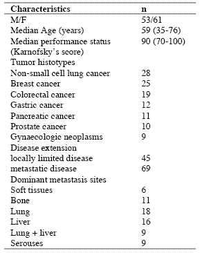
The study included 114 consecutive solid tumor patients with locally limited or metastatic disease, whose clinical characteristics are shown in Table 1. Lung cancer and gastrointestinal tumors were the most frequent neoplasms in our patients. For the immune detections, venous blood samples were collected in the morning after an overnight fast. Operable patients and metastatic patients were investigated before the surgical operation and before the onset of chemotherapy, respectively, in an attempt to exclude the possible influence of the various anticancer therapies on the immune status of patients.

In each sample, we have evaluated total lymphocyte count and the various lymphocyte subpopulations by a flow cytometric assay and monoclonal antibodies, including T helper lymphocytes (CD4), T cytotoxic lymphocytes (CD8), NK cells (CD16CD56), and T regulatory (T-reg) lymphocytes (CD4CD25). Normal values (95% confidence limits) of T-reg observed in our laboratory were below 240/mm3. Moreover, because of its importance in regulating lymphocyte functions and proliferation (Claman, 1998; Sthephens et al, 2004), the circadian rhythm of cortisol was also investigated by collecting blood samples at 8.00 A.M. and at 4.00 P.M., and cortisol serum concentrations were measured in duplicate by using the ECLA method. Data were reported as mean +/- SE, and statistically analyzed by the Student’s t test, the analysis of variance and the chi-square test, as appropriate.

III. Results
As reported in Table 2, an abnormally high number of T-reg was seen in 52/114 (46%) patients. Moreover, the percentage of cases with elevated number of T-reg observed in metastatic patients was significantly higher with respect to that found in non-metastatic patients (44/69 (64%) vs 8/45(18%), p < 0.01). Table 3 shows the mean number of T-reg and the mean percentages of T-reg with respect to both total lymphocytes and T helper (CD4+) lymphocytes observed in cancer patients in relation to their disease extension. The mean number of T-reg observed in metastatic patients was higher with respect to that found in patients with locally limited disease, without, however statistically significant differences. In contrast, the mean percentages of T-reg with respect to that of both lymphocytes and CD4 cells were significantly higher in metastatic patients than in the non-metastatic ones (p< 0.05 and p< 0.001,respectively). Moreover, within the metastatic group, patients with a normal lymphocyte count greater than 1500/mm3 showed a significantly higher mean number of T-reg with respect to the non-metastatic patients, whereas no difference was seen between nonmetastatic patients and metastatic patients with lymphocytopenia, consisting of lymphocyte count lower than 1500/mm3. In contrast, the mean percentages of T-reg with respect to total lymphocytes and CD4+ cells observed in both groups of metastatic patients with normal or low total lymphocyte count were significantly higher than in non-metastatic patients (lymphocytes: p< 0.025, CD4+ cells: p< 0.001).

Table 1. Clinical characteristics of 114 solid tumor patients
090722_clinicalstud1

Table 2. Percentages of abnormally high values of CD4+CD25+ lymphocytes

090722_clinicalstud2

* P < 0.01 vs non-metastatic patients

Table 3. Mean number of CD4+CD25+ lymphocytes and their mean percentages with respect to total lymphocytes and CD4+ lymphocytes in metastatic and non-metastatic patients

090722_clinicalstud3

* p<0.05 vs non-metastatic patients; ** p<0.025 vs non-metastatic patients; *** p<0.01 vs non-metastatic patients; **** p<0.001 vs nonmetastatic patients

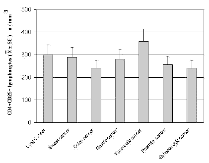
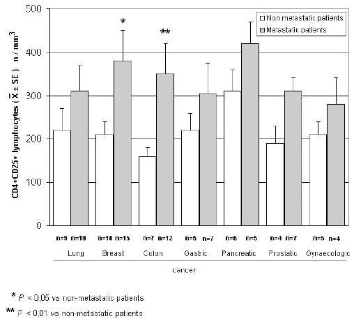
The mean counts of NK and CD8 cells in relation to that of T-reg are reported in Table 4. As shown, no significant difference in the mean number of CD8 lymphocytes was found between patients with normal or abnormally elevated number of T-reg. On the contrary, patients with elevated number of T-reg showed a significantly lower number of NK cells with respect to that found in those with normal T-reg count. Finally, Table 5 shows the circadian rhythm of cortisol in relation to total lymphocytes, CD4+ cells and T-reg mean number. A normal cortisol rhythm, with morning values greater at least than 50% with respect to the values occurring during the afternoon, was found in 85/114 (75%). Total lymphocyte and CD4+ cell mean numbers observed in patients with altered cortisol rhythm were significantly lower than those found in patients with normal cortisol circadianicity (p<0.01), whereas no significant difference was seen in the mean number of T reg. Figure 1 and Figure 2 illustrate T-reg mean numbers in relation to tumor histoptypes in the overall patients and with respect to their disease extension, respectively. No significant difference was seen in relation to tumor histotype. The highest values of T-reg were observed in pancreatic cancer patients, without however significant differences with respect to the overall other histotypes. The metastatic disease was associated with a higher number of T-reg with respect to the non-metastatic group in all tumor histotypes, even though a statistically significant differences occurred for the only breast cancer (p<0.05) and colorectal cancer (p< 0.01).

Table 4. Mean values of NK cells and CD8+ lymphocytes in cancer patients with normal or abnormally high values of CD4+CD25+ lymphocytes
090722_clinicalstud4

* p<0.05 vs normal values of CD4+CD25+ lymphocytes

Table 5. Mean numbers of total lymphocytes, T helper (CD4+) lymphocytes and T regulator lymphocytes (CD4+CD25+) in relation to cortisol circadian secretion in cancer patients
090722_clinicalstud5

* P<0.01 vs patients with altered cortisol rhythm
090722_clinicalstudfig1

Figure 1. CD4+CD25+ lymphocyte mean number in relation to tumor histotype
090722_clinicalstudfig2

Figure 2. CD4+CD25+ lymphocytes in relation to tumor histotype in metastatic and non-metastatic cancer patients

IV. Discussion
According to previous preliminary clinical investigations (Sasada et al, 2003; Cesana et al, 2006), this study confirms in a greater number of cancer patients that the metastatic disease is characterized by the evidence of an abnormally increased percentage of T-reg lymphocytes with respect to both total circulating lymphocytes and CD4+ lymphocytes. This finding does not seem to represent a specific characteristic of some tumor histotypes, then it could constitute a general alteration occurring during the progression of the neoplastic disease, by representing a fundamental immune parameter of cancer-related immunosuppression.

Several immune molecules have appeared to suppress the anticancer immunity, namely IL-6, IL-10, IL-1, TNF-α and TGF-β, but it seems that the common end result of their mechanisms of action may be represented by the stimulation of T-reg generation, with a consequent inhibition of the activation of an effective anticancer immune reaction. On the same way, several immune cells are able to suppress the anticancer immunity, including macrophages, T helper-2 lymphocytes and some myeloidderived suppressor cells, but also in this case they would act in a suppressive way by promoting the generation of Treg.

Then, the detection of T-reg amounts in terms of both absolute number and percentages with respect to total lymphocytes and CD4+ cells could constitute a simple and adequate clinical immune parameter to quantify the whole status of the anticancer immunity in the single cancer patient. Moreover, future clinical studies will be required to establish the possible prognostic significance of changes in T-reg percentage and number in relation to the anticancer efficacy of the various standard antitumor therapies. Moreover, it has to be remarked that T-reg lymphocytes would not represent the only immune cells involved in the suppression of the anticancer immunity. In fact, there is at least another fundamental immunosuppressive system, consisting of the monocytemacrophage cell lineage (Sica and Bronte, 2007). In more detail, it has been observed that the bone marrow may release myeloid precursors provided by suppressive activity on the antitumor immune response and defined as myeloid-derived suppressor cells (MDSC) (Kusmartsev and Gabrilovich, 2005). These cells have appeared to be characterized by the cell surface expression of GR-1, CD11b and CD80 antigens (Anderson et al, 2002; van Ginderachter et al, 2006). The myeloid suppressor cells would promote the generation and activation of T-reg lymphocytes, which at the other side would stimulate MDSC release from the bone marrow and M2 macrophage differentiation (Terabe et al, 2003; Wie et al, 2006).

Moreover, the myeloid suppressive cells would inhibit the anticancer immunity by promoting macrophage differentiation into the M2 sub-type (Ikemoto et al, 2003), which plays a clear inhibitory effect on the anticancer immunity, namely through the release of IL-6 (Ueno et al, 2000), whereas the M1 macrophage sub-type may either stimulate or suppress the antitumor immunity (Mantovani et al, 2004). M1 and M2 macrophage sub-types have appeared to be characterized by a high production of IL-12 or IL-10, respectively (Ueno et al, 2000).

Then, further studies by concomitantly evaluating T reg and MDSC count, will contribute to better define the immune mechanism responsible for the suppression of the anticancer immunity.

References
Anderson CF, Gerber JS, Mosser DM (2002) Modulating macrophage function with IgG immune complexes. J Endotoxin Res 8, 477-81.

Annunziato F, Cosmi L, Liotta F, Lazzeri E, Manetti R, Vanini V, Romagnani P, Maggi E, Romagnani S (2002) Phenotype,localization and mechanism of suppression of CD4+CD25+ human thymocytes. J Exp Med 196, 379-87.

Antony PA, Restito NP (2005) CD4+CD25+ T regulatory cells,immunotherapy of cancer,and interleukin-2. J Immunother 28, 120-8.

Birebent B, Lorho R, Lechartier H, de Guibert S, Alizadeh M,
Vu N, Beauplet A, Robillard N, Semana G (2004) Suppressive properties of human CD4+CD25+regulatory T cells are dependent on CTLA-4 expression. Eur J Immunol 34, 3485-96.

Cesana GC, DeRaffele G, Cohen S, Moroziewicz D, Mitcham J, Stoutenburg J, Cheung K, Hesdorffer C, Kim-Schulze S, Kaufman HL (2006) Characterization of CD4+CD25+ regulatory T cells in patients treated with high-dose interkleukin-2 for metastatic melanoma or renal cell carcinoma. J Clin Oncol 24, 1169-77.

Chen W, Jin W, Hardegen N, Lei KJ, Li L, Marinos N, McGrady G, Wahl SM (2003) Conversion of peripheral CD4+CD25- naïve T cells to CD4+CD25+ regulatory T cells by TGF-β induction of transcription factor Foxp 3. J Exp Med 198, 1875-86.

Claman HN (1998) Corticosteroids and the immune system. Adv Exp Med Biol 245, 203-10.

Dieckmann D, Bruett H, Ploettner H, Lutz MB, Schuler G (2002) Human CD4+CD25+ regulatory contact-dependent T cell induce IL-10 producing,contact-independent type-1-regulatory T cells. J Exp Med 196, 247-53.

Dieckmann D, Plottner H (2001) Ex vivo isolation and characterizationof CD4+CD25+ T cells with regulatory properties from human blood. J Exp Med 193, 1303-10.

Ehrke MJ, Mihich E, Berd D, Mastrangelo MJ (1989) Effects of anticancer drugs on the immune system. Semin Oncol 16, 230-9.

Ghiringhelli F, Larmonier N, Schmitt E, Parcellier A, Cathelin D, Garrido C, Chauffert B, Solary E, Bonnotte B, Martin F (2004) CD4+ CD25+ regulatory T cells suppress tumor immunity but are sensitive to cyclophosphamide which allows Immunotherapy of established tumors to be curative. Eur J Immunol 34, 336-44.

Ikemoto S, Yoshida N, Narita K, Wada S, Kishimoto T, Sugimura K, Nakatani T (2003) Role of tumor-associated macrophages in renal cell carcinoma. Oncol Rep 10, 1843-9.

Jago CB, Yates J, Camara NOS, Lechler RI, Lombardi AG (2004) Differential expression of CTLA-4 among T cell subsets. Clin Exp Immunol 136, 463-71.

Ji HB, Liao G, Faubion WA, Abadía-Molina AC, Cozzo C, Laroux FS, Caton A, Terhorst C (2004) Cutting edge, the natural ligand for glucocorticoid-induced TNF receptorrelated protein abrogates regulatory T cell suppression. J Immunol 172, 5823-7.

Kanamaru F, Youngnak P, Hashiguchi M, Nishioka T, Takahashi T, Sakaguchi S, Ishikawa I, Azuma M (2004) Costimulation via glucocorticoid-induced TNF receptor in both conventional and CD25+ regulatory CD4+ T cells. Immunol 172, 7306-14.

Knutson KL, Disis M, Salazar L (2007) CD4 regulatory T cells in human cancer pathogenesis. Cancer Immunol Immunother 556, 271-85.

Kusmartsev S, Gabrilovich DI (2005) STAT1 signaling regulates tumor-assolciated macrophage-mediate T cell deletion. J Immunol 174, 4880-91.

Lan RY, Ansari AA, Lian ZX, Gershwin ME (2005) Regulatory T cells, development,function,and role in autoimmunity. Autoimmun Rev 4, 351-63.

Leong PP, Mohammad R, Ibrahim N, Ithnin H, Abdullah M, Davis WC, Seow HF (2006) Phenotyping of lymphocytes expressing regulatory and effector markers in infiltrating ductal carcinoma of the breast. Immunol Lett 102, 229-36.

Malek TR, Bayer AL (2004) Tolerance not immunity crucially depends on IL-2. Nat Rev Immunol 4, 665-74.

Mantovani A, Sica A, Sozzani S, Allavena P, Vecchi A, Locati M (2004) The chemokine system in diverse forms of macrophage activation and polarization. Trends Immunol 25, 677-86.

Sasada T, Kimura M, Yoshida Y, Kanai M, Takabayashi A (2003) CD4+CD25+ regulatory T cells in patients with gastrointestinal malignancies, possible involvement of regulatory T cells in disease progression. Cancer 98, 1089-93.

Schwartz RH (2005) Natural regulatory T cells and selftolerance. Nat Immunol 6, 327-30.

Shevach EM (2002) CD4+CD25+ suppressor T cells, more questions than answers. Nat Rev Immunol 2, 389-400.

Sica A, Bronte V (2007) Altered macrophage differentiation and immune dysfunction in tumor development. J Clin Invest 117, 1155-66.

Siddiqui SA, Frigola X, Bonne-Annee S, et al (2007) Tumorinfiltrating Foxp3 CD4+CD25+ T cells predict poor survuival in renal cell carcinoma. Clin Cancer Res 13, 2075-81.

Stephens GL, McHugh RS, Whitters MJ, Young DA, Luxenberg D, Carreno BM, Collins M, Shevach EM (2004) Engagement of glucocorticoid-induced TNFR family-related receptor on effector T cells by its ligand mediates resistance to suppression by CD4+CD25+ T cells. J Immunol 173, 5008-20.

Takahashi T, Tagami T, Yamazaki S, Uede T, Shimizu J, Sakaguchi N, Mak TW, Sakaguchi S (2000) Immunologic and tolerance maintained by CD25+CD4+ regulatory T cells constitutively expressing cytotoxic T lymphocyte-associated antigen. J Exp Med 192, 1285-94.

Terabe M, Matsui S, Park JM, Mamura M, Noben-Trauth N, Donaldson DD, Chen W, Wahl SM, Ledbetter S, Pratt B, Letterio JJ, Paul WE, Berzofsky JA (2003) Transforming growth factor-β production and myeloid cells are an eggector mechanism trhough which CD1d-restricted T cells block cytotoxic T lymphocyte-mediated tumor immunosurveillance, abrogation prevents tumor recurrence. J Exp Med 198, 1741-52.

Thomton AM, Shevach EM (2000) Suppressor effector function of CD4+CD25+ immunoregulatory T cells is antigen nonspecific. J Immunol 164, 183-90.

Ueno T, Toi M, Saji H, Muta M, Bando H, Kuroi K, Koike M, Inadera H, Matsushima K (2000) Significance of macrophage chemo-attractant protein-1 in macrophage recruitment,angiogenesis and survival in human breast cancer. Clin Cancer Res 6, 3282-9.

Van Ginderachter JA, Movahedi K, Hassanzadeh Ghassabeh G, Meerschaut S, Beschin A, Raes G, De Baetselier P (2006) Classical and alternative activation of mononuclear phagocytes, picking the best of both worlds for tumor promotion. Immunobiology 211, 487-501.

Vasu C, Prabhakar BS, Holterman MJ (2004) Targeted CTLA-4 engagement induces CD4+CD25+CTLA-4 high T regulatory cells with target alloantigens specificity. J Immunol 173, 2866-76.

von Boehmer H (2005) Mechanisms of suppression by suppressor T cells. Nat Immunol 6, 338-44.

von Herrath MG, Harrison LC (2003) Regulatory lymphocytes, antigen-induced regulatory T cells in autoimmunity. Nat Rev Immunol 3, 223-32.

Wang XB, Zheng CY, Giscombe R, Lefvert AK (2001) Regulation of surface and intracellularexpression of CTLA-4 on human peripheral T cells. Scand J Immunol 54, 453-8.

Wie S, Kryczeck I, Zou W (2006) Regulatory T-cell compartmentalization and trafficking. Blood 108, 426-31.

Yang R, Cai Z, Zhang Y, Yutzy WH 4th, Roby KF, Roden RB (2006) CD80 in immune suppression by mouse ovarian carcinoma-associated Gr-1+CD11b+ myeloid cells. Cancer Res 66, 6807-15.

Ziegler SF (2006) FOXP3 of mice and men. Annu Rev Immunol 24, 209-26.

Zou W (2006) Regulatory T cells, tumour immunity and immunotherapy. Nat Rev Immunol 6, 295-307.

(download PDF version)


A phase II study of anastrozole plus the pineal anticancer hormone melatonin in the metastatic breast cancer women with poor clinical status

lissoniResearch Article (download PDF version)

Paolo Lissoni1*, Giuseppe Di Fede1, Antonio Battista2, Giusy Messina1, Remo Egardi1, Fernando Brivio3, Franco Rovelli1, Massimo Colciago4, Giuseppe Brera5

1 Institute of Biological Medicine, Milan
2 Azienda Sanitaria locale 2, Avellino;
3 Surgery Division, Bassini Hospital, Cinisello,Milan
4 I.N.R.C.A, Casatenovo, Lecco, Italy
5 Ambrosian University, Milan, Italy

dr. Paolo Lissoni

*Correspondence: Dr. Paolo Lissoni, Divisione di Radioterapia Oncologica, Ospedale S.Gerardo, 20052 Monza, Milano, Italy; Fax: +390392332284, E-mail: p.lissoni@hsgerardo.org
Key words: Anastrozole, breast cancer, melatonin, pineal gland
Abbreviations: melatonin, MLT; estrogen receptor, ER;

Received: 9 March 2009; Revised 1 April 2009;
Accepted: 13 April 2009; electronically published: 28 May 2009

Summary

The recent advances in the psychoneuroendocrinology have suggested the possibility to modulate tumor hormone dependency through a neuroendocrine approach. In particular, it has been proven that the pineal neurohormone melatonin (MLT) may stimulate estrogen receptor (ER) expression in breast cancer cells and inhibit the aromatase activity. On this basis, a study was planned to evacuate the efficacy of a concomitant treatment with the aromatase inhibitor anastrozole plus MLT in metastatic breast cancer. The study included 14 metastatic breast cancer women of poor clinical conditions with ER positive or unknown. Both anastrozole and MLT were given orally at a dose of 1 mg at noon and of 20 mg in the evening, respectively. The clinical response consisted of complete response in 2 and partial response in 6 patients. Then, an objective tumor regression was achieved in 8/14 (57%) patients, with a median duration of 26 months. No neoplastic cachexia occurred on treatment. This preliminary study shows that a neuroendocrine strategy with anastrozole plus the pineal hormone MLT may represent a new effective and well tolerated regimen in the treatment of metastatic breast cancer women, including those with poor clinical status, with therapeutic results apparently superior to those reported in the literature with the only aromatase inhibitor. Then, these results would justify further randomized studies of aromatase inhibitors with or without a concomitant administration of MLT, in an attempt to establish whether the pineal hormone may enhance the efficacy of the aromatase inibibitors in the treatment of human advanced breast cancer.

I. Introduction
Recent experimental studies have demonstrated that the hormone dependency is at least in part under a psychoneuroendocrine regulation (Cos et al, 2008;Grant et al, 2009). In particular, it has been shown that the pineal hormone melatonin (MLT), whose anticancer properties have been well demonstrated (Bartsch et al, 1981; Maestroni, 1993; Reiter et al, 2002), may in vitro stimulate estrogen receptor (ER) expression on breast cancer cell lines (Molis et al, 1995). Therefore, the hormone dependency of breast cancer cells would not depend only on intrinsic characteristics of cancer cells themselves, but also on host neuroendocrine regulation of tumor cell proliferation and differentiation (Bartsch et al, 2000). Moreover, cancer progression has been proven to be associated with pineal alterations, consisting of a progressive decline in MLT nocturnal production. (Maestroni, 1993). Therefore the advanced cancer would require a substitutive endocrine therapy with MLT (Bartsch et al, 1981; Maestroni, 1993). Previous preliminary clinical studies had already suggested that the concomitant administration of the pineal hormone MLT may apparently increase the efficacy of tamoxifen therapy in the treatment of metastatic breast cancer (Lissoni et al, 1995). Moreover, experimental studies have shown that the activity of aromatase enzyme, which is responsible for the peripheral production of estrogens from testosterone (Bagatell et al, 1994), is under a light/dark circadian rhythm (Bhatnagar et al, 1992). Because of the fundamental role of the pineal hormone MLT in the regulation of the daily photoperiod (Bartsch et al, 1981), it is possible to hypothesize that MLT may be involved in the control of the aromatase activity. In fact, recent studies have demonstrated an inhibitory action of MLT on the aromatase activity (Cos et al, 2005). This finding could reserve a prosiming application in the treatment of both early and advanced breast cancer. This statement is justified by the fact that the aromatase inhibitors represent a new class of agents in the endocrine treatment of breast cancer Plourde et al, 1994), with a potential efficacy superior to that achieved by the previous hormonal therapies with anti-estrogens, such as tamoxifene, even though tumor response rate obtained by the aromatase inhibitors are generally not greater than 40%. On this basis, a phase II study was planned in an attempt to evaluate the efficacy of a neuroendocrinotherapeutic regimen consisting of a concomitant administration of the aromatase inhibitor anastrozole and the pineal hormone MLT in metastatic breast cancer women with poor clinical conditions.

II. Materials and methods
The study included 14 consecutive metastatic breast cancer women (median age: 72 years, range 51-82), who were followed at Biological Medicine Institute in Milan, or at Health Local Unit 2 of Avellino, from Feb. 2002 to Sept. 2003. Eligibility criteria were, as follows: histologically proven metastatic breast cancer, measurable lesions, ER positive or unknown, no ability to tolerate chemotherapy because of age, low performance status (PS), important clinical illnesses other than cancer and/or heavy chemotherapeutic pre-treatments, no previous endocrine therapies for the metastatic disease, no double tumor and life expectancy less than 1 year. Previous heavy chemotherapeutic treatment consisting of at least 3 chemotherapeutic lines was made in 11/14 (79 %) patients. Dominant metastasis sites were, as follows: soft tissues:1; bone:1; lung:7 (neoplastic lymphangitis:2); liver:1; lung + liver:1; bone marrow:3. Time-span since first diagnosis of the primary tumor was 44 months (31-66 months). All patients had an acceptable social conditions. The minimum and median follow-up periods were 60 months and 72 months respectively. In all patients, in the case of disease progression, at least to other endocrine therapeutic lines with other aromatase-inhibitors were planned. The experimental protocol, wich was approved by the Health Direction of Biological Medicine Institute of Milan, was explained to each patient and informed consent was obtained. The treatment consisted of anastrozole at a dose of 1 mg/day orally at noon, plus MLT at 20 mg/day orally in the evening, generally half-hour before sleeping, to correct cancer progression-related decline in MLT night secretion (10). Patients were considered to be evaluable when they were treated for at least 3 consecutive months. The clinical response was evaluated according to WHO criteria. Complete response (CR) was the complete disappearance of all neoplastic lesions for at least 1 month. Partial response (PR) was a reduction greater than 50 % of the sum of all neoplastic lesions, for at least 1 month. Stable disesase (SD) was no increase or decrease greater tha 25 % of tumor volume. Progressive Disease (PD) was an increase in tumor volume greater than 25 % or the appearance of new neoplastic lesions. PS was assessed according to Karnofsky’s score, consisting of the evaluation of the quality of life in relation to patient activity and bed-rest period. ER was positive in 10 and unknown in the remaining 4 patients. The median PS was 80% (range 70-100). Data were statistically evaluated by the chi-square test and the Student’s t test, as appropriate.

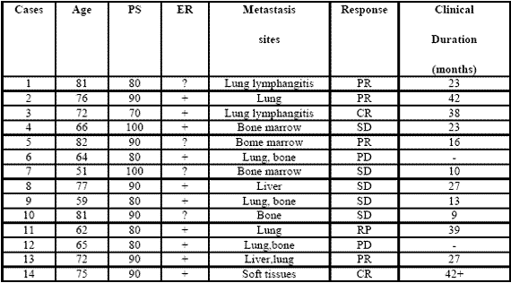
090722_phase2study

Table 1: Clinical characteristics of metastatic breast cancer women and their clinical response (WHO criteria) to a neuroendocrine regimen consisting of anastrozole plus the pineal hormone melatonin.

III. Results
All patients were fully evaluable for the clinical response. The clinical characteristics of patients and their individual clinical response to the treatment are reported in Table 1. As reported, a complete response (CR) was achieved in 2/14 (14%) (soft tissues:1; lung lymphangitis:1). A partial response (PR) was obtained in other 6/14 (43%) (bone:1; lung:3; liver:1; bone marrow:1). Then, an objective tumor response (CR + PR) was reached in 8/14 (57%) patients. The median duration of response was 26 months (range 9-42 months). A stable disease (SD) was seen in other 4/14 (29%), with a median duration of 25 months (range 10-27). Therefore, a disease-control (DC:CR + PR + SD) was achieved in 12/14 (86%) patients, whereas the remaining 2/14 (14%) patients had a progressive disease (PD). No significant difference in tumor response rate was observed between patients with positive or unknown ER ( 6/10(60%) vs 2/4(50%) ). An overall survival at 1 year and at 3 year was achieved in 11/14 (79 %) and in 5/14 (36 %) patients, respectively. Moreover, 3/14 (21%) patients were still alive at 5 years. The treatment was well tolerated in all patients.

Moreover, most patients experienced a relief of asthenia under the treatment and in no patient the neoplastic cachexia occurred. Finally, an evident increase in PS mean values was achieved under treatment, even though it did not reach the statistical significance (86 ±5 vs 93 ± 4, mean ± SE).

IV. Discussion
The results of this preliminary phase II study, by showing a percentage of 1-year survival greater than 70% in patients with live expectancy less than 1 year, would suggest that a neuroendocrine regimen consisting of the aromatase inhibitor anastrozole plus the pineal neurohormone MLT may represent a new effective therapeutic strategy in the treatment of metastatic breast cancer women, also in patients with poor clinical conditions, who would not be able to tolerate the most aggressive therapies. The concomitant administration of the pineal hormone would seem to enhance the efficacy of the aromatase inhibitor in terms of objective tumor regressions with respect to the results commonly reported in the literature with the only aromatase inhibitor (Plourde et al, 1994), which are generally lower than 40%.

The time to progression would seem to be apparently increased by the concomitant treatment with MLT.This finding is not surprising, since MLT could enhance the therapeutic anticancer acitivity of the aromatase inhibitors by either exerting direct antiproliferative antitumor effects (Bartsch et al, 1981; Maestroni, 1993; Reiter et al, 2002), or further inhibiting the aromatase activity by acting on gene and oncogene expression (Molis et al,1995; Cos et al, 2005). In addiction, MLT appeared to stimulate ER expression of breast cancer lines, by transforming ER negative into ER positive breast cancer, as observed in experimental conditions (Danforth et al, 1983).

Since the prognosis of ER positive breast cancers is clearly better than that of ER negative ones, MLT could per se improve the clinical couse of mammary tumors. Finally, because of its interesting therapeutic efficacy as a supportive care (Reiter et al, 2002), MLT would be responsible for the evident improvement in the relief of asthenia and in preventing the occurrence of the neoplastic cachexia. On the other hand, because of the inhibitory effect of MLT (Grant et al, 2009; Reiter et al, 2002) on cancer cell proliferation, the anticancer activity of this polyendocrine regimen would be due not only to an indirect effect, depending on a diminished estrogen production following aromatase enzyme inhibition, but also on a direct inhibition of cancer cell growth, due to MLT itself. Therefore, the results of this preliminary study may justify further clinical randomized investigations with the only aromatase inhibitor versus the concomitant treatment with MLT, in an attempt to confirm the ability of the pineal hormone to enhance the antitumor properties of the aromatase inhibitors in the treatment of metastatic breast cancer women with poor clinical conditions.

References
Bagatell CJ, Dahl KD, Bremner WJ (1994). The direct pituitary effect of testosterone to inhibit gonadotrophin secretion in men is partially mediated by aromatization to estradiol. J Androl 15:15-21.

Bartsch C, Bartsch H (1981) effect of melatonin on experimental tumors under different photoperiods and times of administration. J Neural Transm 52:269-279.

Bartsch H, Buchberger A, Franz H,Bartsch C,Maidonis I,Mecke D, Bayer E (2000) Effect of melatonin and pineal extracts on human ovarian and mammary tumor cells in a chemosensitivity assay. Life Sci 67:2953-2960.

Bhatnagar AS, Muller P, Schenkel L, Trunet PF, Beh I, Schieweck K. (1992). Inhibition of oestrogen biosynthesis and its consequences on gonadotrophin secretion in the male. J of Steroid Biochemistry and Molecular Biology;41:1021-1027

Cos S, Martinez-Campa C,Mediavilla MD, Sanchez-Barcelo E (2005). Melatonin modulates aromatase activity in MCF-7 human breast cancer cells. J Pineal Res 7:136-142.

Cos S, Gonzales A, Martinez-Campa C, Mediavilla MD, Alonzo-Gonzales C, Sanchez-Barcelo EJ (2008): Melatonin as a selective estrogen enzyme modulator. Curr Cancer Drug Targets. Dec 8(8): 691-702.

Danforth DN, Tamarkin L, Lipmann LE: (1983) Melatonin increase oestrogen receptor binding activity of human breast cancer cells. Nature, 305:323-325.

Grant S.G, Melan MA, Latimer JJ, Witt-Enderby PA (2009) : Melatonin and Breast cancer : cellular mechanism, clinical studies and future perspective. Expert Rev Mol Med 11:e5

Lissoni P, Barni S, Meregalli S, Fossati V, Cazzaniga M, Esposti D, Tancini G (1995) Modulation of cancer endocrine therapy by melatonin:a phase II study of tamoxifen plus melatonin in metastatic breast cancer patients progressing under tamoxifen alone. Br J Cancer 71:854-856.

Maestroni GJM (1993) The immunoneuroendocrine role of melatonin. J Pineal Res 14:1-10.

Molis TM, Spriggs LL,Jupiter Y,Hill SM (1995) Melatonin modulation of estrogen-regulated proteins, growth factors,and protooncogenes in human breast cancer. J Pineal Res 18:93-103.

Plourde PV, Dyroff M, Dukes MD (1994) Arimidex: a potent and selective fourth generation aromatase inhibitor. Breast Cancer Res Treat 30:103-111.

Reiter RJ, Tan DX, Sainz RM, Mayo JC, Lopez-Burillo S. (2002) Melatonin:reducing the toxicity and increasing the efficacy of drugs. Pharm Pharmacol 54:1299-1321

(download PDF version)


Evaluation of neopterin and ALCAT test in patients with food intolerance

d.ssa Berardi della IRCCS Policlinico San Matteo di Pavia reparto di dermatologia

Background:
Specific foods and food additives can induce adverse gastrointestinal (GI) and cutaneous reactions, including food allergies that involve an abnormal immunologic reaction to food proteins or food intolerance which is not pathogenetically immune-mediated.

Neopterin is synthesized by human monocyte-derived macrophages upon stimulation with interferon-gamma (IFN-gamma). Measurement of neopterin concentrations is useful for monitoring cell-mediated immune activation as there is a positive direct relationship between neopterin levels and severity of immune-mediated disorders. The aim of this study was to evaluate the neopterin serum levels in a group of patients with food intolerance.

Methods:
A group of forty-six patients (40 females, 6 males, median age 33 years) affected by cutaneous disease (27%) or GI symptoms (73%) consequent to food ingestion were tested by ALCAT and neopterin serum levels were measured.

The ALCAT test utilizes electronic haematology instrumentation and computerized data analysis to measure volumetric shifts in peripheral blood cells following incubation with food antigens.
Results are expressed in terms of percent change for cell volume and number. The degree of reactivity was determined by comparing a baseline distribution curve (of WBC) against the distribution curve generated by the analysis of each test agent/blood sample, and calculating the absolute differences between the curves and the standard deviation (SD). Any reactivity under 2 SD was considered non-reactive (negative) and these foods are allowed in the diet.

The Neopterin-MW EIA kit (DRG Instruments GmbH) employed an enzyme immunoassay technique to measure neopterin in a serum sample and was performed according to the Manufacturer’s Instructions and expressed as ng/mL.

Neopterin values were reported as medians with first and third quartiles (interquartile range). ALCAT test data were described as a percentage of non-reactive foodstuffs. Correlation between neopterin serum levels and percentage of non-reactive food was evaluated by means of the Spearman’s correlation coefficient (r).

Results:
The neopterin median value was 2 ng/mL (25°th-75°th: 1.50-2.25 ng/mL). There was a significant positive relationship between neopterin serum levels and percentage of non-reactive foodstuffs (p=0.05, r=0.302).

Conclusion:
This preliminary study confirms that food intolerance is not provoked by immunological mechanisms.

home page EAACI 2010 >>


Novembre 30, 2012 Newsletter

La gravidanza è uno stato particolare durante il quale tutto il metabolismo della donna va incontro a profonde modificazioni. È bene quindi provvedere con un cambiamento nutrizionale in grado di fornire il corretto apporto calorico e tutti i nutrienti necessari alla mamma e alla crescita del bambino.

Durante le visite di routine, peso e bilancia sembrano essere il fulcro cruciale per gran parte di medici e ginecologi. In realtà, accanto al capitolo del controllo del peso, si apre quello sul corretto apporto di macro e micronutrienti e quello riguardante la prevenzione di sindromi allergiche e intolleranze. Per il benessere di mamma e bambino è importante quanto la donna mangia, ma è fondamentale che cosa mangia. La gestazione deve essere un momento di “educazione alimentare” e non di “restrizione”.

Data la posta in gioco, la gravidanza può essere per la donna un’ottima occasione per acquisire nuove conoscenze in ambito nutrizionale come il corretto frazionamento dei pasti durante la giornata, il corretto apporto di tutti i macronutrienti (carboidrati, proteine, fibre, lipidi) e micronutrienti (sali minerali, vitamine) e la prevenzione di allergie e intolleranze.

L’aumento ponderale in gravidanza è strettamente correlato al peso pre-gravidico e al BMI (Boby Mass Index o Indice di Massa Corporea): un BMI corretto e nella norma lascia di solito ben sperare in un aumento di peso corretto rispetto a un BMI da sottopeso o sovrappeso. Nel primo trimestre, è previsto un aumento di peso di circa 2 kg, dal secondo trimestre il controllo deve essere più oculato e non deve essere superiore ai 500g settimanali onde evitare sofferenza fetale.

In generale evitare gli alcolici e limitare i “nervini” (caffè, tè, cacao) che stimolano la contrazione muscolare e quindi anche quella uterina.

E’ bene frazionare i pasti rispettando lo spuntino di metà mattina e metà pomeriggio così da evitare pasti abbondanti che possono aggravare sintomi quali gonfiore, nausea, vomito. Sempre per gli stessi motivi è consigliato evitare cibi “pesanti” quali fritti, piatti troppo conditi, grassi animali (lardo e pancetta), dolci a base di panna e burro. Nei casi di nausea e vomito evitare piatti in brodo, preferire una colazione salata a quella dolce, prediligere le verdure cotte e condimenti a base di limone (ricordiamo che la presenza di vitamina C favorisce l’assorbimento del ferro presente negli alimenti).

È consigliata la massima igiene alimentare e un’abbondante idratazione (2L di acqua al giorno).

Tenere presente l’aumento del fabbisogno nutrizionale e in particolare di vitamine, calcio, potassio, ferro, fosforo e acido folico; da qui l’importanza di un’alimentazione completa. Meglio sopperire a tale fabbisogno da fonti naturali (gli alimenti) prima che dagli integratori (per quanto riguarda l’acido folico, di cui gli alimenti sono poco ricchi, è bene assumere un integratore secondo indicazione medica). Si consiglia di accompagnare i pasti principali con una buona porzione di verdura (meglio se di stagione) e di assumere due porzioni di frutta al giorno. Frutta, verdura e legumi sono ricchi in micronutrienti. Prediligere, inoltre, i cibi integrali (il pane integrale ad esempio contiene sette volte più ferro di quello bianco).

Veniamo ora all’importanza di un’alimentazione sì equilibrata ma allo stesso modo “preventiva”, come già accennato, nei confronti di allergie e intolleranze alimentari. Ad esempio, nei casi di donne particolarmente allergiche, alcuni studi riportano una minore incidenza di forme allergiche ereditarie se, durante la gravidanza, la donna ha seguito un controllo nutrizionale atto a ridurre tutti i nutrienti che potrebbero far scaturire il problema già nel neonato. Preferire quindi alimenti “anallergici” quali carni bianche, pesce, riso, latte di capra, ecc. riducendo il consumo di latte vaccino e latticini, uova e alimenti molto allergenici (fragole, pesche, frutta secca, cacao, ecc.).

La nutrizione della donna gravida deve essere sì “libera” e a suo gusto secondo le cosiddette “voglie”, ma è comunque giusto attenersi a un profilo nutrizionale molto vario così da evitare, o accentuare, intolleranze alimentari nella mamma e quindi “accumuli” nel nascituro.

Se sospettate di essere intolleranti o se semplicemente volete assicurarvi di seguire un’alimentazione corretta anche per il vostro bambino, una buona idea potrebbe essere quella di eseguire il test ALCAT per le intolleranze alimentari. In seguito, un esperto potrà indicarvi il tipo di alimentazione più idonea per voi, così da evitare spiacevoli sintomi in un momento tanto atteso! L’ALCAT Test, diffuso da più di vent’anni e validato da numerosi studi condotti presso centri di ricerca (Fondazione IRCCS Policlinico San Matteo di Pavia, Laboratorio di Immuno-Allergologia, pubblicazioni sulla rivista ufficiale dell’European Academy of Allergology and Clinical Immunology), permette di seguire una terapia non farmacologica ma nutrizionale. Dopo una prima fase di eliminazione dei cibi risultati positivi al test, si procederà con la reintroduzione graduale e controllata di tutti gli alimenti.

In conclusione, oltre che per il rischio di aumento ponderale, il controllo dell’alimentazione della madre in gravidanza è importante per lo sviluppo del feto e per la prevenzione di allergie, intolleranze alimentari, dermatiti atopiche, bronchioliti, coliti, ecc., sintomi che possono presentarsi già dalle prime settimane di vita del bambino.

a cura della dr.ssa Jessica Barbieri, Biologa Nutrizionista, Consulente IMBIO – Istituto di Medicina Biologica Milano.


Ottobre 30, 2012 Newsletter

Dopo aver presentato pregi e difetti di “zucchero bianco, di canna e fruttosio“, (Come dolcificare nel modo migliore? – parte 1) proseguiamo la panoramica sui dolcificanti naturali e sintetici valutando: saccarina, aspartame, sucralosio e stevia.

SACCARINA: La saccarina è stata il primo dolcificante artificiale. La parola deriva dal latino e significa zucchero. È disponibile in tre forme: Acido Saccarinico, Saccarina di Sodio, Saccarina di Calcio. Quella più usata è la saccarina di sodio.

Ha un potere dolcificante 300 volte superiore al saccarosio, ma presenta un retrogusto leggermente amaro e metallico, generalmente considerato sgradevole specialmente ad alte concentrazioni. A differenza di simili composti di sintesi (es. aspartame), la saccarina è stabile al calore anche in ambiente acido, è inerte rispetto agli altri ingredienti alimentari e non richiede precauzioni di conservazione. Nei paesi in cui l’uso di entrambi i composti è consentito, la saccarina è spesso associata al Ciclammato in proporzioni 1:10 per correggere i citati difetti di retrogusto; è spesso associata anche all’aspartame.

Nel 1878 fu scoperta casualmente in un laboratorio, dove si lavorava il catrame. Fu una scoperta importante, soprattutto per i diabetici. Infatti, la saccarina transita attraverso l’apparato digerente senza alterare i livelli sanguigni d’insulina e in pratica senza fornire alcuna energia all’organismo.

Preoccupazione sulla potenziale nocività e cancerogenicità si espresse massimamente nel 1977, quando fu pubblicato un lavoro che comunicava l’aumento d’incidenza del cancro alla vescica in ratti alimentati con alte dosi di saccarina. Al momento, viste le bassissime quantità di utilizzo, non è stata dimostrata per tali quantità nessuna correlazione. La saccarina è impiegata in una grande varietà di cibi, bevande e cosmetici.

Non è metabolizzata dal nostro organismo; una volta assunta, è rapidamente assorbita (circa 90%) e come tale eliminata con le urine, senza subire modifiche. Non influenza i livelli glicemici e non fornisce alcuna energia all’organismo. Non favorisce la carie e quindi consigliata nelle diete ipocaloriche e nei diabetici. Restano, tuttavia, molti dubbi sulla tossicità, pur esistendo tantissimi studi che ne confermano la sicurezza per dosi di consumo normali. I dubbi circa il coinvolgimento della sostanza nei confronti del cancro alla vescica restano. Molta prudenza per l’uso in gravidanza, data la capacità di attraversare la placenta.

ASPARTAME: L’aspartame è un altro dolcificante artificiale. E’ composto da due aminoacidi, l’acido aspartico e la fenilalanina, più il metanolo che esterifica l’estremità carbossilica della Fenilalanina.

Pur avendo le stesse calorie dello zucchero, il suo potere dolcificante è 200 volte superiore; per questa ragione ne servono piccole quantità per dolcificare cibi e bevande.

Le persone che soffrono di fenilchetonuria (hanno difficoltà nell’assimilare la fenilalanina), devono limitare l’assunzione di questo dolcificante perché è fonte di fenilalanina.
L’utilizzo come dolcificante alimentare, con la sigla E 951, è autorizzato in dose massima giornaliera di 40 mg./Kg di peso. Esistono molte diatribe sul ruolo cancerogeno dell’aspartame (ci sono numerosi studi in merito). Nel 1980, un’inchiesta decretò la mancanza di dati sufficienti a confermare il legame tra aspartame e tumori al cervello.

Tuttavia, fu negata una nuova autorizzazione all’uso, in attesa di nuovi dati. Altri studi evidenziarono l’aumento dell’incidenza di linfomi e leucemie nei topi femmine a seguito dell’assunzione di vari dosaggi di aspartame. Inoltre, uno studio italiano ha confermato questi dati, e ha ipotizzato un legame tra formaldeide (il metabolismo dell’Aspartame, libera anche Metanolo, trasformato prima in Formaldeide e poi in Acido Formico, entrambi tossici) rilasciata dal metabolismo dell’aspartame e l’aumento di tumori cerebrali (Europee Journal of Oncology, del 2005). Entro settembre 2012, l’EFSA, la corrispondente europea dell’FDA americana, dovrà rivalutare la sicurezza dell’aspartame. Particolare importante: qualora sia presente un disturbo (potrebbero esisterne diversi), imputabile all’assunzione di aspartame, sono necessari 60 giorni senza assunzione di aspartame, per far regredire tale sintomatologia.

Uno dei più grossi limiti dell’aspartame è quello della non resistenza alle temperature (superiori ai 30°), quindi anche quella del corpo umano, che attiverebbe i processi che portano alla liberazione delle sostanze tossiche di cui parlavo prima. Per dovere d’informazione, riporto quali sono i punti a favore dell’aspartame che ne spingono l’utilizzo: basso potere calorico (essendo circa 200 volte più dolce del saccarosio) per cui ne servono piccolissime quantità; Non innalza la glicemia, per cui adatto ai diabetici, ma ricordiamo tutti i grossi dubbi ancora esistenti.

SUCRALOSIO: Diffuso da poco tempo, anche se scoperto in Inghilterra già nel 1976, questo dolcificante deriva dal saccarosio e, per le manipolazioni cui è sottoposto lo zucchero, è considerato un dolcificante artificiale.

E’ realizzato con un processo in più fasi, si parte dal classico zucchero da cucina, il saccarosio, cui sono sostituiti tre gruppi ossidrilici (formati da ossigeno e idrogeno) con tre atomi di cloro. Il risultato è un dolcificante stabile, con lo stesso sapore dello zucchero, ma senza calorie.

Dopo questa scoperta, il sucralosio, è stato sottoposto a un programma conclusivo di test sulla sicurezza per un periodo di 20 anni. Oggi il sucralosio, è autorizzato in oltre 80 paesi. Può essere utilizzato da tutti, anche da bambini, durante la gravidanza o l’allattamento, ed anche dai diabetici. Resiste al calore e può essere sottoposto a cottura, anche in forno. E’ molto utile per quanti cercano di ridurre l’assunzione di zucchero e calorie. Aumenta sempre più il numero di alimenti e bevande che sono dolcificate con sucralosio. Quello che rende possibile l’utilizzo in piccolissime quantità, è un potere dolcificante circa 600 volte superiore a quello dello zucchero.
Grosso vantaggio è l’eccellente sapore dolce, molto simile a quello dello zucchero.

Inizialmente vi fu un dubbio sul destino degli atomi di cloro, ma attendibili accertamenti hanno promosso a pieni voti questa sostanza. Infatti, il sucralosio rimane intatto nell’organismo. Il cloro non è liberato, dato che la molecola di sucralosio rimane intatta ed è eliminata quasi totalmente senza modifiche. Altro dato importante è che la molecola non interagisce minimamente con l’alimento che la contiene.

STEVIA: recentemente si è diffuso un nuovo dolcificante, che presenta interessanti caratteristiche: la stevia.

Si tratta di una piccola pianta erbacea arbustiva perenne della famiglia delle composite (lattuga, calendula, cicoria), nativa delle montagne fra Paraguay e Brasile. Ha una buona capacità dolcificante: nella sua forma naturale è circa 10/15 volte più dolce del normale zucchero da tavola. Nella sua forma più comune di polvere bianca, estratta dalle foglie della pianta, arriva a essere dalle 70 alle 400 volte più dolce dello zucchero, pertanto pare sia il dolcificante naturale più potente. Una sola fogliolina fresca dopo qualche istante, trasferisce al palato una forte sensazione dolce, lasciando alla fine un lieve retrogusto di liquirizia. I principi attivi sono lo Stevioside e il Rebaudioside A.

Non causa diabete; non ha calorie; non altera la glicemia; non dovrebbe avere tossicità (al contrario dei dolcificanti sintetici); non provoca carie; non contiene sostanze artificiali; può essere utilizzata per cucinare. Sono stati ipotizzati anche alcuni impieghi in medicina per il diabete e l’obesità. Consente, infatti, di ridurre il consumo di zucchero, specie quello bianco.

Si utilizza ormai su larga scala, in Giappone, per esempio, è già usata per dolcificare la Coca Cola Light.
La sua commercializzazione è stata a lungo ostacolata per via dei numerosi interessi legati alla produzione di altri dolcificanti ( barbabietole da zucchero, canna da zucchero ecc).

Importanti caratteristiche che la connotano sono:

  • Non è fermentabile (come lo zucchero), quindi utile a chi soffre di candida
  • Stabile al calore, almeno fino a 200° C
  • Non è tossica, quindi sicura
  • E’ senza calorie
  • Può essere utilizzata dai Diabetici e nelle diete ipocaloriche

È sicuramente un buon dolcificante naturale, già usato in molti paesi; per il momento i limiti sono dati dai costi, non ancora competitivi per la coltivazione delle piante e per l’estrazione.

Infine, per concludere questa panoramica sui dolcificanti (ricordo la prima parte dell’articolo: Come dolcificare nel modo migliore? – parte 1), vorrei esprimere solo una breve considerazione. Tra i vari dolcificanti, vale sempre la regola delle quantità, ad esempio, il fruttosio, entro limitate quantità, può essere utilizzato tranquillamente, ma aumentando il consumo crescono, in maniera direttamente proporzionale, gli svantaggi. Ricordo inoltre le virtù della stevia e del sucralosio, che veramente restano inerti e senza calorie.

a cura del Dr Francesco Lampugnani, Biologo Nutrizionista, Specialista in Farmacologia


Ottobre 30, 2012 Newsletter

Le proprietà nutritive del latte sono sempre state associate al concetto di salute e crescita, e il suo consumo è parte di una cultura popolare che si tramanda da generazioni.Si tratta, infatti, di un alimento indispensabile per il bambino in crescita. Per gli adulti il discorso è differente e per valutare gli effetti positivi del suo consumo dobbiamo considerare diversi aspetti. Primo fra tutti, la qualità del prodotto.

La gestione dell’allevamento delle mucche da latte è stata modificata nel tempo, con importanti conseguenze nelle proprietà e nella sicurezza dell’alimento: il latte che beviamo oggi ha una qualità inferiore rispetto a quella di tanti anni fa. Perché il latte sia di massima qualità, le mucche dovrebbero poter pascolare e alimentarsi in erba, libere di muoversi per tutto il giorno. Lo scenario può essere invece molto diverso: le mucche sono mantenute in batterie, con mangimi a base di soia, fieno e alimenti derivati. Non possono muoversi liberamente e passano gran parte del tempo a mangiare biada, sottoposte a stress produttivo con la somministrazione di fattori di crescita che aumentano la produzione di latte, anche di 4 o 5 volte in più rispetto alla normalità.

Il latte prodotto dalle mucche al pascolo sarà quindi più ricco di omega tre, antiossidanti naturali, proteine, calcio e minerali, mentre quello prodotto dalle mucche in batteria conterrà questi fattori nutritivi in quantità ridotta.

Altra importante considerazione riguarda la correlazione tra il consumo di latte e latticini nell’adulto e il rischio di contrarre patologie prostatiche, fino al cancro prostatico. [Torniainen S. et al Lactase persistence, dietary intake of milk, and the risk for prostate cancer in Sweden and Finland, Cancer epidemic, Bio makers e prevention, 2007, 16 (5), pp. 956-61].

Dall’altra parte però esiste anche un ruolo protettivo per il tumore al colon, data la presenza nel latte di una sostanza chiamata lattoferricina che deriva dalla digestione enzimatica della lattoferrina, nota per le proprietà anti cancro e immunostimolanti.

Nella donna, un elevato consumo di latte e latticini favorisce la produzione di cisti ovariche, seno fibromatoso e tumore al seno. Ecco perché le linee Guida della prevenzione anticancro, diffuse dai centri di terapia oncologica, sconsigliano il consumo di latte alle donne che hanno avuto un problema oncologico a seno, utero e ovaie.

Inoltre, Il 55% della popolazione mondiale è intollerante al lattosio, il principale zucchero del latte. Tuttavia, esiste una grande variabilità geografica: nei paesi dell’Europa settentrionale il 60-70% delle persone mantiene Ia funzione dell’enzima che digerisce il lattosio. Man mano che si scende dal nord Europa, invece, l’attività della lattasi si riduce: nella nostra penisola e dintorni, bacino del Mediterraneo, il deficit genetico per la Lattasi è molto frequente.

Come tutte in tutte le situazioni confuse, è importante non adottare concetti estremi e prese di posizione inutili. Forse basta una domanda molto semplice: abbiamo bisogno di grandi quantità di latte e prodotti lattiero caseari?
I bambini sì, è chiaro per tutti. Gli adulti no!

Infine, è importante ricordare che uno screening genetico può aiutarci a individuare le persone che possono trarre beneficio da un’alimentazione a base di prodotti lattiero caseari. La personalizzazione del consiglio nutrizionale, e il buon senso, ancora una volta, prevalgono sulle credenze popolari. Bisogna rivolgersi a uno specialista in nutrizione che possa avvalersi dell’indagine genetica, per meglio consigliare e educare non solo il singolo soggetto ma tutta la famiglia.


Ottobre 30, 2011 Newsletter

La resistenza insulinica può essere definita come una condizione in cui una data concentrazione d’insulina produce un effetto minore di quello atteso.

Epidemiologicamente è un fenomeno rilevante con una prevalenza stimata del 3% nella popolazione statunitense e variabile tra il 3 e il 16% nella popolazione occidentale. La minore prevalenza si registra nella popolazione giapponese, dove si attesta al 2% circa.

Secondo le stime dell’OMS, a causa del progressivo aumento della vita media e dell’incremento demografico nei paesi sottosviluppati, la prevalenza mondiale di questa patologia è destinata a modificarsi significativamente soprattutto nella fascia di popolazione tra 45 e 64 anni dove potrebbe arrivare a raddoppiare entro il 2030. Nella fascia di popolazione oltre i 65 anni, data l’aspettativa di vita media della donna superiore a quella dell’uomo, ci si potrebbe aspettare una maggiore prevalenza nel sesso femminile.

La resistenza insulinica predispone all’intolleranza glucidica e con essa allo sviluppo della sindrome metabolica e del Diabete mellito di tipo 2 con le complicanze che ne conseguono.

Oltre che nelle alterazioni del metabolismo glucidico, la resistenza insulinica sembra essere implicata in un concomitante incremento dei trigliceridi e una diminuzione del colesterolo HDL; è importante inoltre rilevare che la predisposizione genetica può aumentare il rapporto trigliceridi / HDL (colesterolo buono).

La diagnosi precoce di tipo genetico che è possibile effettuare presso IMBIO (Istituto di Medicina Biologica di Milano), permette di attuare un programma nutrizionale adeguato, con particolare attenzione al tipo di alimenti che influenzano l’espressione del gene mutato che saranno quindi eliminati o ridotti dall’alimentazione.

Con un piano alimentare personalizzato in base al risultato del test e un’attività fisica moderata e costante si può ridurre, fino ad abbattere, la possibilità di sviluppo del Diabete Mellito di tipo 2.


Marzo 30, 2011 Newsletter

a cura del dr. Francesco Lampugnani

Il problema delle intolleranze alimentari sta diventando sempre più diffuso tra la popolazione e, sicuramente, i pazienti che riescono a confermare la presenza di un’intolleranza alimentare legandola ad una sequela di sintomi lamentati è ancora una grossa minoranza rispetto al grande numero di pazienti che, pur lamentando problemi importanti legati al tipo di vita che si conduce, non identificano un’intolleranza alimentare e di conseguenza un atteggiamento correttivo idoneo, perché o non conoscono l’esistenza della problematica (intolleranza), o perché non hanno ricevuto le giuste informazioni, o perché grosse dosi di scetticismo regnano 110328_alcatancora negli ambienti sanitari.

Fatta questa breve premessa, la mia esperienza in ambulatorio mi permette di confermare quanto riferito, in quanto una grossa percentuale di pazienti si è avvicinata ed ha effettuato il test per le intolleranze alimentari (che successivamente definiremo IA) solo dopo che ha avuto chiare tutte le informazioni necessarie ad associare le diverse sintomatologie manifestate alla presenza di un’IA.

Cosa molto importante è il non sapere, da parte del paziente, che tutta una serie di sintomi manifestati (gonfiore, stanchezza, stipsi, diarrea, cefalea, disturbi dell’umore, ecc) possono essere scatenati dalla presenza di un’IA. Arrivando insieme, durante l’importante momento conoscitivo dell’anamnesi, il paziente si rende conto che tante situazioni, alle quali molte volte non da peso, sono legate all’introduzione di determinati alimenti e che la successiva eliminazione o riduzione potrebbe essere di grande aiuto per il miglioramento della sintomatologia.

Come ormai sappiamo, definiamo Intolleranza Alimentare una reazione ritardata fino a 72 ore dopo l’assunzione di alimenti quotidiani e che si traduce in sintomi molto simili a quelli di un’allergia.

Alla luce della mia esperienza, sicuramente inizio a delineare importanti risultati legati alla presenza di un’IA e le abitudini di vita dei pazienti. Infatti, tra le associazioni più presenti, rientrano sicuramente le ripetitività alimentari: pazienti che ormai da anni assumono sempre gli stessi alimenti, vuoi per pigrizia, vuoi per ristrettezze di gusto.

Altre situazioni che si presentano sono legate alla qualità alimentare. Pazienti con alimentazione poco “salutista”, con utilizzo di alimenti molto sintetici (merendine, alimenti conservati, alimenti con molti grassi ed additivi chimici, ecc) e con pochi alimenti freschi e naturali, hanno manifestato, dopo anni di alimentazione di questo tipo, disturbi che ben indagati e sviscerati, hanno espresso la presenza di un’IA.

Altra situazione molto diffusa è quella legata a pazienti che hanno fatto abbondante uso di antibiotici o che hanno una situazione intestinale non ottimale. Infatti, riuscendo a ristabilire quel famoso equilibrio intestinale che è alla base di una corretta funzionalità, associato al trattamento di alleggerimento verso certi alimenti, si riesce a risolvere diverse situazioni alterate. Questi ultimi riferimenti mi permettono di sottolineare quanto sia importante avere sempre in giusta considerazione l’Eubiosi intestinale. Infatti il primo approccio operativo nei confronti di pazienti con disturbi associabili alla presenza di un’IA è proprio quello di riprendere uno stato di funzionalità intestinale ottimale. Cosa che di solito riesco a fare modificando le abitudini alimentari ed associando sempre Prebiotici e Probiotici, utili anche per una ripresa immunitaria a livello intestinale.

Per quanto riguarda la mia casistica, penso di poter dare un piccolo contributo analizzando un parte di pazienti che hanno fatto il Test per le Intolleranze Alimentari “ALCAT”.

Nello specifico, posso riferire di una casistica di 46 pazienti (31 donne e 15 uomini), con un’età compresa tra i 15 ed 65 anni, con una maggior presenza tra i 46 e 50 anni (17%) ed i 41 e 45 anni (11%) e tra i 56 e 60 anni (11%). Si iniziano anche a delineare dei profili di insorgenza delle IA molto interessanti.
Infatti come dati molto preliminari, saltano all’occhio due importanti risultati: tra gli uomini, l’intolleranza più rappresentata, è stata quella per lo Zucchero di Canna (60% del campione). A seguire Cacao (46% del campione), Caffè (40% del campione), Lievito Chimico (20% del campione) ed a seguire tutte le altre.
Per le donne invece, sempre per il campione analizzato, ho notato questo tipo di risposta: la più rappresentata è stata quella al Pomodoro (48% del campione), a seguire, Caffè, Frumento, Lievito Chimico (per tutte 35%). Discorso a parte merita l’intolleranza al Lattosio, sempre più diffusa (ovviamente), che per i due gruppi ha rappresentato una percentuale tra il 26 ed il 32%.

Molto interessante è una conferma pervenuta da uno studio Americano molto recente che dimostrava l’associazione, nei pazienti studiati, tra Intolleranze al Frumento ed ai Lieviti e la presenza di Tiroiditi. Anche nella mia casistica, ho potuto verificare quasi totalmente tale affermazione, che meriterà logicamente maggiori approfondimenti.

In conclusione, il mio giudizio su questo tipo di test è molto positivo. Ho avuto modo di confrontare i rilevamenti con altre realtà e sicuramente i risultati più importanti li ho avuti con l’Alcat.

Il test, cosa molto importante, mi permette di avere una visione più completa del paziente, che a sua volta riesce a confermare quasi del tutto la presenza dell’Intolleranza ad un alimento molto sospetto, con la controprova del miglioramento della sintomatologia legato alla disintossicazione ed alla riabilitazione verso l’alimento interessato. Inoltre, grazie all’individuazione degli alimenti, riesco a costruire un piano alimentare ritagliato a dovere e permettere tutta una serie di cambiamenti necessari per il paziente (riduzione di peso o aumento di peso; sintomatologia generale; ed infine, cosa non da poco, miglior educazione alimentare).

Penso utile, ai fine dell’informazione, aggiungere che in un certo numero di pazienti anche sportivi ho avuto modo di constatare un miglioramento delle loro prestazioni atletiche, parallelamente al miglioramento della sintomatologia generale, dopo l’individuazione di un’Intolleranza Alimentare.


Dicembre 30, 2010 Newsletter

Nella medicina moderna assumono sempre maggiore importanza le strategie terapeutiche che prevedono la integrazione tra diverse metodologie di cura allo scopo di migliorare il risultato finale. In particolare ciò è vero per i pazienti oncologici nei quali l’approccio con le terapie tradizionali può essere favorevolmente ampliato affiancando altri metodi di cura che possono migliorare il risultato terapeutico.

E’ il caso dell’ipertermia, trattamento conosciuto da molti anni e utilizzato in molti Paesi.

L’indicazione al trattamento viene data dopo una valutazione dei dati clinici e strumentali e spesso viene associata una terapia di supporto intesa non solo come terapia sintomatica ma anche come modalità per cercare di sostenere il metabolismo e il sistema immunitario: ciò significa che oltre alla malattia si cerca di curare soprattutto il paziente, intendendolo come un insieme di organi e apparati che si influenzano vicendevolmente e non, come accade spesso ancora oggi, considerando i vari distretti corporei completamente staccati gli uni dagli altri.

L’ipertermia è un trattamento che utilizza le proprietà terapeutiche del calore: di fatto si sfrutta la minore capacità del tumore di tollerare le temperature oltre i 40 gradi.
Attraverso una somministrazione con tempi e temperature ben determinati si ottiene una valida azione antitumorale con un meccanismo di danno diretto alle cellule tumorali e con la creazione di un ambiente sfavorevole alla loro crescita; può essere favorevolmente applicata in associazione alla chemioterapia e alla radioterapia sia con intento curativo che con intento di miglioramento della qualità di vita e questa associazione è provata da numerosi studi scientifici.

La tolleranza ai trattamenti di ipertermia è generalmente molto buona per cui alla fine della terapia il paziente non lamenta effetti collaterali rilevanti.

Purtroppo in Italia non sono molti i Centri che utilizzano l’ipertermia ed anche nell’ambito oncologico questa terapia è poco conosciuta; date le sue modalità di azione, la sua facile applicabilità e la scarsità di effetti collaterali rilevanti può invece essere un valido supporto nell’iter terapeutico del paziente oncologico.

a cura del dr. Marco Mancuso


Settembre 30, 2009 Newsletter

Attualmente la patologia oncologica rappresenta la seconda causa di mortalità subito dopo le patologie cardiovascolari; sebbene con le terapie classiche – chirurgia-radioterapia e chemioterapia – si possano ottenere buoni risultati, la complessità, la lunghezza e gli effetti indesiderati di queste cure mettono a dura prova i pazienti ed i loro familiari. Non è raro osservare che a causa degli importanti effetti collaterali i pazienti abbandonino le cure o che sia necessario sospendere temporaneamente o definitivamente il programma terapeutico riducendo l’efficacia delle cure stesse.

Sono molti i farmaci che possono contrastare efficacemente i possibili sintomi legati alla cura o alla patologia oncologica; spesso però a loro volta causano altri sintomi che devono essere contrastati con altri farmaci creando quindi una spirale terapeutica che alla lunga stanca; inoltre vengono sovraccaricati i meccanismi di detossificazione e di produzione di energia che in tempo variabile riducono la loro attività indebolendo l’organismo e le sue capacità di resistenza.

Pertanto cercare di mantenere il più possibile la efficienza dell’organismo e le sue capacità di rigenerazione diventa un obiettivo fondamentale che deve far parte dell’approccio oncologico. Ciò è ancora più importante nelle situazioni in cui, data l’impossibilità di giungere alla guarigione, si voglia intraprendere un programma terapeutico che miri a frenare l’evoluzione della malattia, per garantire il più possibile una buona qualità di vita.

Già da alcuni anni presso l’Istituto di Medicina Biologica vengono proposti alcuni schemi terapeutici che affiancano le terapie tradizionali allo scopo di migliorare il risultato terapeutico. Nella nostra esperienza, approcci terapeutici come ipertermia oncologica esterna, microimmunoterapia, integrazioni nutrizionali, oligoelementi, possono efficacemente far parte dell’iter terapeutico globale del paziente oncologico.

Il loro meccanismo di azione, la assenza di effetti collaterali rilevanti e i costi contenuti consentono di proporre un approccio valido ed efficace non in alternativa ma in sinergia con le terapie tradizionali.


Copyright by IMBIO 2017. All rights reserved.