Articoli

Tutto sul nostro settore


Aprile 1, 2015 Newsletter

E’ meglio mangiare pasta o riso?? La pasta ha un contenuto calorico leggermente superiore, è più ricca di lipidi, proteine e fibre mentre è più povera di acqua e carboidrati. Il riso avendo un contenuto proteico inferiore alla pasta, ha un indice chimico superiore che lo rende sotto questo punto di vista equivalente, se non addirittura superiore alla pasta.

Stabilito che mangiare riso fa bene bisogna imparare a cuocerlo!

La cottura del riso è la base per ottenere un risotto eccezionale ed è una delle cose più difficili da fare in cucina. Ci sono tante piccole regole da imparare e qualche suggerimento da seguire per ottenere un piatto perfetto.

Ve ne do 5 di suggerimenti che spero possiate trovare utili:

  • sappiate che per cuocere bene il riso è necessario curarlo, mescolare e aggiungere l’acqua o il brodo al momento giusto e poco a poco. Tuttavia, per chi ha pochissimo tempo a disposizione, suggerisco di provare la pentola pressione. Onestamente la trovo molto comoda: poco tempo (circa 5 minuti) e senza sforzo (potete lasciare anche il riso a se stesso, ci penserà la pentola a cuocerlo). Una soluzione molto pericolosa, però, in termini di resa della cottura. Ci vogliono diversi tentativi per capire gli esatti tempi di cottura e di liquido da inserire in pentola. Insomma, è un metodo da utilizzare solo in caso di necessità e dopo averci preso la mano.
  • altro aspetto da considerare è la qualità del riso, dalla quale dipende non solo il gusto finale del vostro piatto ma anche i tempi di cottura. Controllare i tempi sulla confezione è d’obbligo. Dovete considerare, però, anche un altro aspetto: da quanto tempo avete il riso in dispensa. Se il riso è vecchio, può richiedere un po’ più tempo per cuocere perché una parte dell’umidità in esso contenuta è evaporata.
  • mantecate il riso con olio o formaggio a fuoco spento senza esagerare nel mescolare. Evitare di girare il riso ossessivamente è un suggerimento che vale anche durante la fase di cottura, più lo girate, più la consistenza finale apparirà collosa, perché girando il riso disperdete l’amido. Per questo è importante stare attenti che ci sia sempre abbastanza brodo, non distraetevi e aggiungetelo quando vedete che si sta asciugando, altrimenti il riso si brucerà. Questo è un altro punto a favore della cottura del riso con la pentola a pressione, dove per la natura stessa della pentola, non è possibile intervenire sul riso, che per forza resterà immobile durante tutta la cottura.
  • Per ottenere un risotto “professionale” non dovete tralasciare la tostatura del riso. Buttate il riso sopra il soffritto e fatelo tostare per massimo un minuto. Quando vedete che cambia colore, lo avete tostato alla perfezione e potete farlo sfumare e aggiungere il brodo per la cottura.
  • Infine, un suggerimento che non sempre è preso in considerazione ma che vi assicuro è in grado di modificare il sapore del risotto: lasciatelo riposare qualche minuto prima di servirlo.


Novembre 23, 2014 Newsletter

Prof. Di Fede, perchè Alcat Test è migliore del citotossico? qual’è la differenza?
Sono tutti e due test effettuati mediante prelievo di sangue, ma i risultati dell’Alcat Test sono effettuati da un robot e quindi secondo me più veritieri e sicuri

Il cibo risultato positivo al test va escluso per sempre? 
No! assolutamente ,la sospensione è al massimo (nel caso del Ramge Mpos) di un mese, poi bisogna fare una dieta simile allo svezzamento e reinserire l’alimento gradualmente.
Il disturbo per il quale sono venuto, quando reinserisco l’alimento incriminato ritorna?
No, se si segue correttamente lo “svezzamento”, l’alimento risultato positivo è comunque sempre da non assumere tutti i giorni.

I test va ripetuto ?
No, secondo me, solo quando vi è una positività all’oliva, per verificarne che sia solo un sovraccarico dell’organismo e non un’intolleranza all’olea europea.

Qual’è la differenza con una allergia?
L’allergia crea una reazione immediata dell’organismo, l’intolleranza può svilupparsi anche dopo giorni, mesi, poichè si evidenzia solo quando l’organismo supera il livello di soglia.

Ho fatto un milione di diete, ma senza risultati e ho deciso di fare anche questo perchè una mia amica ha avuto ottimi risultati, e so che era positiva alla Candida, cos’è e perchè ha avuto risultati?
La Candida è un fungo che se supera ilivelli standard dà pesantezza, gonfiore, cattiva digestione e soprattutto non aiuta a perdere peso, quindi bisogna innanzitutto negativizzarla prima di procedere alla dieta di rotazione.L’organismo non risponde perchè va in memoria e meno mangiamo, più va in accumulo di riserve.

[youtube]https://www.youtube.com/watch?v=nzOlx7_fE94&feature=relmfu[/youtube]


Ottobre 28, 2014 Newsletter

Dopo la prima uscita dedicata al Nichel, impariamo a conoscere e combattere a suon di ricette l’intolleranza al lattosio con l’aiuto del Prof. Di Fede, della drssa Jessica Barbieri e da me.

Un aiuto concreto per chi sta vivendo un problema troppo spesso sottovalutato: perché le difficoltà non finiscono al momento della diagnosi. Lo so bene io che ho trasformato un limite in una possibilità, raccogliendo in questo volume tutte le risorse utili a tavola per chi come me soffre della carenza dell’enzima necessario a digerire gli zuccheri presenti nel latte e nei suoi derivati: informazioni, consigli, esperienze e soprattutto… ricette!

Sfogliate e gustate i 99 piatti creati da me, da foodlovers e da tanti amici Chef di fama internazionale coinvolti in un progetto unico: vivere l’intolleranza al lattosio non più come limite, ma come possibilità di sperimentare nuovi sapori.

Non resta che partire insieme in un viaggio nel Mondo delle Intolleranze, con un volume che racchiude in sé il problema e la sua soluzione: nella cucina come nella vita, quando la ami non c’è niente e nessuno che possa indurti a rinunciarvi!

Oltre alle ricette di Tiziana troverete ricette di:

Luca Mauri, Alessandra Barbone, Antonella Rossi, Enrico Bartolini, Andrea Bevilacqua, Claudia Livia Biondini, Massimo Cairati, Antonino Cannavacciuolo, Michele Cannistrano, Giuseppe Capano, Fabio Silva, Isa Mazzocchi, Laura Ghezzi, Massimo Livan, Federico Mattavelli, Matteo Scibilia, Aurora Mazzucchelli, Antonino Messina, Micol Pisa, Milly Callegari, Carlo e Loris Molon, Giancarlo Morelli, Luca Mozzanica, Nunzia Bellomo, Davide Oldani, Armando Palmieri, Roberto Maurizio, Sandro Romano, Almerindo Santucci, Sara Preceruti, Marco Scaglione, Gaetano Simonato,Daniele Zennaro, Robbie Pezzuol

Sono da  da sempre convinta che la cucina sia veicolo di passione e umanità, e non mi sono arresa dopo la diagnosi di intolleranza al nichel e al lattosio. Oggi www.nonnapaperina.it è un blog di riferimento nel mondo delle intolleranze e la mia presenza è richiesta nelle più grandi manifestazioni enogastronomiche italiane. Si perpepisce che il problema delle intolleranze sta crescendo a vista d’occhio.

Il volume dedicato all’intolleranza al lattosio segue il grande successo della sua prima pubblicazione Nichel. L’intolleranza? La cuciniamo!, edito nel 2013.

Siete tutti invitati alla presentazione che si terrà giovedi 13 novembre 2014 all’Hotel de la Ville di Monza alle ore 19.


Novembre 28, 2013 Newsletter

Il glutine è tra gli alimenti più diffusi nella nostra alimentazione, è presente in tutte le farine comunemente disponibili nei supermarket, nella ristorazione, nelle mense scolastiche e lavorative. Insieme all’olio d’oliva, la frutta, la verdura e il pesce rappresenta l’alimento che caratterizza la dieta di tipo mediterraneo. I cereali accompagnano l’alimentazione da diversi anni, ma la composizione del glutine è cambiata negli ultimi decenni.

Tra gli alimenti contenenti glutine abbiamo: orzo, crusca, couscous, farina, pasta, segale, semola, spelta, triticale (ibrido semola e grano tenero), frumento, germe di grano, farina di frumento, crusca di grano, semola di grano.

Negli ultimi anni, si è manifestata – dapprima in maniera subdola fino ad arrivare a interessare una buona parte della popolazione Italiana – una nuova sintomatologia clinica che colpisce l’intestino.

Dopo anni di ricerche e controlli clinici, i ricercatori del Maryland, hanno individuato le possibili cause alla base della sensibilità al glutine, chiamata Gluten Sensitivity.

Ebbene, possiamo distinguere diverse forme di sensibilità al glutine:

Celiachia, dove la componente genetica relativa alla presenza di mutazioni del polimorfismo del DQ2 e DQ8, insieme alla componente autoimmune, determina un’aggressione da parte degli anticorpi anti-gliadina, anti-transglutaminasi e a volte anche gli anti-endomisio, della mucosa intestinale, con sintomi che possono interessare qualsiasi parte dell’organismo. Circa l’1% della popolazione ne è affetta.

  1. Allergia al Glutine, dove la componente allergica è data da una classe ben precisa di anticorpi chiamati IgE, che si attivano contro la proteina del grano. I sintomi sono riconducibili a reazioni allergiche dermatologiche o delle mucose. I test per scoprire un’eventuale allergia al grano e quindi al glutine, si effettuano con i RAST degli alimenti. Presente nel 2-3% della popolazione.
  2. Gluten sensitivity o sensibilità al glutine, di recente scoperta, è stata riconosciuta come nuova patologia nella Consensus Conference di Gastroenterologia, tenutasi a Londra, nel marzo del 2011. La Sensibilità al glutine si presenta come una condizione mista delle due situazioni sopra descritte. La percentuale della popolazione sensibile al glutine si stima sia intorno al 6%, ma i dati clinici a disposizione dovranno essere confermati dai dati di laboratorio attraverso i nuovi esami a disposizione del medico clinico.

Quali possono essere i sintomi legati alla sensibilità al glutine?

I disturbi addominali la fanno da padrone, possono manifestarsi con dolore di tipo colico, gonfiore, stipsi o diarrea, flatulenza; afte e stomatite, disturbi extra addominali che coinvolgono le articolazioni, i muscoli e i legamenti, dolori ossei, fino alla possibile artrite e artrosi. I disturbi possono riguardare anche la pelle, sono possibili manifestazioni simili a orticaria, dermatite secca e pruriginosa e infine un eczema di difficile controllo. La stanchezza cronica e difficoltà di concentrazione e attenzione, facile esauribilità della forza muscolare, anemia da carenza di ferro, abbassamento delle difese immunitarie con esposizioni a virus anche aggressivi come la mononucleosi e le infezioni erpetiche recidivanti. Ma la caratteristica principale della sensibilità al glutine è la persistente lieve infiammazione della mucosa intestinale che nel tempo è in grado di coinvolgere altri distretti del corpo.

Per maggiore chiarezza precisiamo che la malattia celiaca è un’allergia al glutine che coinvolge sistema immunitario con produzione di IgE specifiche, mentre nella Sensibilità al glutine è coinvolto il sistema immunitario innato, rappresentato dai granulociti neutrofili o polimorfo nucleati e linfociti tipo B. La reazione di difesa quindi è data dai granulociti neutrofili, e si tratta pertanto di una reazione di tipo citotossica e non immunologica.

Finora, la diagnosi della sensibilità al glutine si effettuava escludendo le altre due possibilità patologiche, la celiachia e l’allergia. Oggi, grazie all’analisi computerizzata di tipo citotossica standardizzata di ALCAT test, che permette di individuare l’alimento che genera reazione citotossica, è possibile rilevare il livello di sensibilità alla famiglia degli alimenti del glutine, e quindi ottenere un valore diagnostico davvero significativo di patologia di sensibilità al glutine di tipo non celiaco. Il referto ALCAT Test indica i livelli possibili di reattività agli alimenti della famiglia del glutine, che possono variare da un minimo di reattività, reattività media, fino a un massimo rappresentati rispettivamente da 1+, 2+ e 3+.

Cosa fare una volta evidenziata la sensibilità al glutine? Bisogna prima di tutto affidarsi a un nutrizionista che si occupi d’intolleranze alimentari, seguire uno schema nutrizionale personalizzato, che preveda la rotazione degli alimenti, ossia introducendo gradualmente, dopo un periodo di astensione dal glutine, gli alimenti intollerati. Questo tipo di gestione permette il recupero della tolleranza al glutine attraverso un regime alimentare il più vario e bilanciato possibile che include momenti di libertà dalla dieta e favorisce il recupero dello stile di vita comune.

Una diagnosi tempestiva consente di recuperare più velocemente una situazione che se protratta nel tempo si cronicizza, favorendo l’insorgere di disturbi che oltre l’intestino possono interessare altre parti del corpo.


A clinical study of T-regulatory lymphocyte function in cancer patients in relation to tumor histotype, disease extention, lymphocyte subtypes and cortisol secretion

Research Article (download PDF version)

Luigi Vigorè1, Fernando Brivio2, Luca Fumagalli2, Roberto Vezzo1, Giusy Messina6, Franco Rovelli6, Massimo Colciago3, Giovanna D’Amico4, Giuseppe Di Fede5, Paolo Lissoni6

1 Laboratory of Immunomicrobiology, St.Gerardo Hospital, Monza, Milan, Italy
2 Division of Surgery, St.Gerardo Hospital, Monza, Milan, Italy
3 I.N.R.C.A Laboratory of Analyses, Casatenovo, Lecco, Italy
4 Research Center “Fondazione Tettamanti” Clinica Pediatrica Università Milano-Bicocca, Italy
5 Institute of Biological Medicine, Milan, Italy
6 Division of Radiation Oncology, St.Gerardo Hospital, Monza, Milan, Italy

*Correspondence: Dr. Paolo Lissoni, Divisione di Radioterapia Oncologica, Ospedale S.Gerardo, 20052 Monza, Milano, Italy; Fax: +390392332284, E-mail: p.lissoni@hsgerardo.org
Key words: Anticancer immunity, immunosuppression, T regulatory lymphocytes
Abbreviations: cytotoxic T lymphocyte-associated antigen-4, (CTLA-4); glucocorticoid-induced TNF-α receptor, (GITR); myeloidderived suppressor cells, (MDSC); NK cells, (CD16CD56); T cytotoxic lymphocytes, (CD8); T helper lymphocytes, (CD4); Tregulatory lymphocytes, (T-reg)

Received: 24 July 2008; Revised: 11 September 2008
Accepted: 12 September 2008; electronically published: October 2008

Summary

Several clinical investigations showed that the immune status is a prognostic variable in cancer patients, even tough the evaluation of the anticancer immunity is not generally considered in the medical oncology. Several immune parameters, including lymphocyte subsets and cytokine blood concentration, had been proposed to quantify the functional status of the anticancer immunity, but recent discoveries would suggest that the end-result of the various immune interactions is represented by a subtype of CD4 lymphocytes capable of suppressing the antitumor immune reaction, the so called T-regulatory lymphocytes (T-reg). This study was performed to detect T-reg count and percentage in solid tumor patients, in relation to tumor histotype, disease extension, lymphocyte sub-populations and cortisol circadian secretion. The study included 114 consecutive cancer patients affected by the most frequent tumor histotypes, 69 of whom showed a metastatic disease. In each patient we evaluated T-reg cells, identified as CD4+CD25+, in relation to T helper (CD4), T cytotoxic (CD8) and NK (CD16CD56) cells. Abnormally high values of T-reg cells were seen in 52/114 (46%) patients, and the percentage of high values of T-reg was significantly higher in metastatic patients than in non-metastatic ones. In contrast, no significant difference was seen in relation to tumor histotype. Patients with increased T-reg count had a significantly lower NK cell number. Finally no significant difference in T-reg number was seen between patients with altered or normal rithm of cortisol. The study confirmed that, irrespectively of tumor histotype the metastatic disease is associated with a progressive and increased T-reg generation, with a following suppression of anticancer immunity.

I. Introduction
At present, there is no doubt about the existence of a sub-type of T lymphocytes, the so-called T regulatory lymphocytes (T-reg), capable of suppressing the cellular immune responses,including the anticancer immunity (Thomton and Shevach, 2000; Shevach, 2002; von Herrath and Harrison, 2003; Schwartz, 2005; von Boehmer, 2005; Ziegler, 2006; Zou, 2006). However, the exact definition
of T-reg cells in terms of cell surface marker expression still remains controversial, particularly from a clinical point of view. All authors are in agreement to consider Treg lymphocytes as CD4+CD25+ cells, but at present it is still unknown whether the expression of CD4 and CD25 antigens may be sufficient to identify T-reg cells (Thomton and Shevach, 2000; Shevach, 2002; von Herrath and Harrison, 2003; Schwartz, 2005; von Boehmer, 2005; Ziegler, 2006; Zou, 2006), since several authors retain that the intracytoplasmatic expression of the FOX p3 protein is essential for the differentiation into T-reg cells (Ziegler, 2006; Zou, 2006).Recently, however, some preliminary observations would suggest that the cytoplasmatic expression of FOX p3 by CD4+CD25+ cells may be associated at least in some experimental conditions with a diminished, rather than with an enhanced immunosuppressive activity of T-reg cells (Siddiqui et al, 2007). In contrast, all authors agree that the expression of CD152 antigen, also called cytotoxic T lymphocyteassociated antigen-4 (CTLA-4) (Vasu et al, 2004), is fundamental for the immunosuppressive activity of T-reg cells (Takahashi et al, 2000), since the block of its expression by using anti-CTLA-4 monoclonal antibodies may abolish the suppressive activity of T-reg cells, with a following stimulation of the anticancer immunity in cancer patients (Knutson and Disis, 2007) and an enhanced incidence of autoimmune diseases in the healthy subjects (Lan et al, 2005). Therefore, the addition of a third marker, such as CD152 antigen, may allow to define a more homogeneous cell population provided by a regulatory activity with respect to the simple CD4+CD25+ expression (Dieckmann and Plottner, 2001). In fact, the suppressive regulatory action of CD4+CD25+CD152+ has appeared to be clearly higher than that played by the simple CD4+CD25+ T lymphocytes (Leong et al, 2006).This finding is not surprising, since the simple expression of CD25 marker, corresponding to the !-chain of IL-2 receptor, is not an exclusive characteristic of T-reg lymphocytes, but it is a non-specific property of the overall activated T lymphocytes (Thomton and Shevach, 2000; Shevach, 2002; von Herrath and Harrison, 2003; Schwartz, 2005; von Boehmer, 2005; Ziegler, 2006; Zou, 2006). At present, preliminary clinical studies would show that the percent of circulating CD4+CD25+ cells may be about 10% of the all CD4+ lymphocytes, and that of CD4+CD25+CD152+ cells may be about 40% of the total CD4+CD25+ cells, then the expected percent of CD4+CD25+CD152+ in the healthy subjects would be less than 5% of the total circulating CD4+ lymphocytes (Jago et al, 2004). Finally, the expression of glucocorticoidinduced TNF-α receptor (GITR) is also associated with an evident suppressive activity by T-reg lymphocytes (Kanamaru et al, 2004), which in fact are stimulated by
cortisol (Sthephens et al, 2004), that in contrast may inhibit the activity of the most other T lymphocytes, namely that of T helper lymphocytes, with a following diminished production of IL-2 (Claman, 1998). As far as the mechanisms responsible for T-reg-induced suppression of the anticancer immunity are concerned, several experimental observations have shown that T-reg cells may suppress the antitumor immune response through the release of immunosuppressive cytokines, namely IL-10 and TGF-β (Dieckmann et al, 2002), even though other authors would suggest that the suppressive activity of Treg cells on CD4+ and CD8+ lymphocyte activation may be relatively independent from the action of cytokines, by mainly requiring cell surface contact (Birebent et al, 2004). IL-2 has been proven to be essential for T-reg generation and some authors consider IL-2 as the main growth factor of T-reg lymphocytes (Antony and Restito, 2005), but more adequate studies have demonstrated that IL-2 may induce both stimulation and inhibition of T-reg generation and activation (Malek and Bayer, 2004). In fact, IL-2 has appeared to induce and promote T-reg differentiation only in the presence of TGF-β (Chen et al, 2003). Therefore, IL-2 would constitute the main human cytokine in influencing the characteristics of the anticancer immunity, since it may be responsible for both activation and suppression of an effective immune response against cancer cell proliferation and dissemination (Wang et al, 2001), namely depending on the whole status of the cytokine network, in particular on the presence or in the absence of adequate concentrations of TGF-β. In the absence of TGF-β, IL-2 stimulates the anticancer immunity, whereas it counteracts the generation of an effective antitumor immunity in the presence of TGF-β. In other words, IL-2 would physiologically control both tolerance and immunity, depending on the presence of TGF-β and other less known factors (Annunziato et al, 2002). In fact, under cancer immunotherapy with IL-2 the percent of T-reg cells has been shown to decrease in responding patients and to enhance in those with disease progression (Cesana et al, 2006). However, the regulation of T-reg functions does not depend only on immune factors, since it is also under a neuroendocrine control (Ji et al, 2004). In particular, cortisol has appeared to stimulate T-reg cell generation (Ji et al, 2004), with a following enhanced release of IL-10, by representing the main mechanism responsible for cortisol-induced immunosuppression. From a clinical oncological point of view, preliminary observations showed an enhanced percent of circulating CD4+CD25+ lymphocytes in cancer patients, namely in those with advanced disease (Sasada et al, 2003). The present study was performed to better establish which is T-reg behaviour in cancer patients in relation to both tumor histotype and disease extension.

II. Materials and methods
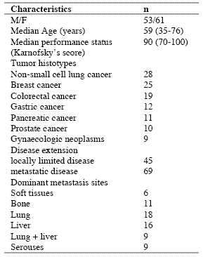
The study included 114 consecutive solid tumor patients with locally limited or metastatic disease, whose clinical characteristics are shown in Table 1. Lung cancer and gastrointestinal tumors were the most frequent neoplasms in our patients. For the immune detections, venous blood samples were collected in the morning after an overnight fast. Operable patients and metastatic patients were investigated before the surgical operation and before the onset of chemotherapy, respectively, in an attempt to exclude the possible influence of the various anticancer therapies on the immune status of patients.

In each sample, we have evaluated total lymphocyte count and the various lymphocyte subpopulations by a flow cytometric assay and monoclonal antibodies, including T helper lymphocytes (CD4), T cytotoxic lymphocytes (CD8), NK cells (CD16CD56), and T regulatory (T-reg) lymphocytes (CD4CD25). Normal values (95% confidence limits) of T-reg observed in our laboratory were below 240/mm3. Moreover, because of its importance in regulating lymphocyte functions and proliferation (Claman, 1998; Sthephens et al, 2004), the circadian rhythm of cortisol was also investigated by collecting blood samples at 8.00 A.M. and at 4.00 P.M., and cortisol serum concentrations were measured in duplicate by using the ECLA method. Data were reported as mean +/- SE, and statistically analyzed by the Student’s t test, the analysis of variance and the chi-square test, as appropriate.

III. Results
As reported in Table 2, an abnormally high number of T-reg was seen in 52/114 (46%) patients. Moreover, the percentage of cases with elevated number of T-reg observed in metastatic patients was significantly higher with respect to that found in non-metastatic patients (44/69 (64%) vs 8/45(18%), p < 0.01). Table 3 shows the mean number of T-reg and the mean percentages of T-reg with respect to both total lymphocytes and T helper (CD4+) lymphocytes observed in cancer patients in relation to their disease extension. The mean number of T-reg observed in metastatic patients was higher with respect to that found in patients with locally limited disease, without, however statistically significant differences. In contrast, the mean percentages of T-reg with respect to that of both lymphocytes and CD4 cells were significantly higher in metastatic patients than in the non-metastatic ones (p< 0.05 and p< 0.001,respectively). Moreover, within the metastatic group, patients with a normal lymphocyte count greater than 1500/mm3 showed a significantly higher mean number of T-reg with respect to the non-metastatic patients, whereas no difference was seen between nonmetastatic patients and metastatic patients with lymphocytopenia, consisting of lymphocyte count lower than 1500/mm3. In contrast, the mean percentages of T-reg with respect to total lymphocytes and CD4+ cells observed in both groups of metastatic patients with normal or low total lymphocyte count were significantly higher than in non-metastatic patients (lymphocytes: p< 0.025, CD4+ cells: p< 0.001).

Table 1. Clinical characteristics of 114 solid tumor patients
090722_clinicalstud1

Table 2. Percentages of abnormally high values of CD4+CD25+ lymphocytes

090722_clinicalstud2

* P < 0.01 vs non-metastatic patients

Table 3. Mean number of CD4+CD25+ lymphocytes and their mean percentages with respect to total lymphocytes and CD4+ lymphocytes in metastatic and non-metastatic patients

090722_clinicalstud3

* p<0.05 vs non-metastatic patients; ** p<0.025 vs non-metastatic patients; *** p<0.01 vs non-metastatic patients; **** p<0.001 vs nonmetastatic patients

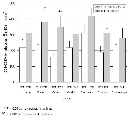
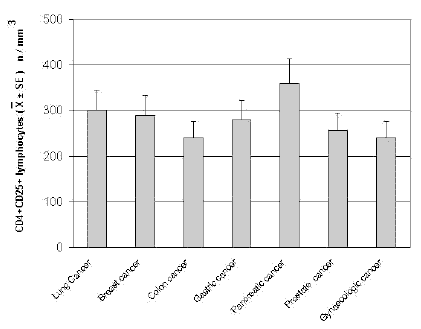
The mean counts of NK and CD8 cells in relation to that of T-reg are reported in Table 4. As shown, no significant difference in the mean number of CD8 lymphocytes was found between patients with normal or abnormally elevated number of T-reg. On the contrary, patients with elevated number of T-reg showed a significantly lower number of NK cells with respect to that found in those with normal T-reg count. Finally, Table 5 shows the circadian rhythm of cortisol in relation to total lymphocytes, CD4+ cells and T-reg mean number. A normal cortisol rhythm, with morning values greater at least than 50% with respect to the values occurring during the afternoon, was found in 85/114 (75%). Total lymphocyte and CD4+ cell mean numbers observed in patients with altered cortisol rhythm were significantly lower than those found in patients with normal cortisol circadianicity (p<0.01), whereas no significant difference was seen in the mean number of T reg. Figure 1 and Figure 2 illustrate T-reg mean numbers in relation to tumor histoptypes in the overall patients and with respect to their disease extension, respectively. No significant difference was seen in relation to tumor histotype. The highest values of T-reg were observed in pancreatic cancer patients, without however significant differences with respect to the overall other histotypes. The metastatic disease was associated with a higher number of T-reg with respect to the non-metastatic group in all tumor histotypes, even though a statistically significant differences occurred for the only breast cancer (p<0.05) and colorectal cancer (p< 0.01).

Table 4. Mean values of NK cells and CD8+ lymphocytes in cancer patients with normal or abnormally high values of CD4+CD25+ lymphocytes

090722_clinicalstud4

* p<0.05 vs normal values of CD4+CD25+ lymphocytes

Table 5. Mean numbers of total lymphocytes, T helper (CD4+) lymphocytes and T regulator lymphocytes (CD4+CD25+) in relation to cortisol circadian secretion in cancer patients
090722_clinicalstud5

* P<0.01 vs patients with altered cortisol rhythm
090722_clinicalstudfig1

Figure 1. CD4+CD25+ lymphocyte mean number in relation to tumor histotype
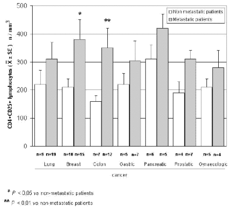
090722_clinicalstudfig2

Figure 2. CD4+CD25+ lymphocytes in relation to tumor histotype in metastatic and non-metastatic cancer patients

IV. Discussion
According to previous preliminary clinical investigations (Sasada et al, 2003; Cesana et al, 2006), this study confirms in a greater number of cancer patients that the metastatic disease is characterized by the evidence of an abnormally increased percentage of T-reg lymphocytes with respect to both total circulating lymphocytes and CD4+ lymphocytes. This finding does not seem to represent a specific characteristic of some tumor histotypes, then it could constitute a general alteration occurring during the progression of the neoplastic disease, by representing a fundamental immune parameter of cancer-related immunosuppression.

Several immune molecules have appeared to suppress the anticancer immunity, namely IL-6, IL-10, IL-1, TNF-α and TGF-β, but it seems that the common end result of their mechanisms of action may be represented by the stimulation of T-reg generation, with a consequent inhibition of the activation of an effective anticancer immune reaction. On the same way, several immune cells are able to suppress the anticancer immunity, including macrophages, T helper-2 lymphocytes and some myeloidderived suppressor cells, but also in this case they would act in a suppressive way by promoting the generation of Treg.

Then, the detection of T-reg amounts in terms of both absolute number and percentages with respect to total lymphocytes and CD4+ cells could constitute a simple and adequate clinical immune parameter to quantify the whole status of the anticancer immunity in the single cancer patient. Moreover, future clinical studies will be required to establish the possible prognostic significance of changes in T-reg percentage and number in relation to the anticancer efficacy of the various standard antitumor therapies. Moreover, it has to be remarked that T-reg lymphocytes would not represent the only immune cells involved in the suppression of the anticancer immunity. In fact, there is at least another fundamental immunosuppressive system, consisting of the monocytemacrophage cell lineage (Sica and Bronte, 2007). In more detail, it has been observed that the bone marrow may release myeloid precursors provided by suppressive activity on the antitumor immune response and defined as myeloid-derived suppressor cells (MDSC) (Kusmartsev and Gabrilovich, 2005). These cells have appeared to be characterized by the cell surface expression of GR-1, CD11b and CD80 antigens (Anderson et al, 2002; van Ginderachter et al, 2006). The myeloid suppressor cells would promote the generation and activation of T-reg lymphocytes, which at the other side would stimulate MDSC release from the bone marrow and M2 macrophage differentiation (Terabe et al, 2003; Wie et al, 2006).

Moreover, the myeloid suppressive cells would inhibit the anticancer immunity by promoting macrophage differentiation into the M2 sub-type (Ikemoto et al, 2003), which plays a clear inhibitory effect on the anticancer immunity, namely through the release of IL-6 (Ueno et al, 2000), whereas the M1 macrophage sub-type may either stimulate or suppress the antitumor immunity (Mantovani et al, 2004). M1 and M2 macrophage sub-types have appeared to be characterized by a high production of IL-12 or IL-10, respectively (Ueno et al, 2000).

Then, further studies by concomitantly evaluating T reg and MDSC count, will contribute to better define the immune mechanism responsible for the suppression of the anticancer immunity.

References
Anderson CF, Gerber JS, Mosser DM (2002) Modulating macrophage function with IgG immune complexes. J Endotoxin Res 8, 477-81.

Annunziato F, Cosmi L, Liotta F, Lazzeri E, Manetti R, Vanini V, Romagnani P, Maggi E, Romagnani S (2002) Phenotype,localization and mechanism of suppression of CD4+CD25+ human thymocytes. J Exp Med 196, 379-87.

Antony PA, Restito NP (2005) CD4+CD25+ T regulatory cells,immunotherapy of cancer,and interleukin-2. J Immunother 28, 120-8.

Birebent B, Lorho R, Lechartier H, de Guibert S, Alizadeh M,
Vu N, Beauplet A, Robillard N, Semana G (2004) Suppressive properties of human CD4+CD25+regulatory T cells are dependent on CTLA-4 expression. Eur J Immunol 34, 3485-96.

Cesana GC, DeRaffele G, Cohen S, Moroziewicz D, Mitcham J, Stoutenburg J, Cheung K, Hesdorffer C, Kim-Schulze S, Kaufman HL (2006) Characterization of CD4+CD25+ regulatory T cells in patients treated with high-dose interkleukin-2 for metastatic melanoma or renal cell carcinoma. J Clin Oncol 24, 1169-77.

Chen W, Jin W, Hardegen N, Lei KJ, Li L, Marinos N, McGrady G, Wahl SM (2003) Conversion of peripheral CD4+CD25- naïve T cells to CD4+CD25+ regulatory T cells by TGF-β induction of transcription factor Foxp 3. J Exp Med 198, 1875-86.

Claman HN (1998) Corticosteroids and the immune system. Adv Exp Med Biol 245, 203-10.

Dieckmann D, Bruett H, Ploettner H, Lutz MB, Schuler G (2002) Human CD4+CD25+ regulatory contact-dependent T cell induce IL-10 producing,contact-independent type-1-regulatory T cells. J Exp Med 196, 247-53.

Dieckmann D, Plottner H (2001) Ex vivo isolation and characterizationof CD4+CD25+ T cells with regulatory properties from human blood. J Exp Med 193, 1303-10.

Ehrke MJ, Mihich E, Berd D, Mastrangelo MJ (1989) Effects of anticancer drugs on the immune system. Semin Oncol 16, 230-9.

Ghiringhelli F, Larmonier N, Schmitt E, Parcellier A, Cathelin D, Garrido C, Chauffert B, Solary E, Bonnotte B, Martin F (2004) CD4+ CD25+ regulatory T cells suppress tumor immunity but are sensitive to cyclophosphamide which allows Immunotherapy of established tumors to be curative. Eur J Immunol 34, 336-44.

Ikemoto S, Yoshida N, Narita K, Wada S, Kishimoto T, Sugimura K, Nakatani T (2003) Role of tumor-associated macrophages in renal cell carcinoma. Oncol Rep 10, 1843-9.

Jago CB, Yates J, Camara NOS, Lechler RI, Lombardi AG (2004) Differential expression of CTLA-4 among T cell subsets. Clin Exp Immunol 136, 463-71.

Ji HB, Liao G, Faubion WA, Abadía-Molina AC, Cozzo C, Laroux FS, Caton A, Terhorst C (2004) Cutting edge, the natural ligand for glucocorticoid-induced TNF receptorrelated protein abrogates regulatory T cell suppression. J Immunol 172, 5823-7.

Kanamaru F, Youngnak P, Hashiguchi M, Nishioka T, Takahashi T, Sakaguchi S, Ishikawa I, Azuma M (2004) Costimulation via glucocorticoid-induced TNF receptor in both conventional and CD25+ regulatory CD4+ T cells. Immunol 172, 7306-14.

Knutson KL, Disis M, Salazar L (2007) CD4 regulatory T cells in human cancer pathogenesis. Cancer Immunol Immunother 556, 271-85.

Kusmartsev S, Gabrilovich DI (2005) STAT1 signaling regulates tumor-assolciated macrophage-mediate T cell deletion. J Immunol 174, 4880-91.

Lan RY, Ansari AA, Lian ZX, Gershwin ME (2005) Regulatory T cells, development,function,and role in autoimmunity. Autoimmun Rev 4, 351-63.

Leong PP, Mohammad R, Ibrahim N, Ithnin H, Abdullah M, Davis WC, Seow HF (2006) Phenotyping of lymphocytes expressing regulatory and effector markers in infiltrating ductal carcinoma of the breast. Immunol Lett 102, 229-36.

Malek TR, Bayer AL (2004) Tolerance not immunity crucially depends on IL-2. Nat Rev Immunol 4, 665-74.

Mantovani A, Sica A, Sozzani S, Allavena P, Vecchi A, Locati M (2004) The chemokine system in diverse forms of macrophage activation and polarization. Trends Immunol 25, 677-86.

Sasada T, Kimura M, Yoshida Y, Kanai M, Takabayashi A (2003) CD4+CD25+ regulatory T cells in patients with gastrointestinal malignancies, possible involvement of regulatory T cells in disease progression. Cancer 98, 1089-93.

Schwartz RH (2005) Natural regulatory T cells and selftolerance. Nat Immunol 6, 327-30.

Shevach EM (2002) CD4+CD25+ suppressor T cells, more questions than answers. Nat Rev Immunol 2, 389-400.

Sica A, Bronte V (2007) Altered macrophage differentiation and immune dysfunction in tumor development. J Clin Invest 117, 1155-66.

Siddiqui SA, Frigola X, Bonne-Annee S, et al (2007) Tumorinfiltrating Foxp3 CD4+CD25+ T cells predict poor survuival in renal cell carcinoma. Clin Cancer Res 13, 2075-81.

Stephens GL, McHugh RS, Whitters MJ, Young DA, Luxenberg D, Carreno BM, Collins M, Shevach EM (2004) Engagement of glucocorticoid-induced TNFR family-related receptor on effector T cells by its ligand mediates resistance to suppression by CD4+CD25+ T cells. J Immunol 173, 5008-20.

Takahashi T, Tagami T, Yamazaki S, Uede T, Shimizu J, Sakaguchi N, Mak TW, Sakaguchi S (2000) Immunologic and tolerance maintained by CD25+CD4+ regulatory T cells constitutively expressing cytotoxic T lymphocyte-associated antigen. J Exp Med 192, 1285-94.

Terabe M, Matsui S, Park JM, Mamura M, Noben-Trauth N, Donaldson DD, Chen W, Wahl SM, Ledbetter S, Pratt B, Letterio JJ, Paul WE, Berzofsky JA (2003) Transforming growth factor-β production and myeloid cells are an eggector mechanism trhough which CD1d-restricted T cells block cytotoxic T lymphocyte-mediated tumor immunosurveillance, abrogation prevents tumor recurrence. J Exp Med 198, 1741-52.

Thomton AM, Shevach EM (2000) Suppressor effector function of CD4+CD25+ immunoregulatory T cells is antigen nonspecific. J Immunol 164, 183-90.

Ueno T, Toi M, Saji H, Muta M, Bando H, Kuroi K, Koike M, Inadera H, Matsushima K (2000) Significance of macrophage chemo-attractant protein-1 in macrophage recruitment,angiogenesis and survival in human breast cancer. Clin Cancer Res 6, 3282-9.

Van Ginderachter JA, Movahedi K, Hassanzadeh Ghassabeh G, Meerschaut S, Beschin A, Raes G, De Baetselier P (2006) Classical and alternative activation of mononuclear phagocytes, picking the best of both worlds for tumor promotion. Immunobiology 211, 487-501.

Vasu C, Prabhakar BS, Holterman MJ (2004) Targeted CTLA-4 engagement induces CD4+CD25+CTLA-4 high T regulatory cells with target alloantigens specificity. J Immunol 173, 2866-76.

von Boehmer H (2005) Mechanisms of suppression by suppressor T cells. Nat Immunol 6, 338-44.

von Herrath MG, Harrison LC (2003) Regulatory lymphocytes, antigen-induced regulatory T cells in autoimmunity. Nat Rev Immunol 3, 223-32.

Wang XB, Zheng CY, Giscombe R, Lefvert AK (2001) Regulation of surface and intracellularexpression of CTLA-4 on human peripheral T cells. Scand J Immunol 54, 453-8.

Wie S, Kryczeck I, Zou W (2006) Regulatory T-cell compartmentalization and trafficking. Blood 108, 426-31.

Yang R, Cai Z, Zhang Y, Yutzy WH 4th, Roby KF, Roden RB (2006) CD80 in immune suppression by mouse ovarian carcinoma-associated Gr-1+CD11b+ myeloid cells. Cancer Res 66, 6807-15.

Ziegler SF (2006) FOXP3 of mice and men. Annu Rev Immunol 24, 209-26.

Zou W (2006) Regulatory T cells, tumour immunity and immunotherapy. Nat Rev Immunol 6, 295-307.

(download PDF version)



A phase II study of anastrozole plus the pineal anticancer hormone melatonin in the metastatic breast cancer women with poor clinical status

lissoniResearch Article (download PDF version)

Paolo Lissoni1*, Giuseppe Di Fede1, Antonio Battista2, Giusy Messina1, Remo Egardi1, Fernando Brivio3, Franco Rovelli1, Massimo Colciago4, Giuseppe Brera5

1 Institute of Biological Medicine, Milan
2 Azienda Sanitaria locale 2, Avellino;
3 Surgery Division, Bassini Hospital, Cinisello,Milan
4 I.N.R.C.A, Casatenovo, Lecco, Italy
5 Ambrosian University, Milan, Italy

dr. Paolo Lissoni

*Correspondence: Dr. Paolo Lissoni, Divisione di Radioterapia Oncologica, Ospedale S.Gerardo, 20052 Monza, Milano, Italy; Fax: +390392332284, E-mail: p.lissoni@hsgerardo.org
Key words: Anastrozole, breast cancer, melatonin, pineal gland
Abbreviations: melatonin, MLT; estrogen receptor, ER;

Received: 9 March 2009; Revised 1 April 2009;
Accepted: 13 April 2009; electronically published: 28 May 2009

Summary

The recent advances in the psychoneuroendocrinology have suggested the possibility to modulate tumor hormone dependency through a neuroendocrine approach. In particular, it has been proven that the pineal neurohormone melatonin (MLT) may stimulate estrogen receptor (ER) expression in breast cancer cells and inhibit the aromatase activity. On this basis, a study was planned to evacuate the efficacy of a concomitant treatment with the aromatase inhibitor anastrozole plus MLT in metastatic breast cancer. The study included 14 metastatic breast cancer women of poor clinical conditions with ER positive or unknown. Both anastrozole and MLT were given orally at a dose of 1 mg at noon and of 20 mg in the evening, respectively. The clinical response consisted of complete response in 2 and partial response in 6 patients. Then, an objective tumor regression was achieved in 8/14 (57%) patients, with a median duration of 26 months. No neoplastic cachexia occurred on treatment. This preliminary study shows that a neuroendocrine strategy with anastrozole plus the pineal hormone MLT may represent a new effective and well tolerated regimen in the treatment of metastatic breast cancer women, including those with poor clinical status, with therapeutic results apparently superior to those reported in the literature with the only aromatase inhibitor. Then, these results would justify further randomized studies of aromatase inhibitors with or without a concomitant administration of MLT, in an attempt to establish whether the pineal hormone may enhance the efficacy of the aromatase inibibitors in the treatment of human advanced breast cancer.

I. Introduction
Recent experimental studies have demonstrated that the hormone dependency is at least in part under a psychoneuroendocrine regulation (Cos et al, 2008;Grant et al, 2009). In particular, it has been shown that the pineal hormone melatonin (MLT), whose anticancer properties have been well demonstrated (Bartsch et al, 1981; Maestroni, 1993; Reiter et al, 2002), may in vitro stimulate estrogen receptor (ER) expression on breast cancer cell lines (Molis et al, 1995). Therefore, the hormone dependency of breast cancer cells would not depend only on intrinsic characteristics of cancer cells themselves, but also on host neuroendocrine regulation of tumor cell proliferation and differentiation (Bartsch et al, 2000). Moreover, cancer progression has been proven to be associated with pineal alterations, consisting of a progressive decline in MLT nocturnal production. (Maestroni, 1993). Therefore the advanced cancer would require a substitutive endocrine therapy with MLT (Bartsch et al, 1981; Maestroni, 1993). Previous preliminary clinical studies had already suggested that the concomitant administration of the pineal hormone MLT may apparently increase the efficacy of tamoxifen therapy in the treatment of metastatic breast cancer (Lissoni et al, 1995). Moreover, experimental studies have shown that the activity of aromatase enzyme, which is responsible for the peripheral production of estrogens from testosterone (Bagatell et al, 1994), is under a light/dark circadian rhythm (Bhatnagar et al, 1992). Because of the fundamental role of the pineal hormone MLT in the regulation of the daily photoperiod (Bartsch et al, 1981), it is possible to hypothesize that MLT may be involved in the control of the aromatase activity. In fact, recent studies have demonstrated an inhibitory action of MLT on the aromatase activity (Cos et al, 2005). This finding could reserve a prosiming application in the treatment of both early and advanced breast cancer. This statement is justified by the fact that the aromatase inhibitors represent a new class of agents in the endocrine treatment of breast cancer Plourde et al, 1994), with a potential efficacy superior to that achieved by the previous hormonal therapies with anti-estrogens, such as tamoxifene, even though tumor response rate obtained by the aromatase inhibitors are generally not greater than 40%. On this basis, a phase II study was planned in an attempt to evaluate the efficacy of a neuroendocrinotherapeutic regimen consisting of a concomitant administration of the aromatase inhibitor anastrozole and the pineal hormone MLT in metastatic breast cancer women with poor clinical conditions.

II. Materials and methods
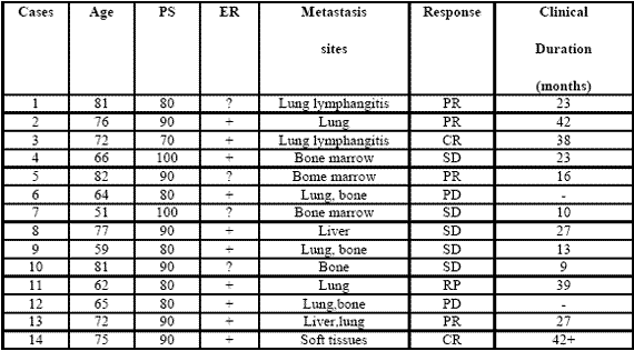
The study included 14 consecutive metastatic breast cancer women (median age: 72 years, range 51-82), who were followed at Biological Medicine Institute in Milan, or at Health Local Unit 2 of Avellino, from Feb. 2002 to Sept. 2003. Eligibility criteria were, as follows: histologically proven metastatic breast cancer, measurable lesions, ER positive or unknown, no ability to tolerate chemotherapy because of age, low performance status (PS), important clinical illnesses other than cancer and/or heavy chemotherapeutic pre-treatments, no previous endocrine therapies for the metastatic disease, no double tumor and life expectancy less than 1 year. Previous heavy chemotherapeutic treatment consisting of at least 3 chemotherapeutic lines was made in 11/14 (79 %) patients. Dominant metastasis sites were, as follows: soft tissues:1; bone:1; lung:7 (neoplastic lymphangitis:2); liver:1; lung + liver:1; bone marrow:3. Time-span since first diagnosis of the primary tumor was 44 months (31-66 months). All patients had an acceptable social conditions. The minimum and median follow-up periods were 60 months and 72 months respectively. In all patients, in the case of disease progression, at least to other endocrine therapeutic lines with other aromatase-inhibitors were planned. The experimental protocol, wich was approved by the Health Direction of Biological Medicine Institute of Milan, was explained to each patient and informed consent was obtained. The treatment consisted of anastrozole at a dose of 1 mg/day orally at noon, plus MLT at 20 mg/day orally in the evening, generally half-hour before sleeping, to correct cancer progression-related decline in MLT night secretion (10). Patients were considered to be evaluable when they were treated for at least 3 consecutive months. The clinical response was evaluated according to WHO criteria. Complete response (CR) was the complete disappearance of all neoplastic lesions for at least 1 month. Partial response (PR) was a reduction greater than 50 % of the sum of all neoplastic lesions, for at least 1 month. Stable disesase (SD) was no increase or decrease greater tha 25 % of tumor volume. Progressive Disease (PD) was an increase in tumor volume greater than 25 % or the appearance of new neoplastic lesions. PS was assessed according to Karnofsky’s score, consisting of the evaluation of the quality of life in relation to patient activity and bed-rest period. ER was positive in 10 and unknown in the remaining 4 patients. The median PS was 80% (range 70-100). Data were statistically evaluated by the chi-square test and the Student’s t test, as appropriate.

090722_phase2study

Table 1: Clinical characteristics of metastatic breast cancer women and their clinical response (WHO criteria) to a neuroendocrine regimen consisting of anastrozole plus the pineal hormone melatonin.

III. Results
All patients were fully evaluable for the clinical response. The clinical characteristics of patients and their individual clinical response to the treatment are reported in Table 1. As reported, a complete response (CR) was achieved in 2/14 (14%) (soft tissues:1; lung lymphangitis:1). A partial response (PR) was obtained in other 6/14 (43%) (bone:1; lung:3; liver:1; bone marrow:1). Then, an objective tumor response (CR + PR) was reached in 8/14 (57%) patients. The median duration of response was 26 months (range 9-42 months). A stable disease (SD) was seen in other 4/14 (29%), with a median duration of 25 months (range 10-27). Therefore, a disease-control (DC:CR + PR + SD) was achieved in 12/14 (86%) patients, whereas the remaining 2/14 (14%) patients had a progressive disease (PD). No significant difference in tumor response rate was observed between patients with positive or unknown ER ( 6/10(60%) vs 2/4(50%) ). An overall survival at 1 year and at 3 year was achieved in 11/14 (79 %) and in 5/14 (36 %) patients, respectively. Moreover, 3/14 (21%) patients were still alive at 5 years. The treatment was well tolerated in all patients.

Moreover, most patients experienced a relief of asthenia under the treatment and in no patient the neoplastic cachexia occurred. Finally, an evident increase in PS mean values was achieved under treatment, even though it did not reach the statistical significance (86 ±5 vs 93 ± 4, mean ± SE).

IV. Discussion
The results of this preliminary phase II study, by showing a percentage of 1-year survival greater than 70% in patients with live expectancy less than 1 year, would suggest that a neuroendocrine regimen consisting of the aromatase inhibitor anastrozole plus the pineal neurohormone MLT may represent a new effective therapeutic strategy in the treatment of metastatic breast cancer women, also in patients with poor clinical conditions, who would not be able to tolerate the most aggressive therapies. The concomitant administration of the pineal hormone would seem to enhance the efficacy of the aromatase inhibitor in terms of objective tumor regressions with respect to the results commonly reported in the literature with the only aromatase inhibitor (Plourde et al, 1994), which are generally lower than 40%.

The time to progression would seem to be apparently increased by the concomitant treatment with MLT.This finding is not surprising, since MLT could enhance the therapeutic anticancer acitivity of the aromatase inhibitors by either exerting direct antiproliferative antitumor effects (Bartsch et al, 1981; Maestroni, 1993; Reiter et al, 2002), or further inhibiting the aromatase activity by acting on gene and oncogene expression (Molis et al,1995; Cos et al, 2005). In addiction, MLT appeared to stimulate ER expression of breast cancer lines, by transforming ER negative into ER positive breast cancer, as observed in experimental conditions (Danforth et al, 1983).

Since the prognosis of ER positive breast cancers is clearly better than that of ER negative ones, MLT could per se improve the clinical couse of mammary tumors. Finally, because of its interesting therapeutic efficacy as a supportive care (Reiter et al, 2002), MLT would be responsible for the evident improvement in the relief of asthenia and in preventing the occurrence of the neoplastic cachexia. On the other hand, because of the inhibitory effect of MLT (Grant et al, 2009; Reiter et al, 2002) on cancer cell proliferation, the anticancer activity of this polyendocrine regimen would be due not only to an indirect effect, depending on a diminished estrogen production following aromatase enzyme inhibition, but also on a direct inhibition of cancer cell growth, due to MLT itself. Therefore, the results of this preliminary study may justify further clinical randomized investigations with the only aromatase inhibitor versus the concomitant treatment with MLT, in an attempt to confirm the ability of the pineal hormone to enhance the antitumor properties of the aromatase inhibitors in the treatment of metastatic breast cancer women with poor clinical conditions.

References
Bagatell CJ, Dahl KD, Bremner WJ (1994). The direct pituitary effect of testosterone to inhibit gonadotrophin secretion in men is partially mediated by aromatization to estradiol. J Androl 15:15-21.

Bartsch C, Bartsch H (1981) effect of melatonin on experimental tumors under different photoperiods and times of administration. J Neural Transm 52:269-279.

Bartsch H, Buchberger A, Franz H,Bartsch C,Maidonis I,Mecke D, Bayer E (2000) Effect of melatonin and pineal extracts on human ovarian and mammary tumor cells in a chemosensitivity assay. Life Sci 67:2953-2960.

Bhatnagar AS, Muller P, Schenkel L, Trunet PF, Beh I, Schieweck K. (1992). Inhibition of oestrogen biosynthesis and its consequences on gonadotrophin secretion in the male. J of Steroid Biochemistry and Molecular Biology;41:1021-1027

Cos S, Martinez-Campa C,Mediavilla MD, Sanchez-Barcelo E (2005). Melatonin modulates aromatase activity in MCF-7 human breast cancer cells. J Pineal Res 7:136-142.

Cos S, Gonzales A, Martinez-Campa C, Mediavilla MD, Alonzo-Gonzales C, Sanchez-Barcelo EJ (2008): Melatonin as a selective estrogen enzyme modulator. Curr Cancer Drug Targets. Dec 8(8): 691-702.

Danforth DN, Tamarkin L, Lipmann LE: (1983) Melatonin increase oestrogen receptor binding activity of human breast cancer cells. Nature, 305:323-325.

Grant S.G, Melan MA, Latimer JJ, Witt-Enderby PA (2009) : Melatonin and Breast cancer : cellular mechanism, clinical studies and future perspective. Expert Rev Mol Med 11:e5

Lissoni P, Barni S, Meregalli S, Fossati V, Cazzaniga M, Esposti D, Tancini G (1995) Modulation of cancer endocrine therapy by melatonin:a phase II study of tamoxifen plus melatonin in metastatic breast cancer patients progressing under tamoxifen alone. Br J Cancer 71:854-856.

Maestroni GJM (1993) The immunoneuroendocrine role of melatonin. J Pineal Res 14:1-10.

Molis TM, Spriggs LL,Jupiter Y,Hill SM (1995) Melatonin modulation of estrogen-regulated proteins, growth factors,and protooncogenes in human breast cancer. J Pineal Res 18:93-103.

Plourde PV, Dyroff M, Dukes MD (1994) Arimidex: a potent and selective fourth generation aromatase inhibitor. Breast Cancer Res Treat 30:103-111.

Reiter RJ, Tan DX, Sainz RM, Mayo JC, Lopez-Burillo S. (2002) Melatonin:reducing the toxicity and increasing the efficacy of drugs. Pharm Pharmacol 54:1299-1321

(download PDF version)


Ottobre 5, 2011 RASSEGNA STAMPA

L’Assessment nutrizionale nello sport. Il giudizio sullo stato nutrizionale di un atleta deve basarsi sulle valutazioni antropometriche, ematochimiche ed ormonali e sullo studio del dispendio energetico La stesura di un piano nutrizionale per uno sportivo amatoriale o di élite deve adattare la richiesta dell’atleta alle esigenze ed al fine che ci vogliamo proporre. In particolare, nella Nutrizione dello Sport, il nutrizionista deve porsi come primo fine la salute degli atleti che a lui si rivolgono e, partendo da questo presupposto, raggiungere quei target che in genere vengono individuati nella Nutrizione dello Sport, vale a dire:

  • Diminuzione della massa grassa, se essa risulta essere in eccesso
  • Mantenimento e/o incremento della Massa Grasso Priva (massa muscolare in particolare)
  • Ottimizzare il dispendio di energia sfruttando al meglio i substrati energetici organici (glicogeno muscolare ed epatico) e quelli introdotti con gli alimenti e/o i supplementi durante la performance
  • Facilitare il recupero dopo lo sforzo
  • Modulare i processi infiammatori che si formano “fisiologicamente” durante l’attività fisica e che possono sia influire sulla performance che favorire in qualche modo gli infortuni muscolari.

Per raggiungere questi obiettivi, ci sembra che sia assolutamente da evitare redigere un piano nutrizionale prescindendo da una serie di valutazioni anamnestiche, ematochimiche e ormonali, antropometriche e di valutazione del dispendio di energia che saranno d’ausilio nel personalizzare al massimo il trattamento sia alimentare che eventualmente di supplementazione/integrazione.

Da un punto di vista metodologico, possiamo individuare i seguenti step:

  • Valutazione anamnestica
  • Indagini ematochimiche
  • Valutazione del dispendio energetico
  • Valutazione antropometrica

Indagine Anamnestica Nutrizionale
L’indagine anamnestica nutrizionale risulta fondamentale per valutare le abitudini alimentari dell’atleta, le sue preferenze alimentari, la quantità e qualità dei macro e micronutrienti (carboidrati, proteine, grassi , vitamine, sali minerali) e se l’atleta utilizza correttamente le forme alimentari in rapporto all’allenamento o alla competizione. In conclusione, se è sensibilizzato sul rapporto alimentazione/performance sportiva. Il colloquio risulterà utile anche nella valutazione su come l’atleta presti attenzione ad una corretta idratazione e servirà al medico anche per individuare la possibilità di cogliere precocemente eventuali sintomi di disturbo dell’alimentazione.
Altra cosa importante sarà quella di valutare alcuni segni e/o sintomi di eventuali intolleranze alimentari, ad esempio reazioni cutanee e/o generali (cefalea, stanchezza, stipsi) relative alla ingestione di un determinato alimento o di gruppi alimentari.

Esami Ematochimici – Ormonali – Stress Ossidativo
Gli esami ematochimici di routine saranno utili per valutare la normalità della glicemia, della funzione epatica tramite le transaminasi e le gamma GT, del colesterolo totale ed HDl, della sideremia, fattore poi determinante della anemia da carenza di ferro frequente negli atleti.

Le valutazioni ormonali non possono prescindere, a nostro avviso, dalla valutazione della funzione tiroidea tramite il dosaggio di FT4 – FT3 – TSH. La funzione tiroidea è indice indiretto dello stato metabolico del nostro organismo, atteggiamenti dietologici non corretti (ad esempio diete restrittive nei primi giorni di ritiro oppure durante l’anno in atleti sovrappeso) possono portare ad una “risposta” della funzione tiroidea di “difesa metabolica”, con diminuzione della produzione degli OT e aumento del TSH e, come dimostrato dalla letteratura scientifica, aumentata produzione di rT3 (ormone di risparmio metabolico). Altro dato da non sottovalutare risulta poi come la patologia tiroidea sia endemica in Italia e, pur se la popolazione da testare è anagraficamente giovane, non si possa escludere la maggiore probabilità di patologie tiroidee (es. tiroiditi autoimmuni) con alterazioni funzionali, come ad esempio un ipotiroidismo che potrebbero influenzare negativamente la performance, oppure l’associazione con altre patologie come le fibromialgie autoimmuni che possono spesso associarsi alle tiroiditi autoimmuni mettendo a rischio l’apparato muscolare. Gli altri ormoni che possono essere utili ai fini di valutare ed eventualmente prevenire lo stato di fatica dell’atleta sono rappresentati dai dosaggi del cortisolo ematico e del testosterone; infatti il rapporto T/C è stato, ed è da sempre, indicato come “indice” funzionale degli stati di over reaching e overtrainig nello sport.

Da sottolineare poi il fatto che la valutazione dell’ormone steroideo può essere effettuata anche nella saliva, evitando dunque il fastidio del prelievo ematico. Inoltre, il dosaggio dell’ormone steroideo sulla saliva permetterebbe di valutare la frazione veramente attiva dell’ormone. La valutazione dell’ormone della crescita (GH) spesso “stressata” dagli articoli on line o su riviste di settore è secondo noi di scarsa utilità pratica in quanto di difficile valutazione a seconda delle diverse fasce di età e potrebbe portare a facili e pericolose ipotesi di deficit di ormone somatotropo (GH) con innesco del fenomeno doping di utilizzo del GH a scopo correttivo di questi presunti deficit, con grave rischio per la salute degli individui sottoposti a tale pratiche illecite (maggior rischio di sviluppo di neoplasie).

I radicali dell’ossigeno ROS sono prodotti fisiologicamente durante i normali processi metabolici e la loro potenziale azione dannosa sulle membrane cellulari viene “tamponata” da un complesso sistema antiossidante messo in atto dal nostro organismo. Nell’attività sportiva si ha indubbiamente un’aumentata produzione di citochine infiammatorie e di radicali liberi legati ai processi metabolici indotti dallo sforzo fisico cui non sempre si contrappone una “risposta antiossidante”. Quando l’equilibrio tra fattori pro-ossidanti e fattori antiossidanti viene alterato a favore dei primi, si crea una situazione fisio-patologica definita “stress ossidativo” che può influire sia sulla performance che sulla predisposizione agli infortuni di natura muscolare. La valutazione dello stress ossidativo non è semplice, pochi sono i test sui liquidi biologici, soprattutto ematici ed urinari ma anche salivari che possono dare informazioni utili ai fini che abbiamo precedentemente esposto.

La valutazione dei radicali liberi nel sangue (DRoms) delle capacità antiossidanti (Oxy Test) e delle riserve antiossidanti (malondialdeide urinaria) può esser proposta come base di valutazione della presenza di uno stress ossidativo ma deve essere a nostro avviso interpretata in senso dinamico con prelievi seriati e programmati durante il periodo di allenamento e gara (stagione agonistica se l’atleta è uno sportivo professionista). Questo per identificare eventuali fenomeni di “adattamento dei sistemi di ossidazione e antiossidazione”, come spesso avviene all’inizio della ripresa dell’attività agonistica, che rendono inutile l’utilizzo ormai usuale di prodotti multivitaminici ad azione antiossidante. In questo caso i droms agirebbero come fenomeni di stimolo alle difese antiossidanti, processo che verrebbe invece inibito dall’uso non razionale dei prodotti antiossidanti.

Si rimanda al prossimo numero la continuazione dell’articolo, con la trattazione delle intolleranze
alimentari.

Fabrizio Angelini


Settembre 30, 2010 Newsletter

Possiamo affermare che l’alimentazione ha un ruolo ben più vasto e importante di quanto si possa immaginare.Gli studi sul DNA iniziati un decennio fa, hanno permesso di identificare alcuni geni che sono attivati dalle sostanze alimentari e da principi attivi in esse contenute.

Le principali predisposizioni studiate dal test Gene e Diet sono lo sviluppo del diabete tipo 2, il controllo dello stress ossidativo e dei radicali liberi, la predisposizione a malattie infiammatorie, artrosi e osteoporosi, la sensibilità allo zucchero, alcol, caffè, grassi, sale, la predisposizione alla celiachia e intolleranza al lattosio.

La conoscenza dell’attività dei nutrienti alimentari su alcuni geni, ci permette di selezionare alimenti che ci aiutano a stare meglio e prevenire le malattie legate all’invecchiamento come la demenza e i tumori.

Conoscere quali geni sono alterati ci permette di attivare i sistemi di prevenzione legati all’alimentazione, attraverso la nutriterapia (curarsi con gli alimenti).

Il futuro della moderna medicina e’ la prevenzione, come affermano gli scienziati di tutto il mondo, che lavorano per la causa delle malattie e la prevenzione.

La prevenzione, la longevità e la salute, passano dalla tavola. Nutriamo bene i nostri geni, la salute si può misurare e mantenere a lungo.

BIBLIOGRAFIA
Jose C. Florez, M.D. Ph. D., Kathleen A. Jablonski, Ph.D. Nick Bayley, B.A.,Toni I. Pollin. Ph.D., Paul I.W. de Bakker, Ph.D., Alan R. Shuldiner, M.D., William C. Knowler, M.D., Dr.P.H., David M.Nathan, M.D., and David Altshuler, M.D., Ph.D., for the Diabetes Prevention Program Research Group. The NEW ENGLAND JOURNAL of MEDICINE TCF7L2 polymorphisms and Progression to Diabetes in the diabetes Prevention Program, July 20,2006

Zheng Ye, Honglin Son. Glutatione s-transferase polymorphism (GSTM1, GSTP1 and GSTT1) and the risk of acute leukaemia:A systematic review and meta-analysis. European Journal of cancer 41, 2005, 980-989

Paolo Palatini, Giudo Ceolotto,FabioRagazzo, Francesca Dorigatti, Francesca Saladini, Italia Papparella, Lucio Mos, -giuseppe Zanata and Massimo Santonastaso. CYP1A2 genotype modifies the association between coffee intake and risk of hypertension. Journal of Hypertension 2009, 27:000-000


Ottobre 5, 2008 RASSEGNA STAMPA

Gli effetti “farmacologici” del cibo sull’organismo sono un argomento quanto mai antico. È invece storia dei nostri giorni l’aumento dei casi di intolleranze alimentari documentato dal numero di persone che si rivolgono a dietologi e specialisti lamentando quadri clinici differenti riconducibili, spesso, all’assunzione di determinate sostanze alimentari. La medicina ufficiale, da sempre attenta alle allergie alimentari, ha orientato il proprio interesse anche alle intolleranze cercando di trovare metodologie diagnostiche sempre più specifiche.

“I limiti di molti test oggi in commercio sono spesso legati alla necessità di una lettura soggettiva dei vetrini da parte del medico”, spiega il dott. Giuseppe Di Fede direttore Sanitario dell’Istituto di Medicina Biologica di Milano. “Quindi, non soltanto i risultati possono essere differenti in base all’esperienza del singolo medico, ma è anche difficile costruire una documentazione oggettiva dei casi. Infatti, mentre per le allergie alimentari è più agevole effettuare la diagnosi clinica e di laboratorio dato che è il paziente stesso a riferire i sintomi all’assunzione di un alimento, per le intolleranze, invece, le reazioni ritardate rispetto all’assunzione degli alimenti rendono di difficile standardizzazione l’approccio diagnostico e clinico.” Per rispondere dunque alle esigenze del mondo medico, negli Stati Uniti è stato introdotto un nuovo metodo di diagnosi chiamato ALCAT® -Antigen Leucocyte Cellular Antibody Test-, che rappresenta un notevole miglioramento nella standardizzazione della metodica diagnostica per le intolleranze alimentari.

Il test usa una metodica “in vitro” su sangue intero che avvicina i risultati alla diagnostica “in vivo”; la reazione con i singoli estratti alimentari in gel (è possibile testare una varietà di alimenti da un minimo di 10 fino ad un massimo di 50 o 100) e non liofilizzati – viene confrontata con aliquote di sangue di controllo non esposte agli estratti alimentari. Dopo la separazione della frazione leucocitaria, ogni aliquota viene analizzata, conteggiata e misurata attraverso uno specifico strumento computerizzato che esprime graficamente la variazione del numero e della dimensione cellulare. I grafici del test, realizzati per singolo alimento, vengono sovrapposti al grafico di controllo determinando classi di positività in base a quanto il campione si allontana dall’istogramma di controllo in termini di deviazioni standard.

Studi clinici Americani, effettuati utilizzando test ALCAT® hanno evidenziato un buon livello di correlazione (83,4%) con il test di provocazione orale a doppio cieco per gli estratti alimentari (dati ricavati da una ricerca che ha coinvolto 19 pazienti, su ognuno dei quali è stata valutata una serie di 50 alimenti), e il 96% di correlazione per gli additivi alimentari (studio condotto su 26 pazienti a cui sono stati effettuati 76 test sugli additivi alimentari, utilizzando il metodo della provocazione orale a doppio cieco con controllo placebo).

Il test Alcat, può valutare la compatibilità ad additivi alimentari, conservanti e sostanze chimiche, antibiotici, antinfiammatori oltre che agli alimenti. Per i bambini sopra i 3 anni è possibile fare il test. La possibilità di evidenziare l’alimento intollerato può aiutare nella risoluzione di un problema che è diventato cronico. È importante evidenziare che il test kit di ALCAT®, l’unico riconosciuto dalla FDA (Food Drug Amministration) sia come allergeni sia come macchina, ha dimostrato la sua affidabilità nei risultati rispetto agli altri test in commercio. Inoltre, si sta procedendo per il riconoscimento CEE, cosa che nessun altro test ha in questo momento.

Garanzia della validità dei risultati è stata la misurazione della potenza proteica (Protein Nitrogen Units). L’inserimento del test nella routine ambulatoriale è immediato grazie alla facilità d’uso da parte del medico e all’attendibilità con cui è in grado di identificare alimenti potenzialmente reattivi nei pazienti con sospetta sensibilizzazione/intolleranza verso alimenti che non si riescono ad identificare agevolmente.

Il Sistema ALCAT TEST
Il SISTEMA ALCAT Test evidenzia una reazione avversa agli alimenti, cioè agli allergeni alimentari (approvati ed autorizzati dalla FDA ). Vengono valutati il numero e le dimensioni dei granulociti neutrofili, prime difese anticorpali dell’organismo, e quando si verifica una loro variazione c’è una reazione avversa a quel determinato alimento.

Il SISTEMA ALCAT Test effettua un dosaggio di qualità secondo parametri ben definiti denominati “criteri di affidabilità”: precisione, sensibilità e specificità. La precisione rappresenta la stima della riproducibilità del test. La sensibilità è la dose minima misurabile di una sostanza (globulo bianco) e il test è risultato sensibile al 96%. La specificità è la capacità di dosare esclusivamente una determinata sostanza (globulo bianco) così da escludere interferenze con sostanze simili e il test è risultato specifico al 92%.
Le modificazioni a carico dei granulociti neutrofili vengono quantificate mediante settori di colori diversi a seconda della gravità della reazione:

settore verde (alimenti non reattivi e correlati);

settore giallo (alimenti con reazione moderata);
settore arancione (alimenti con reazione grave);

settore rosso (alimenti con reazione estrema).

I gradi di reattività non sono necessariamente correlati con la presenza e/o l’intensità delle manifestazioni cliniche. Sarà il medico a vagliare attentamente le risposte del test, in modo da prescrivere una dieta che non privi il paziente dei nutrimenti essenziali e che, nello stesso tempo, riesca a eliminare tutti gli allergeni.
Questa dieta aiuta l’organismo a recuperare nel più breve tempo possibile la tolleranza nei confronti degli alimenti non tollerati; a non riconoscere più gli alimenti verso cui si era intollerante come estranei e nocivi, ma ad accettarli senza provocare in modo diretto o indiretto alterazioni a carico di qualsiasi organo, apparato o sistema.

Per ulteriori informazioni sull’interpretazione di “Alcat test” si prega di consultare la documentazione messa a disposizione dal distributore (IMGeP – tel. 02.58300376).


Marzo 30, 2008 Newsletter

5° Congresso AMEB

Associazione Medicina Estetica Benessere

Magazzini del Cotone
Genova, 15-16 marzo 2008

Il 5° Congresso AMEB (Associazione Medicina Estetica Benessere), aperto a Medici di base, in primis, ma altresì a Dermatologi, Chirurghi plastici e vascolari annovera un alto numero di presenze. Sono state oltre cinquecento nelle prime quattro edizioni e tendente all’ aumento nella quinta edizione, tanto da richiedere il passaggio da una realtà storica ma piccola come Palazzo Ducale, sede della 4° edizione, ad una realtà più ampia come il Cotone Centro Congressi.

L’afflusso, nel tempo, è risultato essere esteso a Regioni limitrofe quali il Piemonte e la Lombardia ma l’interesse sarebbe quello di allargare l’ utenza alle altre regioni.
La 4° edizione del Congresso dell’Associazione Medicina Estetica Benessere vuole essere esteso a tutti gli operatori del settore estetico prevedendo spazi distinti per Medici e tutti gli altri operatori del settore (centri estetici, centri benessere, centri fitness etc.), nell’ intento di divulgare al massimo il concetto di benessere anche ad operatori non medici.
Il congresso è riconosciuto e verrà accreditato dal Ministero della Salute sotto la direzione organizzativa della AMEB (Associazione Medicina Estetica e Benessere – Comitato Scientifico: Presidente Dr. Paolo Rossi).

Segreteria Scientifica
AMEB Associazione Medicina Estetica e Benessere
Corso Tardy e Benech 13/2, Savona – Email: info@meb-it.org
Resp. Scientifico: dott. Paolo Rossi – Segreteria: dott.ssa Susanna Camurati

Segreteria Organizzativa
M.A.F. Servizi srl – Torino/ Genova www.mafservizi.it
Sede di Genova Sede di Torino
Via XX Settembre, 23 Corso Svizzera,185
tel. 010.5954304 tel. 011.505900
fax 010.5954304 fax 011.505976
Project Leader-Sponsor Dott.ssa Anna Botto botto@mafservizi.it
Resp. Iscrizioni Sigra Elena Vitali vitali@mafservizi.it

Iscrizioni
La partecipazione ai Corsi è gratuita. Dato il numero limitato di iscrizioni ad un massimo di persone per ogni corso, è indispensabile la pre-registrazione. Le iscrizioni, da inviare alla Segreteria Organizzativa, verranno accettate in base all’ordine cronologico di arrivo della scheda di iscrizione sino ad esaurimento posti.

La Segreteria Organizzativa provvederà ad inviare la conferma di iscrizione all’indirizzo e-mail o al numero di fax segnalato sulla scheda. Le schede sprovviste del numero di fax e/o email non verranno accettate.

Scheda di Registrazione
La scheda di iscrizione è disponibile a questo link oppure inviando una email di richiesta all’indirizzo botto@mafserviz.it

Crediti formativi E.C.M.
Sabato, 15 marzo 2008

N. 5 crediti Massimo n. 100 partecipanti – “Medicina Generale, Dermatologia e Chirurgia Plastica:protocolli comuni nella prevenzione di patologie e nel follow-up di trattamenti chirurgici”
Professione – Medico Chirurgo
Discipline – MMG – Chirurgia plastica e ricostruttiva – Dermatologia e venereologia

N. 4 crediti Massimo n. 100 partecipanti – “Ruolo dell’insegnamento della corretta alimentazione e del corretto stile di vita nell’ambulatorio di medicina generale”
Professione – Medico Chirurgo
Discipline – MMG – Malattie metaboliche e diabetologia – Scienza dell’alimentazione e dietetica

Domenica, 16 marzo 2008

N. 3 crediti Massimo n. 70 partecipanti- “Fitoterapici: Modalità di preparazione ed utilizzi terapeutici”
Professione – Farmacista
Discipline – Area interdisciplinare
Professione – Medico Chirurgo
Discipline – MMG – Malattie metaboliche e diabetologia – Scienza dell’alimentazione e dietetica

Non accreditato E.C.M. Massimo n. 70 partecipanti – “Termalismo, SPA e trattamenti ancillari”


Programma

15 MARZO 2008 – Sala Ponente
Medicina Generale, Dermatologia e Chirurgia Plastica: protocolli comuni nella prevenzione di patologie e nel follow-up di trattamenti chirurgici

Prima sessione: Dermatologia
Moderatori Giuseppe Cannata, Alessandro Farris, Aurora Parodi, Marina Romagnoli
09,45 Rosacea – Aurora Parodi
10,20 Terapie laser – chirurgiche delle teleangectasie – Marina Romagnoli
10,40 Laser epilazione – Annalisa Beatini
11,00 Pausa

Seconda sessione: medicina estetica
Moderatori Piermario Isola, Piergiorgio Mandelli, Stefano Picasso, Laura Simona
11,20 Fillers del volto – Tiziana Lazzari
11,40 Ozonoterapia – Mario Sirito
12,00 Neocollagenogenesi: rigenerazione cutanea – Vittorio Gennaro
12,00 Chirurgia superficiale con SELP – Giorgio Fippi
12,30 WORKSHOP Medigest (non accreditato ECM) – Giorgio Fippi
13,30 Pausa

Terza Sessione: Chirurgia Plastica
Moderatori Antonio Grande, Sonia Balestrero, Pierluigi Santi
14,00 Ricostruzione mammaria – Pierluigi Santi
14,20 Rinoplastica – Giorgio Lavagnino, Raffaella Sommariva
14,40 Peelings combinati – Lucio Miori
15,00 Mesoterapia omotossicologia – Massimo De Bellis
15,20 Biorivitalizzazione combinata – Massimo Ladisi
15,40 Rimodellamento non chirurgico del profilo nasale – Aldo Bauducco
16,00 Fosfatidilcolina – Rosanna Dalla Costa
16,20 Trattamento chirurgico dei nei – Paola Barabino
16,40 Pausa

Quarta Sessione: Flebo linfologia
Moderatori Sergio Capurro, Giorgio Forgione, Giovanni Perdonò
17,00 Analisi strumentale della circolazione venosa – Giovanni Perdonò
17,15 Scleroteapia tridimensionale – Sergio Capurro
17,30 Crossectomia e stripping – Eddy Zepponi
17,45 Focus sulla chirurgia vascolare degli arti inferiori – Giorgio Forgione
18,00 Scleroterapia – Pietro Forfori
18,15 Flebologia – Alberto Macciò
19,00 Compilazione Test – Chiusura lavori


15 MARZO 2008 – Sala Levante
Ruolo dell’insegnamento della corretta alimentazione e del corretto stile di vita nell’ambulatorio di Medicina Generale – con il Patrocinio di ASSIMEFAC

Prima Sessione: Corretta alimentazione
Moderatori Renato Giusto Vice-Presidente Ordine dei Medici di Savona
Andrea Stimamiglio Coordinatore FIMMG Genova
09,00 Consigli dietologici nel diabetico – Caterina Bordone
09,30 Consigli dietetici per patologie – Fabrizio Gallo
11,00 Corretta alimentazione, sovrappeso e sottopeso – Giuseppe Daino
12,00 Importanza della deglutizione nell’alimentazione – Diego Cossu
13,00 Pausa

Seconda Sessione: Nuove metodologie di operative nel campo dietologico
Moderatori Luca Lione Diabetologo – Emanuele Russo M.M.G.
14,00 Alimentazioni proteiche: vizi e virtù – Mario Pucci
15,00 Ruolo dell’analisi strumentale nella dieta – Pietro Morini
16,00 WORKSHOP I.M.Ge.P. (non accreditato ECM)
Focus on: intolleranze alimentari Attilio Speciani

Tavola Rotonda – Moderatore Giuseppe Daino
17,30 Tavola rotonda
18,30 Compilazione Test – Chiusura lavori


16 MARZO 2008 – Sala Ponente
Termalismo, SPA e trattamenti estetici ancillari

Prima Sessione Terme e SPA
Moderatori Antonio Basso – Giuseppe Nappi Presidente nazionale Medicina Termale
Francesco Berti Riboli Confindustria Genova
09,10 Gestione e marketing di una SPA – Paolo Torazza Confindustria
09,30 Patologie orl e cure termali – Federico Baricalla
10,00 Metodologie elettromeccaniche nelle SPA – Raul Novelli
10,20 Clinica e terapia dello stress nelle SPA – Alberto Massirone
10,40 Riabilitazione in centro termale – Fiorenza Di Matteo
11,00 Pausa

Seconda Sessione: Medicina dello sport ed anti-aging
Moderatori Damiano Galimberti Presidente nazionale AMIA – Alberto Massirone Presidente Agorà
Gianraffaele Politi
11,10 Nutriceutica (alimentazione ed invecchiamento) – Damiano Galimberti
11,25 Ormoni e prevenzione dell’ invecchiamento – Ascanio Polimeni
11,40 Cura della pelle e prevenzione anti-aging – Aldo Ciambellotti
11,55 Terapie anti stress ossidativo – Eugenio Luigi Iorio
12,10 Attività anaerobica ed anti-aging – Franceso Balducci
12,30 Gestione di un centro sportivo – Enrico Carmagnani
13,00 Pausa

Terza Sessione: estetica aggiornamenti teorici e pratici
Moderatori Guido Botto – Luca Costi Segretario Generale Confartigianato Liguria
Olimpio Marcello Delegato Confartigianato Genova
14,00 Estetista e servizio alla persona – Francesca Di Terlizzi , Nennella Santelli
14,20 Cosmeceutici anti age – Umberto Borellini
14,40 Cosmetici da cabina e domiciliari – Davide Antichi,Alberto Pavesi
15,00 Trucco semi-permanente – Marco Altamura
15,15 Dimostrazione dal vivo di trucco – moda – Marco Piccinini
16,00 Pausa
16,15 Ultrasuoni e Fosfatidilcolina:teoria e pratica – Danila Crevani
17,00 Tricologia – Diego Dal Sacco
17,15 Tricologia olistica – Ronda Gloriana
17,30 Estetica Olistica: teoria e pratica – Gabriello Ferrero, Enza Zuffo
19,00 Chiusura lavori


16 MARZO 2008 – Sala Levante
Fitoterapici: Modalità di preparazione ed utilizzi terapeutici

Prima Sessione
Moderatori Edilio Lancellotti – Felice Ribaldone Presidente dell’Ordine dei Farmacisti
Sergio Mendelevich Regione Liguria
09,00 Uso degli omega tre e del riso rosso sull’ipercolesterolemia – Livia Pisciotta
10,00 Fitoterapia per patologie – Gianguglielmo Bergamaschi
10,30 Ayurveda – Bruno Grassi
11,00 Pausa3

Seconda Sessione
Moderatori Paolo Corte Presidente AGIFAR Savona – Paolo Piaggio Presidente AGIFAR Genova
11,20 Uso dei fitoderivati nelle alterazioni circolatorie – Antonello Sannia
12,00 WORKSHOP I.M.B. (non accreditato ECM)
Rapporto omega tre-omega 6 come marker dello stato di salute – Giuseppe Di Fede
13,30 Pausa

Terza Sessione
Moderatori Maurizio Devasini Presidente regionale UNERBE – Augusta Nazari – Antonello Sannia
14,00 Normative e formulazione dei fitoderivati – Marina Boscarini
15,00 Definizione ed azione cutanea della fitomelatonina – Francesca Ferri
16,00 Focus sulle intolleranze alimentari – Claudio Santi
15,30 Pausa

Quarta Sessione
Moderatori Umberto Cosce – Claudio Santi
16,00 Agopuntura – Umberto Cosce
16,30 Alga Klamath ed altre innovazioni – Marco Fornaciari
18,30 Compilazione Test – Chiusura lavori


Copyright by IMBIO 2017. All rights reserved.