Articoli

Tutto sul nostro settore


Dicembre 17, 2013 Newsletter

Alcuni geni mutati presenti nel genoma delle persone tendenti all’obesità potrebbero essere responsabili di un modello meno sano di consumo alimentare.

Secondo lo studio effettuato da alcuni ricercatori Canadesi, che hanno analizzato il patrimonio genetico di un campione di bambini di 4 anni, a seconda del genotipo riscontrato, si evidenziano variazioni e interazioni per l’assunzione di alimenti grassi e/o proteine.

Sono stati selezionati 150 bambini di 4 anni con una variante genetica che predispone al consumo di alimenti particolarmente calorici, vs bambini negativi per la variante genetica analizzata. È stato fatto un’ulteriore controllo tramite la variazione del BMI nel tempo (indice di massa corporea ).

Ai bambini è stata proposta una merenda con lo scopo di confrontare i consumi e le calorie dei pasti totali e individuali e il tipo di alimento consumato.

Dallo studio è emerso che la variante genetica e soprattutto il sesso femminile erano più predisposti  a consumare pasti calorici e grassi, rispetto ai maschietti e ai non portatori. Quindi le bambine consumavano cibi più calorici e gelato, rispetto a verdure e frutta, noci e pane integrale.

Concludendo, è possibile che i portatori della variante genetica, siano sin dall’infanzia maggiormente predisposti a scelte alimentari poco sane e che siano destinati a diventare obesi da grandi.

Grazie alla genetica preventiva, è possibile individuare i soggetti che necessitano di una attenzione maggiore, e pianificare fin da subito un percorso di prevenzione che nasce da una corretta alimentazione e stile di vita.


Novembre 28, 2013 Newsletter

Si è tenuto recentemente a Genova il Congresso Internazionale Highlights in Allergy and Respiratory Diseases, l’appuntamento dedicato alle malattie allergiche e respiratorie, nel corso del quale è emerso che circa il 40% della popolazione mondiale soffre di una o più patologie allergiche.

L’iniziativa, che ha raccolto i contributi di 500 specialisti, è stata l’occasione per uno scambio di opinioni ed esperienze a livello internazionale, grazie alla partecipazione di diversi continenti: dal Giappone all’India, dagli Stati Uniti, dalla Germania, dalla Scozia e dall’America Latina.

Sia nei paesi occidentali sia in quelli in via di sviluppo le malattie allergiche – asma, rinite, anafilassi, allergia a farmaci, allergia alimentare, allergia al veleno d’insetti, eczema, orticaria e angioedema – sono in drastico aumento. Una quota elevata di quest’aumento riguarda i giovani; per cui quando questa popolazione giovane raggiungerà l’età adulta, la prevalenza delle malattie allergiche sarà ancora maggiore.

Secondo i dati forniti dall’Organizzazione Mondiale della Sanità (OMS), centinaia di milioni di soggetti nel mondo soffrono di rinite allergica, si stima che 300 milioni di questi siano affetti anche da asma.

Aumentano anche i casi più complessi di allergia che comportano la polisensibilizzazione e interessano molteplici organi, con un’alta morbidità e un aumento della domanda dei servizi di assistenza sanitaria. Si stima che i problemi allergici aumenteranno ulteriormente dato l’inquinamento atmosferico e l’aumento del riscaldamento globale.

Questi cambiamenti ambientali influenzeranno la conta pollinica, la presenza o l’assenza di insetti pungenti e di muffe legate alle malattie allergiche.

In questo scenario, credo sia doveroso occuparci anche di salute e prevenzione delle malattie allergiche con un controllo che dovrebbe essere rivolto non solo alle allergie, ma anche e direi soprattutto, all’alimentazione. Sappiamo che una reazione avversa agli alimenti può essere uno stimolo in più per rafforzare una reazione allergica. Le reazioni allergiche crociate si possono controllare attraverso lo studio del sistema immunitario nativo, quello che ci difende dai virus e batteri, oltre che “controllare” le reazioni d’intolleranza o tolleranza verso un alimento.

In questo scenario, il test ALCAT è l’unico valido sistema di controllo, finora disponibile, per la ricerca di reazioni avverse agli alimenti, di origine non allergica, approvato dalla FDA Americana e utilizzato presso l’ospedale San Matteo di Pavia (immunologia clinica pediatrica). L’analisi permette di stabilire con un’alta specificità e sensibilità quali sono i cibi più reattivi, che disturbano l’organismo e che influenzano e/o accentuano una reazione crociata verso allergie note.


Novembre 28, 2013 Newsletter

In occasione del IXX Congresso Nazionale della FISMAD (Federazione Italiana Società Malattie Apparato Digerente) è stato presentato per la prima volta uno studio retrospettivo dal titolo “Non-Celiac Wheat Sensitivity Diagnosed by Double-Blind Placebo-Controlled Challenge: exploring a New Clinical Entity”, pubblicato recentemente sul American Journal of Gastroenterology. Lo studio, realizzato su oltre 250 pazienti, aveva l’obiettivo di indagare la sensibilità al glutine non celiaca e ottenere le prime indicazioni sui marker diagnostici, sierologici ed istologici per questa patologia. Per questo sono state riviste retrospettivamente le cartelle di pazienti messi, in cieco, a dieta senza glutine e, in seguito, riesposti a questa molecola. I partecipanti non dovevano essere celiaci e dovevano avere disturbi ascritti alla sindrome del colon irritabile (IBS).

I risultati confermano l’esistenza della sensibilità al glutine non celiaca e la connotano come una condizione eterogenea: circa 1/3 dei pazienti con IBS sono risultati sensibili al glutine.

In realtà dato che il challenge in doppio cieco è stato eseguito con farina di frumento e non con glutine puro sarebbe meglio parlare di ipersensibilità al frumento. Dal punto di vista sierologico è stato confermato che gli anticorpi antigliadina IgG e IgA sono spesso elevati, cioè hanno una sensibilità intorno al 60%. Dal punto di vista istologico, lo studio evidenzia la presenza, nei soggetti con sospetto di sensibilità al glutine/frumento, di una maggiore quantità di granulociti eosinofili nel duodeno e nel colon.

Spiega il Prof. Carroccio, coordinatore dello studio: per quanto riguarda la sensibilità al glutine non celiaca, sebbene tuttora sia possibile fare una diagnosi soltanto sul piano clinico per esclusione, tutti questi elementi che stiamo aggiungendo rappresentano ulteriori passi avanti quali indicatori di sospetto che ci permetteranno di avere dei motivi sempre più solidi per approfondire l’anamnesi.

La possibilità di eseguire un test genetico salivare che riveli la predisposizione a sviluppare una sensibilità al glutine o una celiachia, permette di anticipare i disturbi correlati all’intolleranza e di prevenire patologie e condizioni cliniche legate a tale intolleranza.

Carroccio A, et al. Am J Gastroenterol 2012; 107(12):1898-906; doi: 10.1038/ajg.2012.236.


Novembre 28, 2013 Newsletter

Il glutine è tra gli alimenti più diffusi nella nostra alimentazione, è presente in tutte le farine comunemente disponibili nei supermarket, nella ristorazione, nelle mense scolastiche e lavorative. Insieme all’olio d’oliva, la frutta, la verdura e il pesce rappresenta l’alimento che caratterizza la dieta di tipo mediterraneo. I cereali accompagnano l’alimentazione da diversi anni, ma la composizione del glutine è cambiata negli ultimi decenni.

Tra gli alimenti contenenti glutine abbiamo: orzo, crusca, couscous, farina, pasta, segale, semola, spelta, triticale (ibrido semola e grano tenero), frumento, germe di grano, farina di frumento, crusca di grano, semola di grano.

Negli ultimi anni, si è manifestata – dapprima in maniera subdola fino ad arrivare a interessare una buona parte della popolazione Italiana – una nuova sintomatologia clinica che colpisce l’intestino.

Dopo anni di ricerche e controlli clinici, i ricercatori del Maryland, hanno individuato le possibili cause alla base della sensibilità al glutine, chiamata Gluten Sensitivity.

Ebbene, possiamo distinguere diverse forme di sensibilità al glutine:

Celiachia, dove la componente genetica relativa alla presenza di mutazioni del polimorfismo del DQ2 e DQ8, insieme alla componente autoimmune, determina un’aggressione da parte degli anticorpi anti-gliadina, anti-transglutaminasi e a volte anche gli anti-endomisio, della mucosa intestinale, con sintomi che possono interessare qualsiasi parte dell’organismo. Circa l’1% della popolazione ne è affetta.

  1. Allergia al Glutine, dove la componente allergica è data da una classe ben precisa di anticorpi chiamati IgE, che si attivano contro la proteina del grano. I sintomi sono riconducibili a reazioni allergiche dermatologiche o delle mucose. I test per scoprire un’eventuale allergia al grano e quindi al glutine, si effettuano con i RAST degli alimenti. Presente nel 2-3% della popolazione.
  2. Gluten sensitivity o sensibilità al glutine, di recente scoperta, è stata riconosciuta come nuova patologia nella Consensus Conference di Gastroenterologia, tenutasi a Londra, nel marzo del 2011. La Sensibilità al glutine si presenta come una condizione mista delle due situazioni sopra descritte. La percentuale della popolazione sensibile al glutine si stima sia intorno al 6%, ma i dati clinici a disposizione dovranno essere confermati dai dati di laboratorio attraverso i nuovi esami a disposizione del medico clinico.

Quali possono essere i sintomi legati alla sensibilità al glutine?

I disturbi addominali la fanno da padrone, possono manifestarsi con dolore di tipo colico, gonfiore, stipsi o diarrea, flatulenza; afte e stomatite, disturbi extra addominali che coinvolgono le articolazioni, i muscoli e i legamenti, dolori ossei, fino alla possibile artrite e artrosi. I disturbi possono riguardare anche la pelle, sono possibili manifestazioni simili a orticaria, dermatite secca e pruriginosa e infine un eczema di difficile controllo. La stanchezza cronica e difficoltà di concentrazione e attenzione, facile esauribilità della forza muscolare, anemia da carenza di ferro, abbassamento delle difese immunitarie con esposizioni a virus anche aggressivi come la mononucleosi e le infezioni erpetiche recidivanti. Ma la caratteristica principale della sensibilità al glutine è la persistente lieve infiammazione della mucosa intestinale che nel tempo è in grado di coinvolgere altri distretti del corpo.

Per maggiore chiarezza precisiamo che la malattia celiaca è un’allergia al glutine che coinvolge sistema immunitario con produzione di IgE specifiche, mentre nella Sensibilità al glutine è coinvolto il sistema immunitario innato, rappresentato dai granulociti neutrofili o polimorfo nucleati e linfociti tipo B. La reazione di difesa quindi è data dai granulociti neutrofili, e si tratta pertanto di una reazione di tipo citotossica e non immunologica.

Finora, la diagnosi della sensibilità al glutine si effettuava escludendo le altre due possibilità patologiche, la celiachia e l’allergia. Oggi, grazie all’analisi computerizzata di tipo citotossica standardizzata di ALCAT test, che permette di individuare l’alimento che genera reazione citotossica, è possibile rilevare il livello di sensibilità alla famiglia degli alimenti del glutine, e quindi ottenere un valore diagnostico davvero significativo di patologia di sensibilità al glutine di tipo non celiaco. Il referto ALCAT Test indica i livelli possibili di reattività agli alimenti della famiglia del glutine, che possono variare da un minimo di reattività, reattività media, fino a un massimo rappresentati rispettivamente da 1+, 2+ e 3+.

Cosa fare una volta evidenziata la sensibilità al glutine? Bisogna prima di tutto affidarsi a un nutrizionista che si occupi d’intolleranze alimentari, seguire uno schema nutrizionale personalizzato, che preveda la rotazione degli alimenti, ossia introducendo gradualmente, dopo un periodo di astensione dal glutine, gli alimenti intollerati. Questo tipo di gestione permette il recupero della tolleranza al glutine attraverso un regime alimentare il più vario e bilanciato possibile che include momenti di libertà dalla dieta e favorisce il recupero dello stile di vita comune.

Una diagnosi tempestiva consente di recuperare più velocemente una situazione che se protratta nel tempo si cronicizza, favorendo l’insorgere di disturbi che oltre l’intestino possono interessare altre parti del corpo.


Ottobre 5, 2013 RASSEGNA STAMPA

Per valutare lo stato nutrizionale di un atleta è bene considerare anche le possibili intolleranze alimentari e il dispendio energetico.

Continuiamo e concludiamo la trattazione della tematica “l’assessment nutrizionale nello sport”, iniziata nel numero 34 de La Palestra, prendendo in esame le intolleranze alimentari e il dispendio energetico dell’atleta. Le reazioni di sensibilizzazione e/o intolleranza a componenti di alimenti, muffe e agenti chimici sono difficili da individuare singolarmente ma esse indubbiamente provocano una serie di alterazioni delle componenti cellulari (in particolare linfociti e piastrine) che
possono essere individuate con specifici Test Ematochimici.

Il test da noi utilizzato è il Test Alcat®, test ematico di valutazione delle intolleranze alimentari.

A cosa serve

Il test ALCAT (ANTIGEN LEUKOCYTE CELLULAR ANTIBODY TEST) permette di dare una soluzione al problema, spesso sottovalutato, delle intolleranze alimentari che si manifestano con una sintomatologia di tipo psico-fisico molto variegata. Cefalea, depressione, scarsa concentrazione, nausea, diarrea, colite epigastralgia, dermatite, eczema, dolore articolare, congiuntivite, edemi e sovrappeso possono spesso esserne le manifestazioni più eclatanti e regredire quasi contemporaneamente all’eliminazione degli alimenti non tollerati.

Le cause delle “intolleranze alimentari” che interessano almeno cinque persone su dieci sono state individuate nello stato di “disbiosi” o alterazione della flora batterica intestinale e nei numerosi “agenti stressanti” che indeboliscono il sistema immunitario intestinale.

Come funziona
Il Test ALCAT si esegue mediante un normale prelievo di sangue che viene poi messo a contatto con cinquanta alimenti di cui si sospetta essere intolleranti. Uno strumento di laboratorio evidenzia una reazione avversa agli alimenti, valutando il numero e le dimensioni di particolari globuli bianchi (granulociti neutrofili).

Quando si verifica una variazione del numero e delle dimensioni dei granulociti neutrofili c’è una reazione avversa a quel determinato alimento. L’esecuzione di questo Test permette a nostro avviso di individualizzare ancora di più il piano nutrizionale dell’atleta andando ad agire soprattutto sui fenomeni infiammatori o su quei sintomi (meteorismo intestinale, cefalea, gastrite, alitosi, sensazioni di gonfiore, dolori muscolari aspecifici) che possono non permettere all’atleta di svolgere al meglio la preparazione all’evento agonistico oppure aumentare una infiammazione Low-Grade negativa anche per la predisposizione all’infortunio, soprattutto di natura muscolare. L’interpretazione del Test non deve essere a nostro avviso troppo rigida e schematica né portare a diete di esclusione ma a diete di rotazione che prevedranno una limitazione dell’assunzione degli alimenti verso cui l’atleta risulta intollerante nel corso della settimana. Durante i ritiri pre partita e nel post partita l’atleta seguirà invece l’alimentazione del team senza particolari limitazioni.

La valutazione del dispendio energetico
La valutazione del dispendio energetico nel calciatore può essere fatta sia con misurazioni di base come la calorimetria indiretta sia a riposo che sotto sforzo che con Holter metabolici che, applicati all’atleta durante lo sforzo fisico, possono dare una valutazione anche settimanale del dispendio di energia . Questi dati devono comunque essere sempre valutati con molta elasticità poiché chiunque si occupi di nutrizione sa quanto è difficile valutare l’effettivo dispendio energetico di un individuo sia a riposo che sotto sforzo.

La composizione corporea
Uno dei metodi da utilizzare assolutamente per il monitoraggio dello stato di benessere dell’atleta è la misurazione della composizione corporea, utilizzabile, tra l’altro, come specchio fedele di un corretto stato nutrizionale sia per quanto riguarda la Massa Grassa (FM) e la Massa Grasso_Priva (FFM), lo stato di idratazione, e gli indici metabolici muscolari (BCM). Risulta intuitivo quanto sia (FFM), lo stato di idratazione, e gli indici metabolici muscolari (BCM). Risulta intuitivo quanto sia importante avere la possibilità di misurare la massa grassa e quella magra per poter definire esattamente lo stato di forma di un atleta, per avere, inoltre, la capacità di intervenire in modo mirato allo scopo, eventualmente, di aumentare la massa magra e diminuire la massa grassa, attualmente le metodiche più utilizzate sono la Bioimpedenziometria Corporea (BIA) e la DEXA.

La BIA sfrutta il diverso comportamento, misurabile, del comparto extracellulare e di quello intracellulare al passaggio di una corrente elettrica alternata. Questi valori, espressi come impedenza, reattanza ed angolo di fase, entrano a far parte di diverse formule matematiche in grado di determinare il valore dell’acqua totale corporea (TBW) scomposta nelle due forme di acqua extracellulare (ECW) ed intracellulare (ICW), inoltre si può calcolare la fondamentale (in particolare per un atleta) quantità di “massa cellulare attiva” (BCM). La BIA vettoriale (o BIVA, da “Bioelectrical Impedance Vector Analysis”) è una metodologia che consente di valutare la composizione corporea dell’atleta dalla misura diretta del vettore impedenza e non dipende né da equazioni né da modelli matematici. Il metodo opera indipendentemente dalla conoscenza del peso corporeo ed è influenzato soltanto dall’errore di misura dell’impedenza e dalla variabilità biologica dei soggetti. La BIVA consente un approccio basato sull’analisi vettoriale delle misure di resistenza e di reattanza fornite dal sensore e divise per l’altezza in metri del soggetto esaminato, in assenza di vincoli a modelli compartimentali o assunzioni sull’idratazione. L’analisi vettoriale è in grado di individuare variazioni dei tessuti molli (massa cellulare) e/o dello stato di idratazione, anche se non offre tutte le informazioni riguardo ai compartimenti di massa grassa e massa magra e non può misurare i volumi dei fluidi corporei. La massa cellulare riflette il tessuto metabolicamente attivo (organi) e quello contrattile (muscolo) e contiene le proteine viscerali e muscolari.

La DXA (Dual Energy X ray Assessement) è una tecnica con cui, utilizzando l’emissione a due livelli energetici di fasci di raggi X a bassissima dose di radiazioni, si possono misurare delle “attenuazioni” proporzionali alla composizione (massa grassa e massa magra) del soggetto ed ottenere, addirittura, la composizione corporea segmentale, cioè dei singoli distretti. La scansione viene eseguita in tempi relativamente brevi, il che, unitamente alla bassissima dose di radiazioni, la rende particolarmente adatta per gli atleti di tutte le età. I valori misurati con metodica DXA per la massa ossea, massa alipidica e massa lipidica sono stati comparati con misure effettuate con altre tecniche.

Conclusioni
Come abbiamo cercato brevemente di illustrare, la valutazione dello stato nutrizionale di un atleta non può prescindere sia dalle valutazioni antropometriche classiche che strumentali, dalla valutazione del dispendio energetico e da una serie di valutazioni ematochimiche ed ormonali.

La valutazione dello stress ossidativo e delle intolleranze alimentari completerà un approccio metodologico che potrà consentire al nutrizionista e allo staff medico di cercare di valutare lo stato nutrizionale dell’atleta ed il suo “livello d’infiammazione” che è indotto dall’attività fisica ad alta intensità e ripetuta nel tempo, permettendo la stesura di un piano nutrizionale e di supplementazione/integrazione, che hanno come primo obiettivo la preservazione dello stato di salute dell’atleta stesso, l’ottimizzazione dei rapporti tra massa grassa e massa magra, il mantenimento di un corretto stato d’idratazione e il controllo dello stato infiammatorio; la conseguenza di questo approccio è il mettere l’atleta nelle migliori condizioni per esprimere le proprie qualità psico-fisiche nella performance, di garantire un buon recupero muscolare dopo lo sforzo, di controllare i livelli di stress e di mantenere, durante l’attività, una buona lucidità psicofisica.

Fabrizio Angelini

scarichi il pdf (37 kb)

Fonte: La Palestra.net – 31 luglio 2011


Ottobre 5, 2013 RASSEGNA STAMPA

Consigli pratici e 111 ricette per unire la salute al gusto: Intervista a Tiziana Colombo e domande/risposte introduttive al professor Giuseppe di Fede – Direttore Sanitario di IMBIO, Direttore Sanitario di I.M.Ge.P. (Istituto di Medicina Genetica, Preventiva e Personalizzata), prof. a.c. in Nutrigenomica presso l’Università di Pavia e vicepresidente di ARTOI (Associazione Ricerca Terapie Oncologiche Integrate), specialista in nutrizione e dietetica clinica, in medicina genetica preventiva, in ipertermia oncologica e immunoterapia

Milano, 21 maggio 2013: Intervista a Tiziana Colombo e domande/risposte introduttive al professor Di Fede

Prof. Giuseppe di Fede, ultimamente si sente spesso parlare di intolleranze….che cosa sono, e che differenze ci sono con le allergie

Per intolleranza alimentare si intende una reazione avversa dell’organismo verso alcuni alimenti o additivi, conservanti alimentari, lenta nel tempo e che puo’ coinvolgere diverse parti dell’organismo come intestino, stomaco, pelle.
Mentre nell’allergia c’è una reazione immediata, dopo aver ingerito un alimento,(esempio mangio una fragola e ho un immediata reazione con bolle sulla pelle), nell’ intolleranza invece c’è una reazione avversa più prolungata
nel tempo verso un alimento o un gruppo di alimenti che possono appartenere alla stessa famiglia alimentare (ad. esempio le solanacee: pomodoro, melanzana, peperone, patata), lenta nel tempo e soggetta ad accumulo, con introduzione frequente anche quotidiana dei cibi.
Può dare quindi fastidi continui, a volte silenti, o a volte con chiari sintomi di malessere come: coliti, stipsi, cefalea, mal di stomaco, dermatiti e cistiti o candidosi ricorrenti.

Ci sono analisi specifiche per testare e individuare sia le allergie che le intolleranze alimentari?
Le allergie alimentari si possono testare con i Rast degli alimenti.
Per le intolleranze alimentari invece c’è molta confusione, in quanto i test oramai li fanno tutti, dalla farmacia alla erboristeria, con mezzi a dir poco
fantasiosi senza dare un servizio concreto e affidabile all’utente che, del tutto sprovveduto, accetta l’esito del test eseguito e nella maggioranza dei casi, non riceve le spiegazioni adeguate al risultato generando delle serie problematiche di gestione dell’alimentazione.
Un solo test, riconosciuto sia come metodica di analisi che per gli estratti alimentari, è il test Alcat.
Noi usiamo ALcat test che è l’unico test riconosciuto dalla Food and Drug amministration Americana, l’unico ente che riconosce sia i test che le apparecchiature ed anche lo stesso sistema. Alcat è un test citotossico automatizzato che si effettua utilizzando una apposita macchina da laboratorio dedicata e si esegue tramite un prelievo di sangue venoso.
La possibilità di eseguire i test su alimenti, conservanti, additivi, funghi e muffe offre un certo vantaggio nell’indagine diagnostica una volta che i test tradizionali non hanno dato la risposta al problema clinico. Alcat esiste da oltre 15 anni, ed è utilizzato in 52 paesi del mondo.
Noi abbiamo una media di circa 2500 test all’anno effettuati su pazienti che cercano nel test Alcat la risposta e la risoluzione al loro problema.

Quali sono le intolleranze che riscontrate maggiormente negli ultimi tempi?
Ogni giorno, su un campione medio di circa una quindicina di persone, almeno 2 su 5 risultano intolleranti al frumento e al glutine, poi seguono intolleranza al latte e derivati, e una parte risulta intollerante al nichel.

Come è nata la vostra collaborazione con Tiziana Colombo meglio conosciuta nel web come “nonna paperina”?
Tiziana è una mia paziente e visto che, con la mia compagna Paola Carassai, erano anni che cercavamo una collaboratrice che ci portasse avanti il progetto delle ricette di cucina adatte per chi soffre di intolleranze alimentari, e non solo, parlando con lei ci siamo trovati subito in linea. Abbiamo gli
stessi pensieri, la stessa volontà e soprattutto la serietà nel dare una corretta informazione alla gente, che ha problemi di intolleranze alimentari in famiglia e quindi nella cucina di tutti i giorni.

Ora chiediamo direttamente a Tiziana perché ha deciso di scrivere questo libro.
Tiziana:
Questo libro nasce per una ragione molto semplice: è un’insieme di informazioni che non sono mai riuscita a trovare quando mi servivano. Cercavo ricette appetitose e immagini attraenti, dalle quali trasparisse empatia verso le persone con intolleranze alimentari e le loro difficoltà
quotidiane, cercavo idee per proporre dei piatti che tutti potessero condividere, senza neanche rendersi conto che erano “per malati” privi di nichel.
Cercavo informazioni sui prodotti nuovi che dovevo cominciare a mangiare e avevano per me nomi assurdi: quinoa, amaranto, soia, gomasio, etc.

Cercavo notizie su questo problema, consigli e tanto altro e saltavo come una palla rimbalzina da un sito all’altro. Allora è nato il progetto ambizioso di provare a riunire più informazioni, dati e ricette possibili per aiutare chiunque ha il mio stesso problema.
Il percorso che mi ha portata a prendere coscienza di quanto le allergie e le intolleranze incidano sulla vita delle persone e come conviverci al meglio è simile a quello di molti altri.

Quando mi è stata riscontrata l’intolleranza al nichel prima, e al lattosio dopo, mi è crollato il mondo addosso.
L’idea che alimenti generalmente innocui potessero essere dannosi per il mio organismo mi faceva stare peggio.
All’improvviso, le pietanze preferite si sono trasformate in un percorso minato. Le persone, lo si percepisce, sono realmente spaventate all’idea di invitarci a casa loro. Ho impiegato parecchio a capire ciò che dovevo sapere e come fare per spiegarlo agli altri, in particolare a mia suocera e a mio figlio Stefano. L’unica persona che mi ha sostenuto moralmente fin dall’inizio è stata mia sorella Maria Grazia a cui dedico questo risultato.

Non era un’artista in cucina, ma si impegnava a preparare piattini invitanti e gustosi da mangiare proprio per farmi capire che il mondo non era finito con la mia intolleranza…
Tuttavia, mi sono ben presto resa conto che non sono sola. Ho scoperto quanti “nichelini” hanno il mio stesso problema. Dalla condivisione delle mie esperienze sono emerse problematiche simili, non ultima la difficoltà di spiegare agli altri quali siano i problemi e come chiedere il loro aiuto.

Dalla mia esperienza fatta prima di giungere alla diagnosi dell’intolleranza, e dalla conseguente necessità di cominciare ad alimentarsi in maniera diversa, ho capito quanto ogni momento, all’apparenza drammatico, che ti si presenta nel cammino della vita, sia un’occasione per mettersi in gioco, per conoscere i punti forti del proprio carattere.
Ho pensato spesso di non farcela, ma dopo il percorso di conoscenza costruito, dopo gli studi effettuati in materia e dopo tutti i consigli ottenuti dal “mio Prof”, ho ricominciato a credere in me e ora mi sento capace di affrontare tutto.
È’ stata un’esperienza che mi dato modo di riamare la vita perché mi ha concesso la scoperta di questa forza interiore e di questa convinzione di
me, questo amore per la vita. I miei ricordi di momenti tristi che sono passati mi hanno insegnato molto.
Devo dire che un aiuto l’ho avuto anche dal sito di cucina http://www.nonnapaperina.it che ho aperto anni fa. Quando mi hanno riscontrato l’intolleranza non pubblicavo più ricette, mangiavo solo riso in bianco e bistecche ai ferri con insalata. Poi un giorno mio marito mi ha convinto a
provare a cucinare qualche ricetta adattandola al mio problema, altrimenti cosa avrebbero potuto pensare gli affezionati fan di Nonna Paperina? E così ho fatto!
Comunque ora riesco a mangiare bene, e stare bene con me stessa e gli altri, e questo è importante! Ho scoperto nel mio percorso ricette davvero deliziose e quando dicevo, perché ora non lo faccio più, “non so se può piacervi” oppure “l’ho fatto per poterlo mangiare anche io” mi guardavano come un’extraterrestre…
La cucina o la ami o la odi. E quando la ami non c’è nulla e niente che ti può far rinunciare a lei. In tutto ciò che fai, in tutti i momenti liberi, trovo sempre un motivo e un’occasione per pensare a un piatto, un ingrediente, una ricetta.
Ogni momento libero che ho è un’occasione per creare, sperimentare, provare una nuova ricetta.
E il momento più bello è quando ciò che ho creato, aggiungendoci sempre quell’ingrediente magico che è l’amore, lo condivido con gli altri.
Aspetto con ansia il loro giudizio, le loro parole, con la paura di aver fallito, di non essere riuscita a soddisfare le loro aspettative.
Sì, perché non si cucina mai un piatto per sé stessi, ma per poterlo condividere con gli altri, per riuscire a donare a chi ti stima un po’ del tuo amore. La mia idea è che chi mangia i piatti che preparo possa avere una percezione assoluta della passione con cui preparo le mie ricette e con cui vengono servite.
Questa filosofia è condivisa anche da Paola e Giuseppe, che mi hanno convinto a mettere insieme le mie idee e cucinare i piatti da divulgare a chi ha lo stesso mio problema.
Le nozioni e le ricette contenute in questo libro sono frutto di informazioni derivate da studi e testi specializzati. Non hanno lo scopo di diagnosticare o fare prescrizioni per patologie mediche o psicologiche, né sostengono o pretendono di prevenire, trattare, mitigare o guarire tali condizioni.
Per un parere medico o altre informazioni specifiche, consultate uno specialista, meglio se vi recate presso l’Istituto di Medicina Biologica (Imbio) dove è possibile diagnosticare l’intolleranza al nichel tramite ALCAT, il test che consente anche di verificare la sensibilità agli additivi alimentari compreso il nichel solfato.
I risultati di ALCAT Test permettono, inoltre, di mettere a punto un iter terapeutico personalizzato, che prevede un regime alimentare adeguato, e
una terapia di supporto a scopo desensibilizzante e detossificante.

IL LIBRO DI TIZIANA COLOMBO
L’intolleranza? La cuciniamo!
Consigli pratici e 111 ricette per unire la salute al gusto
di Tiziana Colombo
collana La cucina di Tiziana
90 illustrazioni a colori
cartonato
EAN 9788836626618
€ 20,00

Per informazioni:
www.silvanaeditoriale.it


Ottobre 5, 2013 RASSEGNA STAMPA

IL LIBRO TRUCCHI E RICETTE PER CHI SOFFRE DI QUESTA INTOLLERANZA POCO NOTA

Nonna blogger insegna a vivere senza nichel – scarichi l’immagine dell’articolo (68 kb)

CASALINGA, madre, nonna. E ora scrittrice. La prima in Italia a pubblicare un libro di ricette per chi soffre di intolleranza al nichel. Tiziana Colombo, 53enne di Cavenago, è una piccola celebrità: da qualche tempo gira l’Italia per raccontare la sua esperienza, per spiegare agli alunni delle scuole alberghiere come preparare piatti gustosi depurandoli dell’odiato metallo.

130411_colomboCONFERENZE a Bari, Roma, Bolzano, Piacenza, Catania, Milano. Dunque conferenziera, appunto scrittrice, in partenza blogger. Perché tutto nasce dal suo blog «nonnapaperina» che fa il pieno di contatti raccontando che è possibile mangiare bene, anzi benissimo, dribblando allergie e intolleranze. Troppo appassionata della buona cucina, Tiziana, per arrendersi. E così comincia a studiare, a confrontarsi con i medici specialisti, ad ascoltare il vissuto di chi è stato costretto a modificare le proprie abitudini. «Ho sofferto per anni di disturbi di cui nessuno aveva capito l’origine: solo nel 2009 ho saputo di essere intollerante al nichel». Invece di scoraggiarsi, inizia la battaglia: «Da allora mi sono impegnata per sensibilizzare la popolazione e le istituzioni. Non c’è abbastanza considerazione, tant’è che l’esame specifico non viene nemmeno passato dalla mutua».

DA INTERNET alla carta il passo è stato breve: nel volume pubblicato da Silvana Editoriale, intitolato «Nichel. L’intolleranza? La cuciniamo», c’è un vasto campionario di ricette, almeno 115, e un lungo elenco di consigli sui metodi di cottura e le pentole da utilizzare, sui detersivi o i cosmetici da evitare. «Un libro così è una novità in Italia perché non esiste una sensibilità diffusa verso questa intolleranza. Ma gli incontri in giro per l’Italia testimoniano che un piccolo interesse sta nascendo. Anche perché l’intolleranza al nichel è sempre più frequente e colpisce soprattutto le donne. E i disturbi sono gravi, dalle crisi d’asma ai mal di testa ricorrenti». Il ricavato servirà a finanziare il sodalizio nato per sensibilizzare le istituzioni: «Sono riuscita a ricostruire la mia quotidianità, ora voglio aiutare altre persone che vivono questa condizione».

Rassegna stampa: La Stampa – 27 maggio 2013 – di MARCO DOZIO


Ottobre 5, 2013 RASSEGNA STAMPA

Nutraceutica e nutrigenomica, rappresentano ormai la frontiera degli studi e delle terapie che riguardano la nutrizione, da anni uno dei temi più discussi nel mondo medico e farmaceutico. Per ribadirne l’importanza potremmo partire da un dato, che vuole il 37 per cento dei tumori generato da alimenti. Forse in molti avrebbero scommesso sulla relazione, piuttosto intuitiva, tra cibo e patologie oncologiche, ma ancora in pochi conoscono le innovazioni nel campo: studi e relative tecniche che hanno come obiettivo proprio individuare l’alimentazione migliore per scongiurare anche le malattie più gravi. A entrare nel dettaglio di queste nuove discipline è il professor Giuseppe Di Fede, direttore sanitario dell’Istituto di Medicina Biologica (Imbio) e dell’Istituto di Medicina Genetica Preventiva di Milano, entrambi fondati con la dottoressa Paola Carassai.

«Un terzo delle malattie oncologiche – insiste Di Fede – ha origine da un’alimentazione scorretta protratta negli anni. La prima soluzione sta nella prevenzione, che nasce a tavola e inizia da piccoli. La medicina, negli ultimi anni, ha fatto molti passi avanti fino alla definizione, per esempio, della nutraceutica, parola derivante dai termini nutrizione e farmaceutica. In questa prospettiva, il cibo non è più solo un alimento, ma è considerato alla stregua di un farmaco,che nutre le nostre cellule attraverso le sostanze in esso contenute.
Grazie alla nutraceutica, analizzando la targa genetica di un soggetto, si favorisce l’assunzione degli alimenti ai quali è ben predisposto, eliminando o riducendo quelli negativi».

Ma come identificare le patologie legate agli alimenti e quali opportunità di cura esistono?
«Sia le allergie sia le intolleranze – risponde Di Fede –, nonostante le notevoli differenze, sono rilevate e diagnosticate tramite esami effettuati sul sangue.
Tra questi il test più valido per metodica e attendibilità dei risultati è l’Alcat Test, utilizzato in tutto il mondo. L’Alcat Test è oggi lo strumento più utile perla diagnosi delle intolleranze alimentarie perla corretta impostazione di una terapia individuale che guidi verso il recupero della tolleranza immunologica.Il test consente, inoltre, di valutare la sensibilità agli additivi alimentari, ai conservanti, ai coloranti, ai contaminanti ambientali, agli antibiotici e agli anti infiammatori».

Per insegnare un nuovo modo di cucinare, seguendo le indicazioni emerse dal referto di un Alcat Test, Di Fede e Carassai hanno collaborato alla realizzazione del libro di Tiziana Colombo “Nichel. L’intolleranza? La Cuciniamo!”.

«Il libro – spiega il professore – contiene, oltre alle ricette, tutte le informazioni necessarie per affrontare i disturbi connessi all’intolleranza al nichel, passando in rassegna i singoli alimenti, i metodi di cottura, le pentole da utilizzare, e fornendo indicazioni anche in tema di cosmetici e di detersivi».

Ultimamente si è scoperto che alcuni alimenti sono capaci di stimolare in modo positivo l’espressione delle proteine prodotte dal Dna. «Come del resto – continua Di Fede – alcuni alimenti sono invece dannosi per la salute, nel senso che ci fanno invecchiare più velocemente. Nuovi test genomici, sono in grado di aiutarci in un percorso preventivo. In particolare lo studio delle malattie degenerative cerebrovascolari, artrosiche e nutrizionali, ha permesso di tracciare nuovi profili genomici di predisposizione e relative misure preventive personalizzate». In questo quadro Imbio si pone come un istituto che offre visite e terapie per diverse patologie e promuove la diffusione di strumenti diagnostici per un’azione di medicina predittiva e preventiva. «Il principio su cui si basa l’Istituto è la prevenzione e la cura creando per ogni singolo paziente un check up personalizzato. Imbio, inoltre, offre diverse terapie tra cui quelle sulle intolleranze alimentari, quelle di ipertermia preventiva e oncologica, l’immunoterapia biologica e la terapia antiossidante».

Rassegna stampa: Sanissimi- maggio 2013 – periodico di medicina, politiche sanitarie e ricerca, allegato mensile al quotidiano nazionale IL GIORNALE. di Renato Ferretti

GIUSEPPEDI FEDE PARLA DELLE NUOVE DISCIPLINE GRAZIE ALLE QUALI L’ALIMENTAZIONE ASSUME UNA NUOVA PROSPETTIVA «ANCHE CONTRO LE MALATTIE PIÙ GRAVI»

scarichi l’articolo (pdf 197 kb)


Modulation of the anticancer immunity by natural agents: inhibition of T regulatory lymphocyte generation by arabinoxylan in patients with locally limited or metastatic solid tumors

lissoniResearch Article (download PDF version)

Paolo Lissoni1,*, Giusy Messina1, Fernando Brivio2, Luca Fumagalli2, Luigi Vigoré3, Franco Rovelli3, Luisa Maruelli4, Mauro Miceli4, Paolo Marchiori4, Giorgio Porro1, Michael Held5, Giuseppe di Fede6, Toshi Uchiyamada7

1 Division of Radiation Oncology, San Gerardo Hospital, Milan, Italy
2 Division of Surgery, San Gerardo Hospital, Milan, Italy
3 Laboratory of Immunomicrobiology, San Gerardo Hospital, Milan, Italy
4 Natur-Spiritual, Milan, Italy
5 Biological Medicine Center, Rome, Italy
6 Institute of Biological Medicine, Milan, Italy
7 Daiwa Pharmaceuticals, Tokyo, Japan

dr. Paolo Lissoni

*Correspondence: Dr. Paolo Lissoni, Divisione di Radioterapia Oncologica, Ospedale S.Gerardo, 20052 Monza, Milano, Italy; Fax: +390392332284, e-mail: p.lissoni@hsgerardo.org
Key words: Anticancer immunity, arabinoxylan, immunostimulation, T regulatory lymphocytes
Abbreviations: interleukin 10, (IL-10); interleukin 6, (IL-6); interleukin-2, (IL-2); interleukn 12, (IL-12); NK cells, (CD16+CD56+); T cytotoxic lymphocytes, (CD8+); T helper lymphocytes, (TH), (CD4+); T lymphocites, (CD3+); Transforming growth factor beta, (TGF-β) T-regulatory lymphocytes, (T-reg), (CD4+CD25+)

Received: 30 September 2008; Revised: 1 November 2008
Accepted: 17 November 2008; electronically published: December 2008

Summary

In the last years, several immunomodulating antitumor agents have demonstrated in the nature, particularly from Aloe plant and rice bran. However, the major problem concerning the natural antitumor agents is to define their immune mechanisms of action in relation to the more recent advances in tumor immunobiology. At present, the main cause responsible for the lack of an effective antitumor response in advanced cancer patients is belived to be represented by the generation of a subtype of T helper lymphocytes (CD4+) with suppressive activity on anticancer immunity, the so-called T regulatory lymphocytes (T reg), which may be clinically identified as CD4+CD25+ cells. On this basis, a study was planned to evaluate the effect of rice bran extract arabinoxylan on T reg cell count and percentage in solid tumor patients in relation to the various lymphocyte subpopulations. The study included 22 evaluable cancer patients, 16 of whom had an untreatable metastatic solid tumor. Arabinoxylan was given orally at a dose of 2000 mg/day for the first month, followed by a dose of 1000 mg/day for the next month. In each patient we evaluated by monoclonal antibodies the absolute number of lymphocytes, T lymphocytes (CD3+), T helper (TH) lymphocytes (CD4+), T cytotoxic lymphocytes (CD8+), NK cells (CD16+CD56+), T reg lymphocytes (CD4+CD25+) and TH/T reg ratio before and after 2 months of therapy. No substantial change occurred on therapy in the mean number of lymphocytes, CD3+, CD8+ and NK cells. On the other hand, the mean number of TH cells increased, whereas that of T reg cell decreased on treatment, even though none of these differences was statistically significant. On the contrary, TH/T reg mean ratio significantly enhanced after arabinoxylan therapy. In addition to its previously demonstrated stimulatory action on NK function, this study shows that arabinoxylan may inhibit the production of T reg cells, which are responsible for cancer-related immunosuppression, with a following improvement in the anticancer immunity. If further studies will confirm these results, arabinoxylan could be successfully associated with chemotherapy to induce not only a cytotoxic destruction of cancer cells, but also an improvement in the immune status.

I. Introduction
The recent advances in the definition of the mechanisms responsible for tumor progression have suggested the possibility to control cancer growth not only trough chemotherapy-induced cancer cell destruction, but also by stimulating the anticancer immunity. In addiction to the exisence of endogenous antitumor molecules, several agents capable of stimulating the anticancer immunity have alsso isolated from plants. However, the immunomodulatory effects of most natural immunomodulating agents need to be better investigated in an attempt to establish their mechanisms of action in relation to the most recent discoveries concerning the physiopathology of the anticancer immunity. At present, Aloe extracts (Lissoni et al, 1998) and arabinoxylan extract from rice bran (Ghoneum and Jewett, 2000) would represent some of the potential natural agents which could be utilized in the complementary therapy of human neoplasms. Today, it is known that the antitumor immune response is the end-result of several interactions involving cytokines and immune cells, provided by stimulatory or suppressive effects on the anticancer immunity (Atzpodien and Kirchner, 1990; Rosenberg, 1992). Therefore, the lack of an effective anticancer immune response in most cancer patients with advanced disease would simply depend on the prevalence of immunosuppressive mechansisms with respect to the immunostimulatory ones (Atzpodien and Kirchner, 1990). The anticancer immunity is mainly activated by T helper-type 1 lymphocytes by releasing IL-2 (Whittington and Faulds, 1993), and by dentritic cells, which act as antigen-presenting cells producing IL-12 (Banks et al, 1995), T cytotoxic lymphocytes and NK-LAK system, which are involved in the induction of the antigen-dependent and antigen-independent cytotoxicity, respectively (Atzpodien and Kirchner, 1990). Therefore, IL-2 and IL-12 would represent the main anticancer cytokines in humans. On the contrary, the suppression of the anticancer immune response is mediated by several cytokines, namely IL-10 (Moore et al, 1993), IL-6 (Matsuda and Hirano, 1990) and TGF-β (Shevach, 2002). Recently, however, it has been demonstrated that the various endogenous suppressive factors would exert their inhibitory immune effect through a common end-mechanism, consisting of the generation of a subtype of T helper lymphocytes (CD4+cells), provided by a fundamental suppressive activity on the anticancer immunity, the so-called T regulatory lymphocyte (T reg) (Dieckmann et al, 2001), which at present seems to constitute the main mechanism responsible for cancer-related immunosuppressive status. T reg cells may be identified by the simultaneous expression of the alpha-chain of IL-2 receptor (CD25) and CD4 antigen (Dieckmann et al, 2001). Then, T reg cells may be clinically recognized as CD4+CD25+ lymphocytes. Therefore, each eventual natural immunomodulating agent would have to be investigated in relation to its possible effect on T reg generation since, at least from a theoretical point of view, each natural agent capable of counteracting T reg activity could positively influence the prognosis of the neoplastic disease by improving the efficacy of the anticancer immune response. Moreover, our previous preliminary studies have suggested that the percentage of T reg cells with respect to the total number of T helper cells, as expressed as CD4/CD4CD25 ratio, may represent an optimal synthetic immune index to investigate the functional status of the anticancer immunity in the single cancer patient, by representing the synthesis of the actions of the great number of immunostimulating and immunosuppressive factors involved in the modulation of the anticancer immunity (Dieckmann et al, 2001). Within the great number of natural agents derived from plants and potentially usefull to be employed in the complementary therapy of cancer, arabinoxylan would seem to represent one of the potential natural agent, because of its efficacy in improving the clinical status of cancer patients (Ghoneum and Jewett, 2000; Ghoneum and Gollapudi, 2005; Markus et al, 2006; Ghoneum et al, 2007). The immunomodulating properties of this nautral substances extracted from plants have been confirmed by experimental studies, but unfortunately most experiments have been limited to the investigations of they effects on non-specific immune parameters for the anticancer immunity, such as NK cell cytotoxicity. In contrast, since reg cells play a fundamental role in suppressing the generation of the anticancer immunty, each potential antitumor immunomodulatory natural substances, would have to be investigated also in relation to their eventual influence on T reg cell system. On the basis of the recent discoveries in tumor immunobiology (Dieckmann et al, 2001; Shevach, 2002), a study was planned to investigate the possible influence of arabinoxylan on both absolute number of T reg cells and their ratio with respect to the total CD4+ T cells in a group of solid tumor patients, affected by locally limited or metastatic disease.

II. Materials and methods
The study included 24 consecutive patients, 18 of whom had a metastatic solid tumor, which did not respond to the conventional anticancer chemotherapies and for whom no other effective standard treatment was available, while the remaining 6 patients had been surgically treated for a locally limited neoplasm. Patients were followed at Biological Medical Institute of Milan and the protocol was approved by the Director of the Institute. Eligibility criteria were, as follows:histologically proven locally limited or metastatic solid tumor, no double tumor, no chronic therapy with corticosteroids because of their immunosuppressive effects and no concomitant treatment with other immunomodulating agents,such as interferons,interleukins and monoclonal antibodies. At the time of the start of arabinoxylan therapy, patients with untreatable metastatic cancer were under treatment with the only supportive care, consisting of anti-inflammatory agents for pain, anti-dopaminergic drugs for nausea and vomiting and with the pineal hormone melatonin for the therapy of the neoplastic cachexia (Banks et al, 1995). Patients were considered as fully evaluable when they had received arabinoxylan therapy for at least 2 consecutive months. Arabinoxylan was given orally at a dose of 1000 mg twice/day for the first month, followed by a dose of 1000 mg/day for the next month. Arabinoxylan was supplied by DAIWA Pharmaceutical (Tokyo, Japan). It was derived from rice bran treated enzymatically with an extract of the shiitake mushrooms. It is a polysaccharide containing β-1,4-xylopironase hemicellulose, commercially available and known as Biobran. For the immune investigation, venous blood samples were collected in the morning after an overnight fast before the onset of arabinoxylan therapy and after 2 consecutive months of treatment. In each blood sample, we evaluated the absolute number of total lymphocytes, T lymphocytes (CD3+), T helper (TH) lymphocytes (CD4+), T cytotoxic lymphocytes (CD8+), NK cells (CD16+ CD56+ and T regulatory (T reg) lymphocytes (CD4+ CD25+). The different lymphocyte subsets were measured with a flow cytometric assay by using specific monoclonal antibodies supplied by Becton-Dickinson (Milan, Italy). Moreover, because of the importance not only of their absolute number, but also of their percentage with respect to the other lymphocyte subsets, namely to that of CD4+ cells, CD4/CD4CD25 ratio, corresponding to TH/T reg ratio, was also determined before and after therapy. Normal values (95% confidence limits) of T reg number and TH/T reg ratio observed in our laboratory were below 240/mm3 and above 4.0, respectively. Data were reported as mean +/- SE and statistically analyzed by the Student’s t test, the analysis of variance and the chi-square test, as appropriate.

III. Results
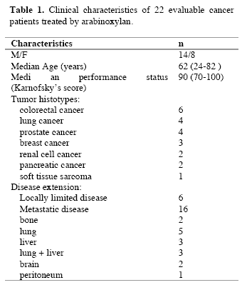
Evaluable patients were 22/24, while the remaining 2 patients, both affected by untreatable disseminated liver metastases due to colorectal cancer, rapidly died for disease progression before concluding the two planned months of arabinoxylan therapy. The clinical characteristics of the evaluable patients are reported in Table 1. Figure 1 illustrates changes in the mean number of total lymphocytes, T lymphocytes, T cytotoxic lymphocytes and NK cells occurring after 2 months of arabinoxylan therapy. No substantial variation was found in the mean number of lymphocytes, T lymphocytes, T cytotoxic lymphocytes and NK cells under arabinoxylan treatment. In contrast, as illustrated in Figure 2, TH and T reg mean numbers increased and decreased, respectively, after arabinoxylan therapy, without, however statistically significant differences with respect to the values seen prior to therapy. On the contrary, a statistically significant increase in TH/T reg mean ratio was achieved after arabinoxylan therapy (p<0.025). The increase in TH/T reg ratio under arabinoxylan therapy was more pronounced in patients with an abnormally low ratio prior to therapy with respect to that occurring in those with normal pre-treatment ratio, however without statistically significant differences ( 2.3 +/- 0.4 vs 1.7 +/- 0.5). In more detail,
090722_linfocitiT_table1
090722_linfocitiT_1

Figure 1. Changes in the number of lymphocytes, Tlymphocytes (CD3), T cytotoxic lymphocytes (CD8) and NK cells (CD16 CD56) after 2 months of arabinoxylan therapy.
090722_linfocitiT_2

Figure 2. Changes in the mean number of T helper (TH) lymphocytes (CD4) and T regulatory lymphocytes (cd4 cd24) and in TH/T reg mean ratio.

before arabinoxylan therapy, an abnormally low TH/T reg ratio was present in 12/22 (55%) evaluable patients. Arabinoxylan treatment induced a normalization of TH/T reg ratio in 5/12 (42%) patients with an abnormally low ratio prior to therapy. The percentage of arabinoxylan-induced TH/T reg normalization obtained in lymphocytopenic patients was not significantly different from that achieved in patients with normal pre-treatment lymphocyte count ( 3/7(43%) vs 2/5(40%) ). No toxicity was observed under arabinoxylan treatment, which was well tolerated in all patients. Asthenia was present in 8/22 (36%) evaluable patients. An evident relief of asthenia, as assessed by a specific patient report, was obtained under arabinoxylan therapy in 5/8 (63%) patients.

IV. Discussion
Previous experimental studies had already demonstrated some immunomodulating properties of arabinoxylan, in particular consisting of stimulation of NK cytotoxic function (Ghoneum, 1998), whereas NK cell number did not seem to be influenced by arabinoxylan administration. However, it has to be remarked that NK cells were belived to be fundamental in the antitumor immunity until some years ago, before the discovery of the essential role played by the antitumor cytokines, such as IL-2 and IL-12 (Whittington and Faulds, 1993) and dendritic cells, because of their function as antigen-presenting cells (Banks et al, 1995). In fact, it has to be considered that the cytotoxic activity of NK cells is effective only against artificial laboratory cancer cell lines, whose biological malignant properties are different from those presented by fresh human tumor cells (Whittington and Faulds, 1993). In addition, NK cells have been proven to be also able to destroy fresh human cancer cells only after the activation of their cytotoxic function by IL-2 (Atzpodien and Kirchner, 1990). From this point of view, arabinoxylan had been already proven to amplify the stimulatory effect of IL-2 on NK-mediated antitumor cytotoxicity (Ghoneum and Jewett, 2000). In contrast, no study has been performed up to now to evaluate the possible influence of arabinoxylan not only on the mechanisms responsible for the generation of an effective anticancer immune response, but also on those involved in the suppression of anticancer immunity. The results of this preliminary study, carried out to evaluate the influence of arabinoxylan on T reg cells, which represent the most important cells involved in the suppression of the antitumor cytotoxic immune response, demonstrates that arabinoxylan may counteract T reg cell generation by reducing their number and percentage with respect to the total amounts of CD4+ cells and circulating lymphocytes. Since NK cell function is inhibited by T reg activation (Shevach, 2002), the previously demonstrated arabinoxylan-induced stimulation of NK cell cytotoxic function might depend at least in part on its capacity of counteracting T reg generation (Dieckmann et al, 2001). Moreover, this study would suggest that the inhibitory action of arabinoxylan on T reg generation is more pronounced in patients with an abnormally high percentage of T reg cells prior to therapy, with a following pre-treatment abnormally low TH/T reg ratio before therapy, whereas its effect was less evident in patients with a pre-treatment value of TH/T reg ratio within the normal range. Therefore, the influence of arabinoxylan on T reg generation would consist of a modulatory action rather than an inhibitory activity. This finding could explain a potential favourable immunomodulatory effect of arabinoxylan also in patients with autoimmune diseases (Ghoneum, 1998), who in contrast to cancer patients would tend to present abnormally low amounts of T reg cells. In any case, the importance of the inhibition of T reg generation in the induction of an effective anticancer immune response has been recently confirmed by the evidence that the block of T reg activity by specific monoclonal antibodies may induce objective tumor regressions in humans (Yang et al, 2007). Obviously, the major problem is the exact identification of he T reg cell population. Even though T reg cells may express other immune markers, namely FOX-p2 cytoplasmatic antigen, most clinicians are in agreement to identify the CD4+CD25+ cells as T reg lymphocytes (12). In any case, further studies, by evaluating other immune markers, will be required to better identify T reg cells population, namely FOX-p3, even though recently some Authors have shown that FOX-p3 expression by T reg cells is associated with a lower suppressive activity (Dieckmann et al, 2001; Shevach, 2002). Moreover, it has to be remarked that several patients included in the present study were concomitantly under palliative therapy with the anti-cachectic pineal hormone melatonin (Brzezinski, 1997), which may also play immunomodulating effects (Maestroni, 1993). Therefore, further randomized studies with arabinoxylan alone versus arabinoxylan plus melatonin will be required to better define the immunomodulating action of arabinoxylan. If further clinical and experimental studies will confirm the inhibitory action of arabinoxylan on T reg cell system, it could be included in cytokine-based immunotherapies to enhance their efficacy by counteracting T reg cell generation.

References
Atzpodien J, Kirchner H (1990) Cancer, cytokines and cytotoxic cells:interleukin-2 in the immunotherapy of human neoplasms. Klin Wochenschr 14, 1-10.
Banks RE, Patel PM, Selby PJ (1995) Interleukin-12:a novel clinical player in cytokine therapy. Br J cancer 71, 655-659.
Brzezinski A (1997) Melatonin in humans. N Engl J Med 336, 185-195.
Dieckmann D, Plottner H, Berchtold S, Berger T, Schuler G (2001) Ex vivo isolation and characterization of CD4+CD25+ T cells with regulatory properties from human blood. J Exp Med 193, 1303-1310.
Ghoneum M (1998) Enhancement of human natural killer cell activity by modified arabinoxylane fro rice bran(MGN-3). Int J Immunother 14, 89-99.
Ghoneum M, Gollapudi S (2005) Synergistic of arabinoxilan rice bran (MGN-3/Biobran in S. Cerevisiae-induced apoptosis of monolayer breast cancer MFC-7 cells. Anticancer Res 25(6B), 4187-96.
Ghoneum M, Brown J, Gollapudi S (2007) Yeast therapy for the treatment of cancer and its enhancement by MGN-3/Biobran, an arabinoxylan rice bran. Cellular Signaling and Apoptosis Research (Ed. Alex R. Demasi) Cap IV: 185-200.
Ghoneum M, Jewett A (2000) Production of tumor necrosis factor-alpha and interferon-gamma from human peripheral blood lymphocytes by MGN-3, a modified arabinoxylan from rice bran, and its synergy with interleukin-2 in vitro. Cancer Detect Prevent 24, 314-324.
Lissoni P, Giani L, Zerbini S, Trabattoni P, Rovelli F (1998) Biotherapy with the pineal immunomodulating hormone melatonin versus melatonin plus Aloe vera in untreatable advanced solid neoplasms. Nat Immun 16, 27-33.
Maestroni JGM (1993) The immunoneuroendocrine role of melatonin. J Pineal Res 14, 1-10.
Markus J, Miller A, Smith M, Orengo I (2006) Metastatic hemangiopericytoma of the skin treated with wide local excision and MGN-3. Dermatol Surg 32, 145-147.
Matsuda T, Hirano T (1990) Interleukin-6 (IL-6). Biotherapy 2, 363-371.
Moore KW, O’Garra A, De Waal-Malefyt R (1993) Interleukin-10. Ann Rev Immunol 11, 165-174.
Rosenberg SA (1992) The immunotherapy and gene therapy of cancer. J Clin Oncol 10, 181-191.
Shevach EM (2002) CD4+CD25+ suppressor T cells:more questions than answers. Nat Rev Immunol 2, 389-400.
Whittington R, Faulds D (1993) Interleukin-2. Drugs 46, 446-514.
Yang JC, Hughes M, Kammula U, Royal R, Sherry RM, Topalian SL, Suri KB, Levy C, Allen T, Mavroukakis, Lowy I, White DE, Rosenberg SA (2007) Ipilimubab (anti-CTLA4 antibody)causes regression of metastatic renal cell cancer associated with enteritis and hypophysitis. J Immunother 30, 825-830.

(download PDF version)


A clinical study of T-regulatory lymphocyte function in cancer patients in relation to tumor histotype, disease extention, lymphocyte subtypes and cortisol secretion

Research Article (download PDF version)

Luigi Vigorè1, Fernando Brivio2, Luca Fumagalli2, Roberto Vezzo1, Giusy Messina6, Franco Rovelli6, Massimo Colciago3, Giovanna D’Amico4, Giuseppe Di Fede5, Paolo Lissoni6

1 Laboratory of Immunomicrobiology, St.Gerardo Hospital, Monza, Milan, Italy
2 Division of Surgery, St.Gerardo Hospital, Monza, Milan, Italy
3 I.N.R.C.A Laboratory of Analyses, Casatenovo, Lecco, Italy
4 Research Center “Fondazione Tettamanti” Clinica Pediatrica Università Milano-Bicocca, Italy
5 Institute of Biological Medicine, Milan, Italy
6 Division of Radiation Oncology, St.Gerardo Hospital, Monza, Milan, Italy

*Correspondence: Dr. Paolo Lissoni, Divisione di Radioterapia Oncologica, Ospedale S.Gerardo, 20052 Monza, Milano, Italy; Fax: +390392332284, E-mail: p.lissoni@hsgerardo.org
Key words: Anticancer immunity, immunosuppression, T regulatory lymphocytes
Abbreviations: cytotoxic T lymphocyte-associated antigen-4, (CTLA-4); glucocorticoid-induced TNF-α receptor, (GITR); myeloidderived suppressor cells, (MDSC); NK cells, (CD16CD56); T cytotoxic lymphocytes, (CD8); T helper lymphocytes, (CD4); Tregulatory lymphocytes, (T-reg)

Received: 24 July 2008; Revised: 11 September 2008
Accepted: 12 September 2008; electronically published: October 2008

Summary

Several clinical investigations showed that the immune status is a prognostic variable in cancer patients, even tough the evaluation of the anticancer immunity is not generally considered in the medical oncology. Several immune parameters, including lymphocyte subsets and cytokine blood concentration, had been proposed to quantify the functional status of the anticancer immunity, but recent discoveries would suggest that the end-result of the various immune interactions is represented by a subtype of CD4 lymphocytes capable of suppressing the antitumor immune reaction, the so called T-regulatory lymphocytes (T-reg). This study was performed to detect T-reg count and percentage in solid tumor patients, in relation to tumor histotype, disease extension, lymphocyte sub-populations and cortisol circadian secretion. The study included 114 consecutive cancer patients affected by the most frequent tumor histotypes, 69 of whom showed a metastatic disease. In each patient we evaluated T-reg cells, identified as CD4+CD25+, in relation to T helper (CD4), T cytotoxic (CD8) and NK (CD16CD56) cells. Abnormally high values of T-reg cells were seen in 52/114 (46%) patients, and the percentage of high values of T-reg was significantly higher in metastatic patients than in non-metastatic ones. In contrast, no significant difference was seen in relation to tumor histotype. Patients with increased T-reg count had a significantly lower NK cell number. Finally no significant difference in T-reg number was seen between patients with altered or normal rithm of cortisol. The study confirmed that, irrespectively of tumor histotype the metastatic disease is associated with a progressive and increased T-reg generation, with a following suppression of anticancer immunity.

I. Introduction
At present, there is no doubt about the existence of a sub-type of T lymphocytes, the so-called T regulatory lymphocytes (T-reg), capable of suppressing the cellular immune responses,including the anticancer immunity (Thomton and Shevach, 2000; Shevach, 2002; von Herrath and Harrison, 2003; Schwartz, 2005; von Boehmer, 2005; Ziegler, 2006; Zou, 2006). However, the exact definition
of T-reg cells in terms of cell surface marker expression still remains controversial, particularly from a clinical point of view. All authors are in agreement to consider Treg lymphocytes as CD4+CD25+ cells, but at present it is still unknown whether the expression of CD4 and CD25 antigens may be sufficient to identify T-reg cells (Thomton and Shevach, 2000; Shevach, 2002; von Herrath and Harrison, 2003; Schwartz, 2005; von Boehmer, 2005; Ziegler, 2006; Zou, 2006), since several authors retain that the intracytoplasmatic expression of the FOX p3 protein is essential for the differentiation into T-reg cells (Ziegler, 2006; Zou, 2006).Recently, however, some preliminary observations would suggest that the cytoplasmatic expression of FOX p3 by CD4+CD25+ cells may be associated at least in some experimental conditions with a diminished, rather than with an enhanced immunosuppressive activity of T-reg cells (Siddiqui et al, 2007). In contrast, all authors agree that the expression of CD152 antigen, also called cytotoxic T lymphocyteassociated antigen-4 (CTLA-4) (Vasu et al, 2004), is fundamental for the immunosuppressive activity of T-reg cells (Takahashi et al, 2000), since the block of its expression by using anti-CTLA-4 monoclonal antibodies may abolish the suppressive activity of T-reg cells, with a following stimulation of the anticancer immunity in cancer patients (Knutson and Disis, 2007) and an enhanced incidence of autoimmune diseases in the healthy subjects (Lan et al, 2005). Therefore, the addition of a third marker, such as CD152 antigen, may allow to define a more homogeneous cell population provided by a regulatory activity with respect to the simple CD4+CD25+ expression (Dieckmann and Plottner, 2001). In fact, the suppressive regulatory action of CD4+CD25+CD152+ has appeared to be clearly higher than that played by the simple CD4+CD25+ T lymphocytes (Leong et al, 2006).This finding is not surprising, since the simple expression of CD25 marker, corresponding to the !-chain of IL-2 receptor, is not an exclusive characteristic of T-reg lymphocytes, but it is a non-specific property of the overall activated T lymphocytes (Thomton and Shevach, 2000; Shevach, 2002; von Herrath and Harrison, 2003; Schwartz, 2005; von Boehmer, 2005; Ziegler, 2006; Zou, 2006). At present, preliminary clinical studies would show that the percent of circulating CD4+CD25+ cells may be about 10% of the all CD4+ lymphocytes, and that of CD4+CD25+CD152+ cells may be about 40% of the total CD4+CD25+ cells, then the expected percent of CD4+CD25+CD152+ in the healthy subjects would be less than 5% of the total circulating CD4+ lymphocytes (Jago et al, 2004). Finally, the expression of glucocorticoidinduced TNF-α receptor (GITR) is also associated with an evident suppressive activity by T-reg lymphocytes (Kanamaru et al, 2004), which in fact are stimulated by
cortisol (Sthephens et al, 2004), that in contrast may inhibit the activity of the most other T lymphocytes, namely that of T helper lymphocytes, with a following diminished production of IL-2 (Claman, 1998). As far as the mechanisms responsible for T-reg-induced suppression of the anticancer immunity are concerned, several experimental observations have shown that T-reg cells may suppress the antitumor immune response through the release of immunosuppressive cytokines, namely IL-10 and TGF-β (Dieckmann et al, 2002), even though other authors would suggest that the suppressive activity of Treg cells on CD4+ and CD8+ lymphocyte activation may be relatively independent from the action of cytokines, by mainly requiring cell surface contact (Birebent et al, 2004). IL-2 has been proven to be essential for T-reg generation and some authors consider IL-2 as the main growth factor of T-reg lymphocytes (Antony and Restito, 2005), but more adequate studies have demonstrated that IL-2 may induce both stimulation and inhibition of T-reg generation and activation (Malek and Bayer, 2004). In fact, IL-2 has appeared to induce and promote T-reg differentiation only in the presence of TGF-β (Chen et al, 2003). Therefore, IL-2 would constitute the main human cytokine in influencing the characteristics of the anticancer immunity, since it may be responsible for both activation and suppression of an effective immune response against cancer cell proliferation and dissemination (Wang et al, 2001), namely depending on the whole status of the cytokine network, in particular on the presence or in the absence of adequate concentrations of TGF-β. In the absence of TGF-β, IL-2 stimulates the anticancer immunity, whereas it counteracts the generation of an effective antitumor immunity in the presence of TGF-β. In other words, IL-2 would physiologically control both tolerance and immunity, depending on the presence of TGF-β and other less known factors (Annunziato et al, 2002). In fact, under cancer immunotherapy with IL-2 the percent of T-reg cells has been shown to decrease in responding patients and to enhance in those with disease progression (Cesana et al, 2006). However, the regulation of T-reg functions does not depend only on immune factors, since it is also under a neuroendocrine control (Ji et al, 2004). In particular, cortisol has appeared to stimulate T-reg cell generation (Ji et al, 2004), with a following enhanced release of IL-10, by representing the main mechanism responsible for cortisol-induced immunosuppression. From a clinical oncological point of view, preliminary observations showed an enhanced percent of circulating CD4+CD25+ lymphocytes in cancer patients, namely in those with advanced disease (Sasada et al, 2003). The present study was performed to better establish which is T-reg behaviour in cancer patients in relation to both tumor histotype and disease extension.

II. Materials and methods
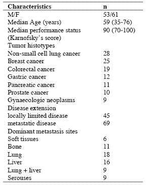
The study included 114 consecutive solid tumor patients with locally limited or metastatic disease, whose clinical characteristics are shown in Table 1. Lung cancer and gastrointestinal tumors were the most frequent neoplasms in our patients. For the immune detections, venous blood samples were collected in the morning after an overnight fast. Operable patients and metastatic patients were investigated before the surgical operation and before the onset of chemotherapy, respectively, in an attempt to exclude the possible influence of the various anticancer therapies on the immune status of patients.

In each sample, we have evaluated total lymphocyte count and the various lymphocyte subpopulations by a flow cytometric assay and monoclonal antibodies, including T helper lymphocytes (CD4), T cytotoxic lymphocytes (CD8), NK cells (CD16CD56), and T regulatory (T-reg) lymphocytes (CD4CD25). Normal values (95% confidence limits) of T-reg observed in our laboratory were below 240/mm3. Moreover, because of its importance in regulating lymphocyte functions and proliferation (Claman, 1998; Sthephens et al, 2004), the circadian rhythm of cortisol was also investigated by collecting blood samples at 8.00 A.M. and at 4.00 P.M., and cortisol serum concentrations were measured in duplicate by using the ECLA method. Data were reported as mean +/- SE, and statistically analyzed by the Student’s t test, the analysis of variance and the chi-square test, as appropriate.

III. Results
As reported in Table 2, an abnormally high number of T-reg was seen in 52/114 (46%) patients. Moreover, the percentage of cases with elevated number of T-reg observed in metastatic patients was significantly higher with respect to that found in non-metastatic patients (44/69 (64%) vs 8/45(18%), p < 0.01). Table 3 shows the mean number of T-reg and the mean percentages of T-reg with respect to both total lymphocytes and T helper (CD4+) lymphocytes observed in cancer patients in relation to their disease extension. The mean number of T-reg observed in metastatic patients was higher with respect to that found in patients with locally limited disease, without, however statistically significant differences. In contrast, the mean percentages of T-reg with respect to that of both lymphocytes and CD4 cells were significantly higher in metastatic patients than in the non-metastatic ones (p< 0.05 and p< 0.001,respectively). Moreover, within the metastatic group, patients with a normal lymphocyte count greater than 1500/mm3 showed a significantly higher mean number of T-reg with respect to the non-metastatic patients, whereas no difference was seen between nonmetastatic patients and metastatic patients with lymphocytopenia, consisting of lymphocyte count lower than 1500/mm3. In contrast, the mean percentages of T-reg with respect to total lymphocytes and CD4+ cells observed in both groups of metastatic patients with normal or low total lymphocyte count were significantly higher than in non-metastatic patients (lymphocytes: p< 0.025, CD4+ cells: p< 0.001).

Table 1. Clinical characteristics of 114 solid tumor patients
090722_clinicalstud1

Table 2. Percentages of abnormally high values of CD4+CD25+ lymphocytes

090722_clinicalstud2

* P < 0.01 vs non-metastatic patients

Table 3. Mean number of CD4+CD25+ lymphocytes and their mean percentages with respect to total lymphocytes and CD4+ lymphocytes in metastatic and non-metastatic patients

090722_clinicalstud3

* p<0.05 vs non-metastatic patients; ** p<0.025 vs non-metastatic patients; *** p<0.01 vs non-metastatic patients; **** p<0.001 vs nonmetastatic patients

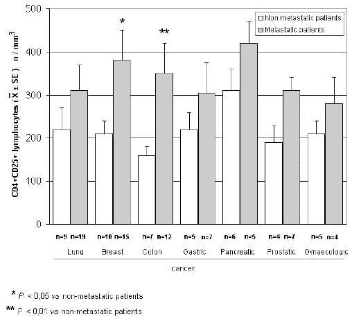
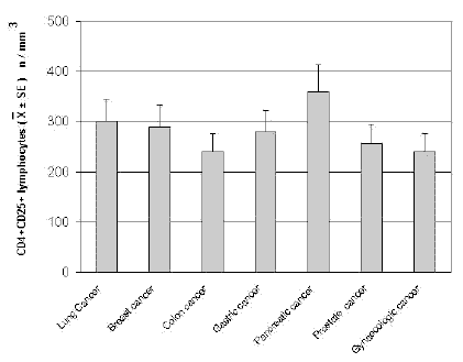
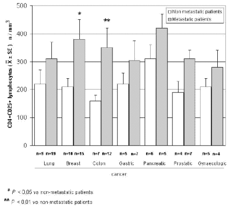
The mean counts of NK and CD8 cells in relation to that of T-reg are reported in Table 4. As shown, no significant difference in the mean number of CD8 lymphocytes was found between patients with normal or abnormally elevated number of T-reg. On the contrary, patients with elevated number of T-reg showed a significantly lower number of NK cells with respect to that found in those with normal T-reg count. Finally, Table 5 shows the circadian rhythm of cortisol in relation to total lymphocytes, CD4+ cells and T-reg mean number. A normal cortisol rhythm, with morning values greater at least than 50% with respect to the values occurring during the afternoon, was found in 85/114 (75%). Total lymphocyte and CD4+ cell mean numbers observed in patients with altered cortisol rhythm were significantly lower than those found in patients with normal cortisol circadianicity (p<0.01), whereas no significant difference was seen in the mean number of T reg. Figure 1 and Figure 2 illustrate T-reg mean numbers in relation to tumor histoptypes in the overall patients and with respect to their disease extension, respectively. No significant difference was seen in relation to tumor histotype. The highest values of T-reg were observed in pancreatic cancer patients, without however significant differences with respect to the overall other histotypes. The metastatic disease was associated with a higher number of T-reg with respect to the non-metastatic group in all tumor histotypes, even though a statistically significant differences occurred for the only breast cancer (p<0.05) and colorectal cancer (p< 0.01).

Table 4. Mean values of NK cells and CD8+ lymphocytes in cancer patients with normal or abnormally high values of CD4+CD25+ lymphocytes

090722_clinicalstud4

* p<0.05 vs normal values of CD4+CD25+ lymphocytes

Table 5. Mean numbers of total lymphocytes, T helper (CD4+) lymphocytes and T regulator lymphocytes (CD4+CD25+) in relation to cortisol circadian secretion in cancer patients
090722_clinicalstud5

* P<0.01 vs patients with altered cortisol rhythm
090722_clinicalstudfig1

Figure 1. CD4+CD25+ lymphocyte mean number in relation to tumor histotype
090722_clinicalstudfig2

Figure 2. CD4+CD25+ lymphocytes in relation to tumor histotype in metastatic and non-metastatic cancer patients

IV. Discussion
According to previous preliminary clinical investigations (Sasada et al, 2003; Cesana et al, 2006), this study confirms in a greater number of cancer patients that the metastatic disease is characterized by the evidence of an abnormally increased percentage of T-reg lymphocytes with respect to both total circulating lymphocytes and CD4+ lymphocytes. This finding does not seem to represent a specific characteristic of some tumor histotypes, then it could constitute a general alteration occurring during the progression of the neoplastic disease, by representing a fundamental immune parameter of cancer-related immunosuppression.

Several immune molecules have appeared to suppress the anticancer immunity, namely IL-6, IL-10, IL-1, TNF-α and TGF-β, but it seems that the common end result of their mechanisms of action may be represented by the stimulation of T-reg generation, with a consequent inhibition of the activation of an effective anticancer immune reaction. On the same way, several immune cells are able to suppress the anticancer immunity, including macrophages, T helper-2 lymphocytes and some myeloidderived suppressor cells, but also in this case they would act in a suppressive way by promoting the generation of Treg.

Then, the detection of T-reg amounts in terms of both absolute number and percentages with respect to total lymphocytes and CD4+ cells could constitute a simple and adequate clinical immune parameter to quantify the whole status of the anticancer immunity in the single cancer patient. Moreover, future clinical studies will be required to establish the possible prognostic significance of changes in T-reg percentage and number in relation to the anticancer efficacy of the various standard antitumor therapies. Moreover, it has to be remarked that T-reg lymphocytes would not represent the only immune cells involved in the suppression of the anticancer immunity. In fact, there is at least another fundamental immunosuppressive system, consisting of the monocytemacrophage cell lineage (Sica and Bronte, 2007). In more detail, it has been observed that the bone marrow may release myeloid precursors provided by suppressive activity on the antitumor immune response and defined as myeloid-derived suppressor cells (MDSC) (Kusmartsev and Gabrilovich, 2005). These cells have appeared to be characterized by the cell surface expression of GR-1, CD11b and CD80 antigens (Anderson et al, 2002; van Ginderachter et al, 2006). The myeloid suppressor cells would promote the generation and activation of T-reg lymphocytes, which at the other side would stimulate MDSC release from the bone marrow and M2 macrophage differentiation (Terabe et al, 2003; Wie et al, 2006).

Moreover, the myeloid suppressive cells would inhibit the anticancer immunity by promoting macrophage differentiation into the M2 sub-type (Ikemoto et al, 2003), which plays a clear inhibitory effect on the anticancer immunity, namely through the release of IL-6 (Ueno et al, 2000), whereas the M1 macrophage sub-type may either stimulate or suppress the antitumor immunity (Mantovani et al, 2004). M1 and M2 macrophage sub-types have appeared to be characterized by a high production of IL-12 or IL-10, respectively (Ueno et al, 2000).

Then, further studies by concomitantly evaluating T reg and MDSC count, will contribute to better define the immune mechanism responsible for the suppression of the anticancer immunity.

References
Anderson CF, Gerber JS, Mosser DM (2002) Modulating macrophage function with IgG immune complexes. J Endotoxin Res 8, 477-81.

Annunziato F, Cosmi L, Liotta F, Lazzeri E, Manetti R, Vanini V, Romagnani P, Maggi E, Romagnani S (2002) Phenotype,localization and mechanism of suppression of CD4+CD25+ human thymocytes. J Exp Med 196, 379-87.

Antony PA, Restito NP (2005) CD4+CD25+ T regulatory cells,immunotherapy of cancer,and interleukin-2. J Immunother 28, 120-8.

Birebent B, Lorho R, Lechartier H, de Guibert S, Alizadeh M,
Vu N, Beauplet A, Robillard N, Semana G (2004) Suppressive properties of human CD4+CD25+regulatory T cells are dependent on CTLA-4 expression. Eur J Immunol 34, 3485-96.

Cesana GC, DeRaffele G, Cohen S, Moroziewicz D, Mitcham J, Stoutenburg J, Cheung K, Hesdorffer C, Kim-Schulze S, Kaufman HL (2006) Characterization of CD4+CD25+ regulatory T cells in patients treated with high-dose interkleukin-2 for metastatic melanoma or renal cell carcinoma. J Clin Oncol 24, 1169-77.

Chen W, Jin W, Hardegen N, Lei KJ, Li L, Marinos N, McGrady G, Wahl SM (2003) Conversion of peripheral CD4+CD25- naïve T cells to CD4+CD25+ regulatory T cells by TGF-β induction of transcription factor Foxp 3. J Exp Med 198, 1875-86.

Claman HN (1998) Corticosteroids and the immune system. Adv Exp Med Biol 245, 203-10.

Dieckmann D, Bruett H, Ploettner H, Lutz MB, Schuler G (2002) Human CD4+CD25+ regulatory contact-dependent T cell induce IL-10 producing,contact-independent type-1-regulatory T cells. J Exp Med 196, 247-53.

Dieckmann D, Plottner H (2001) Ex vivo isolation and characterizationof CD4+CD25+ T cells with regulatory properties from human blood. J Exp Med 193, 1303-10.

Ehrke MJ, Mihich E, Berd D, Mastrangelo MJ (1989) Effects of anticancer drugs on the immune system. Semin Oncol 16, 230-9.

Ghiringhelli F, Larmonier N, Schmitt E, Parcellier A, Cathelin D, Garrido C, Chauffert B, Solary E, Bonnotte B, Martin F (2004) CD4+ CD25+ regulatory T cells suppress tumor immunity but are sensitive to cyclophosphamide which allows Immunotherapy of established tumors to be curative. Eur J Immunol 34, 336-44.

Ikemoto S, Yoshida N, Narita K, Wada S, Kishimoto T, Sugimura K, Nakatani T (2003) Role of tumor-associated macrophages in renal cell carcinoma. Oncol Rep 10, 1843-9.

Jago CB, Yates J, Camara NOS, Lechler RI, Lombardi AG (2004) Differential expression of CTLA-4 among T cell subsets. Clin Exp Immunol 136, 463-71.

Ji HB, Liao G, Faubion WA, Abadía-Molina AC, Cozzo C, Laroux FS, Caton A, Terhorst C (2004) Cutting edge, the natural ligand for glucocorticoid-induced TNF receptorrelated protein abrogates regulatory T cell suppression. J Immunol 172, 5823-7.

Kanamaru F, Youngnak P, Hashiguchi M, Nishioka T, Takahashi T, Sakaguchi S, Ishikawa I, Azuma M (2004) Costimulation via glucocorticoid-induced TNF receptor in both conventional and CD25+ regulatory CD4+ T cells. Immunol 172, 7306-14.

Knutson KL, Disis M, Salazar L (2007) CD4 regulatory T cells in human cancer pathogenesis. Cancer Immunol Immunother 556, 271-85.

Kusmartsev S, Gabrilovich DI (2005) STAT1 signaling regulates tumor-assolciated macrophage-mediate T cell deletion. J Immunol 174, 4880-91.

Lan RY, Ansari AA, Lian ZX, Gershwin ME (2005) Regulatory T cells, development,function,and role in autoimmunity. Autoimmun Rev 4, 351-63.

Leong PP, Mohammad R, Ibrahim N, Ithnin H, Abdullah M, Davis WC, Seow HF (2006) Phenotyping of lymphocytes expressing regulatory and effector markers in infiltrating ductal carcinoma of the breast. Immunol Lett 102, 229-36.

Malek TR, Bayer AL (2004) Tolerance not immunity crucially depends on IL-2. Nat Rev Immunol 4, 665-74.

Mantovani A, Sica A, Sozzani S, Allavena P, Vecchi A, Locati M (2004) The chemokine system in diverse forms of macrophage activation and polarization. Trends Immunol 25, 677-86.

Sasada T, Kimura M, Yoshida Y, Kanai M, Takabayashi A (2003) CD4+CD25+ regulatory T cells in patients with gastrointestinal malignancies, possible involvement of regulatory T cells in disease progression. Cancer 98, 1089-93.

Schwartz RH (2005) Natural regulatory T cells and selftolerance. Nat Immunol 6, 327-30.

Shevach EM (2002) CD4+CD25+ suppressor T cells, more questions than answers. Nat Rev Immunol 2, 389-400.

Sica A, Bronte V (2007) Altered macrophage differentiation and immune dysfunction in tumor development. J Clin Invest 117, 1155-66.

Siddiqui SA, Frigola X, Bonne-Annee S, et al (2007) Tumorinfiltrating Foxp3 CD4+CD25+ T cells predict poor survuival in renal cell carcinoma. Clin Cancer Res 13, 2075-81.

Stephens GL, McHugh RS, Whitters MJ, Young DA, Luxenberg D, Carreno BM, Collins M, Shevach EM (2004) Engagement of glucocorticoid-induced TNFR family-related receptor on effector T cells by its ligand mediates resistance to suppression by CD4+CD25+ T cells. J Immunol 173, 5008-20.

Takahashi T, Tagami T, Yamazaki S, Uede T, Shimizu J, Sakaguchi N, Mak TW, Sakaguchi S (2000) Immunologic and tolerance maintained by CD25+CD4+ regulatory T cells constitutively expressing cytotoxic T lymphocyte-associated antigen. J Exp Med 192, 1285-94.

Terabe M, Matsui S, Park JM, Mamura M, Noben-Trauth N, Donaldson DD, Chen W, Wahl SM, Ledbetter S, Pratt B, Letterio JJ, Paul WE, Berzofsky JA (2003) Transforming growth factor-β production and myeloid cells are an eggector mechanism trhough which CD1d-restricted T cells block cytotoxic T lymphocyte-mediated tumor immunosurveillance, abrogation prevents tumor recurrence. J Exp Med 198, 1741-52.

Thomton AM, Shevach EM (2000) Suppressor effector function of CD4+CD25+ immunoregulatory T cells is antigen nonspecific. J Immunol 164, 183-90.

Ueno T, Toi M, Saji H, Muta M, Bando H, Kuroi K, Koike M, Inadera H, Matsushima K (2000) Significance of macrophage chemo-attractant protein-1 in macrophage recruitment,angiogenesis and survival in human breast cancer. Clin Cancer Res 6, 3282-9.

Van Ginderachter JA, Movahedi K, Hassanzadeh Ghassabeh G, Meerschaut S, Beschin A, Raes G, De Baetselier P (2006) Classical and alternative activation of mononuclear phagocytes, picking the best of both worlds for tumor promotion. Immunobiology 211, 487-501.

Vasu C, Prabhakar BS, Holterman MJ (2004) Targeted CTLA-4 engagement induces CD4+CD25+CTLA-4 high T regulatory cells with target alloantigens specificity. J Immunol 173, 2866-76.

von Boehmer H (2005) Mechanisms of suppression by suppressor T cells. Nat Immunol 6, 338-44.

von Herrath MG, Harrison LC (2003) Regulatory lymphocytes, antigen-induced regulatory T cells in autoimmunity. Nat Rev Immunol 3, 223-32.

Wang XB, Zheng CY, Giscombe R, Lefvert AK (2001) Regulation of surface and intracellularexpression of CTLA-4 on human peripheral T cells. Scand J Immunol 54, 453-8.

Wie S, Kryczeck I, Zou W (2006) Regulatory T-cell compartmentalization and trafficking. Blood 108, 426-31.

Yang R, Cai Z, Zhang Y, Yutzy WH 4th, Roby KF, Roden RB (2006) CD80 in immune suppression by mouse ovarian carcinoma-associated Gr-1+CD11b+ myeloid cells. Cancer Res 66, 6807-15.

Ziegler SF (2006) FOXP3 of mice and men. Annu Rev Immunol 24, 209-26.

Zou W (2006) Regulatory T cells, tumour immunity and immunotherapy. Nat Rev Immunol 6, 295-307.

(download PDF version)


Copyright by IMBIO 2017. All rights reserved.