Articoli

Tutto sul nostro settore


Febbraio 25, 2015 Newsletter
Riportiamo e commentiamo l’intervista fatta al Dr. Alberto Mantovani, direttore di reparto dip. sanità pubblica veterinaria e sicurezza alimentare (Spvsa) – tossicologia alimentare e veterinaria, Istituto superiore di sanità.
Che cosa sono gli interferenti endocrini (Ie) e quali i loro meccanismi d’azione?

Sono un gruppo eterogeneo di sostanze in grado di alterare il funzionamento del sistema endocrino con meccanismi diversi (recettoriali, metabolici ecc.). I principali bersagli dei loro effetti sono l’omeostasi degli steroidi sessuali e della tiroide, dunque la salute riproduttiva e lo sviluppo.

Quali sono i principali Ie e quali sono le fonti di assunzione per l’uomo?
Molte sostanze di origine antropica sono Ie: composti alogenati che si bioaccumulano e interferiscono con gli steroidi sessuali, come le diossine, che hanno un effetto antiestrogeno e i policlorobifenili (PCB). Poi ancora diversi pesticidi e biocidi (per esempio etilene-bisditiocarbammati) e composti industriali (come ritardanti di fiamma polibromurati) che hanno un’azione tireostatica.
Sono Ie anche alcune sostanze naturali: lo zearalenone, una micotossina contaminante dei cereali, ha un’azione estrogenica simile. Lascia ancora dei dubbi l’esposizione a dosi elevate di fitoestrogeni come isoflavoni e lignani, durante la gravidanza o la prima infanzia: sono infatti deboli agonisti del recettore estrogeno beta e sono presenti anche nella soia, ingrediente base di integratori e latti artificiali.
Una delle fonti di esposizione per l’uomo sono le matrici alimentari, soprattutto se ricche di grassi (per es., certi tipi di pesci), dove i vari Interferenti Endocrini (IE) si possono accumulare.

Quali sono le categorie più a rischio e quali sono le patologie associate?
La gravidanza e l’infanzia sono le fasi più a rischio, anche se spesso gli effetti si manifestano a lungo termine, e possono variare con il sesso e l’età in cui è avvenuta l’esposizione. Il WHO segnala che l’esposizione può aumentare il rischio di patologie riproduttive, disturbi comportamentali nell’infanzia, fino a diabete e alcuni tipi di cancro (testicolo, mammella).

Va poi considerato che dosi molto basse di diversi IE con la stessa azione, possono sommare i loro effetti fino ad indurre un effetto tossico significativo.

Quali sono i comportamenti corretti per ridurre il rischio?
Secondo il Decalogo per i cittadini sugli IE pubblicato dall’ISS e dal Ministero Ambiente è opportuno minimizzare l’esposizione alle fonti: usare contenitori in plastica o stoviglie integri, secondo le modalità previste dal produttore; limitare il consumo di alimenti che possono accumulare IE e il contatto dei cibi con carta oleata o pellicole. Inoltre bisogna fare attenzioni ai metodi di cottura (eliminare parti carbonizzate che contengono idrocarburi policiclici aromatici); limitare il consumo di cibi affumicati ed evitare il ristagno dei fumi in cucina e della polvere negli ambienti chiusi; per i bambini, variare la dieta e assicurare un apporto adeguato di vitamine e microelementi con effetto protettivo.

Aggiungiamo che un controllo sul qualità della composizione corporea, eseguendo un test sui capelli, mineralogramma, ci indicherà la percentuale di minerali presenti nei tessuti, la possibilità di riscontro di metallo pesanti potenzialmente tossici.

Un controllo sulla qualità della flora batterica dell’intestino, ci orienterà sul grado di Disbiosi intestinale, e sulla possibilità di correggere l’alimentazione.

Il test su possibili intolleranze alimentari ( ALCAT Test) segnalerà le possibili incompatibilità di alcuni alimenti indirizzando il nutrizionista specialista sul tipo di dieta da seguire per evitare infiammazioni e possibili malattie derivanti da contaminati alimentari. Il test ALCAT analizza anche intolleranze a sostanze chimiche presenti negli alimenti oltre che a conservanti e additivi alimentari.

Valutazione e integrazione sono alla base dei percorsi di prevenzione individuale e personalizzata.


Febbraio 23, 2015 Newsletter

Era il 2000 quando i primi risultati sul sequenziale di tutto il DNA umano fu concluso. Furono svelati i segreti di come siamo fatti, a quali malattie siamo predisposti è come possiamo curarci. Ma leggere la sequenza di basi del nostro materiale genetico racconta solo una parte di chi siamo e a cosa siamo predisposti ad avere nel tempo.

È allo stesso modo importante anche sapere come vengono espressi (come funzionano i geni).Oggi sappiamo qualcosa di più al riguardo grazie al Roadmap Epigenomics Project, i cui risultati sono stati pubblicati su riviste del gruppo Nature.

Gli studi in questione descrivono un fattore molto importante che influenza l’attività dei geni, l’epigenomica, ovvero mostrano come, in diversi tipi di cellule umane, il DNA viene letto, il modo cioè in cui una stessa informazione genetica (tutte le cellule di uno stesso organismo hanno lo stesso dna) è usata (espressa) in modo diverso da cellula a cellula.

Ma cos’è l’epigenetica? È come funziona? A cosa serve?

Per epigenetica si intende l’insieme dei fenomeni che stanno al di fuori e al di sopra del DNA e quindi dei geni, che in qualche modo hanno una influenza sull’espressione dell’attività dei geni. Il come è codificato da tutte le modifiche che non alterano la sequenza genetica stessa ma stanno, per così dire, sopra (da qui la parola epigenetica).

Sono modifiche epigenetiche per esempio la metilazione del DNA (l’aggiunta di gruppi metilici alle basi di citosina) o il trasferimento di un gruppo acetile sugli istoni (proteine che legano il dna).
Per rendere meglio l’idea, ogni cellula può esprimere il proprio dnai n modo diverso a seconda del proprio lavoro (se deve essere una cellula cardiaca sarà diversa da un neurone), proprio come una stessa orchestra può suonare un pezzo in molti modi diversi.

I numerosi risultati pubblicati su Nature raccontano quindi di queste diverse sinfonie, suonate dalle cellule, scoperte analizzando campioni di tessuto embrionali (in fase di sviluppo e differenziazione) e adulto. Si scopre così, per esempio, che la trasformazione da staminali a cellule neuronali è orchestrata da specifici processi di metilazione, e più in generale la differenziazione delle cellule staminali è influenzata dalle modificazioni della cromatina (l’insieme del dna e delle proteine attaccate a esso), che influenza a sua volta il pattern di espressione genica.

Ma l’analisi epigenomica condotta dal gruppo di ricercatori, ha anche mostrato che l’epigenoma di una cellula cancerosa si porta dietro le impronte del tipo cellulare da cui ha avuto origine. O ancora: alterazioni epigenomiche nelle cellule del giro cingolato (un gruppo di cellule nervose che formano una regione specifica del cervello) potrebbero avere un ruolo nella sindrome da deficit di attenzione e iperattività.

Lo studio dell’epigenomica quindi potrebbe aprire le porte alla comprensione dell’origine delle malattie, e al tempo stesso alla comprensione di come gli stili di vita influenzino il nostro genoma.Le modifiche epigenetiche infatti sono in parte ereditarie, come i geni stessi, in parte dipendono dalle condizioni ambientali, quali la dieta delle mamme durante la gravidanza.

Concludendo, da queste osservazioni e conferme da parte dei ricercatori, possiamo affermare che siamo di fronte ad un punto di svolta per quanto riguarda la nutrizione e come questa sia in grado di influenzare l’attività dei geni, che a Loro volta si metteranno al lavoro, influenzando la salute o la possibile Malattia.

La nutrigenomica, da anni è al sevizio della medicina il cui scopo è quello di attivare programmi nutrizionali personalizzati che possono influenzare la salute e preventive le malattie.


Febbraio 16, 2015 Newsletter

Il riso Oryza Sativa, pianta erbacea annuale della famiglia delle Gramineae, di origine asiatica che insieme alla Oryza Glaberrima coltivata in Africa, è una delle due varietà vegetali da cui si produce il riso.

L’Oryza Sativa costituisce la maggior parte della produzione, in quanto coltivata su circa il 95% della superficie mondiale seminata a riso. Cereale, dall’incerta origine, si ritiene che le varietà più antiche siano comparse oltre quindicimila anni fa lungo le pendici dell’Himalaya.  Le statistiche documentano che il riso costituisca il cibo principale per circa la metà della popolazione mondiale e che venga coltivato in quasi tutti i paesi del globo.

Ne esistono tre tipologie:

Indica: tipica dei climi tropicali dall’alto valore di mercato, cariosside lunga e sottile. Tipologia dalla produttività media tipica dell’India, Cina meridionale, Filippine, America Latina, Italia, Brasile;

Japonica: tipica dei climi temperati, basso valore di mercato, cariosside corta e arrotondata, nota per l’alta produzione è coltivata in Giappone, Corea, Cina settentrionale, USA, Egitto, Italia;

Javanica: tipologia di minore importanza.

La Japonica è la sottospecie maggiormente coltivata in Italia, tipicamente usata per i risotti. A sua volta si divide in 4 tipologie:

Risi comuni (tondi e piccoli)

Risi semifini (tondi di media lunghezza)

Risi fini (affusolati e lunghi)

Risi superfini (grossi e lunghi)

L’Italia, con 1,44 milioni di tonnellate di riso prodotti nel 2005, rappresenta il principale produttore europeo e il ventisettesimo a livello mondiale. La coltivazione è concentrata principalmente nelle regioni Piemonte e Lombardia, nel triangolo Vercelli, Novara, Pavia. Viene inoltre coltivato in provincia di Mantova, in Emilia-Romagna in particolare nel basso ferrarese, in Veneto in particolare nella bassa veronese (Isola della Scala), nel Vicentino centrale (Grumolo delle Abbadesse), in Sardegna nella valle del Tirso e in Calabria nella Piana di Sibari.

Oggi però parliamo del riso rosso fermentato o Monascus Purpureus, un valido aiuto contro il colesterolo, nato dalla fermentazione del comune riso da cucina (Oryza Sativa), grazie ad un particolare lievito, chiamato Monascus Purpureus o lievito rosso.Questo riso, che deve il suo nome alla caratteristica colorazione, rappresenta una componente tradizionale della fitoterapia cinese ma è molto conosciuto anche in occidente per le preziose virtù ipolipidemizzanti, cioè coadiuvanti alla riduzione delle quantità di lipidi nel sangue.

Questo notevole interesse nei confronti del riso rosso è dovuto alla presenza di Monascus Purpureus, lievito che durante la sua attività fermentatrice, si arricchisce di un gruppo di sostanze, denominate monacoline, a cui è stata scientificamente attribuita un’elevata attività ipocolesterolemizzante, cioè che contribuisce ad abbassare il livello di colesterolo.

Appare dunque evidente che il riso rosso fermentato riduca il rischio cardiovascolare grazie ad azioni antiaterosclerotiche di altro tipo (effetto antinfiammatorio, vasodilatante e riduttivo sui livelli di lipoproteina A).   L’impiego di estratti di riso rosso fermentato è ammesso anche nella produzione di integratori alimentari, purché rimanga entro certi limiti fissati dal Ministero.

Tutto ciò ha contribuito ad alimentare il già fiorente business del riso rosso, spesso pubblicizzato in maniera eccessiva.D’altra parte, l’acquirente medio è attirato dalla possibilità di perdere peso ricorrendo ad un prodotto naturale, non elaborato sinteticamente e che per di più funge da valida alternativa alimentare per coloro che sono costretti ad assumere quotidianamente le statine, svolgendo al tempo stesso un ruolo preventivo futuro.


Ottobre 28, 2014 Newsletter

Dopo la prima uscita dedicata al Nichel, impariamo a conoscere e combattere a suon di ricette l’intolleranza al lattosio con l’aiuto del Prof. Di Fede, della drssa Jessica Barbieri e da me.

Un aiuto concreto per chi sta vivendo un problema troppo spesso sottovalutato: perché le difficoltà non finiscono al momento della diagnosi. Lo so bene io che ho trasformato un limite in una possibilità, raccogliendo in questo volume tutte le risorse utili a tavola per chi come me soffre della carenza dell’enzima necessario a digerire gli zuccheri presenti nel latte e nei suoi derivati: informazioni, consigli, esperienze e soprattutto… ricette!

Sfogliate e gustate i 99 piatti creati da me, da foodlovers e da tanti amici Chef di fama internazionale coinvolti in un progetto unico: vivere l’intolleranza al lattosio non più come limite, ma come possibilità di sperimentare nuovi sapori.

Non resta che partire insieme in un viaggio nel Mondo delle Intolleranze, con un volume che racchiude in sé il problema e la sua soluzione: nella cucina come nella vita, quando la ami non c’è niente e nessuno che possa indurti a rinunciarvi!

Oltre alle ricette di Tiziana troverete ricette di:

Luca Mauri, Alessandra Barbone, Antonella Rossi, Enrico Bartolini, Andrea Bevilacqua, Claudia Livia Biondini, Massimo Cairati, Antonino Cannavacciuolo, Michele Cannistrano, Giuseppe Capano, Fabio Silva, Isa Mazzocchi, Laura Ghezzi, Massimo Livan, Federico Mattavelli, Matteo Scibilia, Aurora Mazzucchelli, Antonino Messina, Micol Pisa, Milly Callegari, Carlo e Loris Molon, Giancarlo Morelli, Luca Mozzanica, Nunzia Bellomo, Davide Oldani, Armando Palmieri, Roberto Maurizio, Sandro Romano, Almerindo Santucci, Sara Preceruti, Marco Scaglione, Gaetano Simonato,Daniele Zennaro, Robbie Pezzuol

Sono da  da sempre convinta che la cucina sia veicolo di passione e umanità, e non mi sono arresa dopo la diagnosi di intolleranza al nichel e al lattosio. Oggi www.nonnapaperina.it è un blog di riferimento nel mondo delle intolleranze e la mia presenza è richiesta nelle più grandi manifestazioni enogastronomiche italiane. Si perpepisce che il problema delle intolleranze sta crescendo a vista d’occhio.

Il volume dedicato all’intolleranza al lattosio segue il grande successo della sua prima pubblicazione Nichel. L’intolleranza? La cuciniamo!, edito nel 2013.

Siete tutti invitati alla presentazione che si terrà giovedi 13 novembre 2014 all’Hotel de la Ville di Monza alle ore 19.


Ottobre 20, 2014 Newsletter

Il VI Congresso ARTOI. Oncologia Integrata e Nutrizione: il futuro nella Integrazione e nella Tradizione si terrà a Roma il 07 e 08 novembre 2014,  presso l’Aula Magna- “Università G. Marconi “, in via Vittoria Colonna 11alle ore  8:30.

Venerdi 07/11/2014  seduta plenaria.  Ore 20,30, cena di gala.

Sabato 08/11/2014  mattina: casistiche, ricerche, prevenzione, nutrizione.

Sabato 08/11/2014  pomeriggio: Workshop di Ipertermia Oncologica, organizzato da Andromedic.

Il costo è di  €50 per i soci ARTOI, €80 per i non soci ed € 50 per i non soci ARTOI no ECM

Il corso rilascerà crediti ECM per gli aventi diritto.

I posti sono limitati pertanto ti consigliamo di registrarti subito.

La data ultima per la registrazione e il pagamento è il primo novembre 2014.

La Deadline di invio abstract per la ” Sessione Poster” è stata prorogata al 30/10/2014


Giugno 19, 2014 Newsletter

Numerose evidenze scientifiche pubblicate nel corso degli anni hanno ampliato la conoscenza delle potenzialità metaboliche dell’intestino, conferendogli un’importanza fondamentale per il buon funzionamento del nostro sistema immunitario, del nostro sistema endocrino e della nostra sfera psichica. Oggi il nostro intestino deve essere considerato un organo di fondamentale importanza per la salute ed il benessere di tutto l’organismo.

A livello intestinale si riscontrano una notevole quantità di germi e batteri . La cosa più importante è che questa la flora intestinale  “fisiologica” sia in simbiosi con l’organismo. Questa condizione di equilibrio è definita eubiosi.

Quando questo equilibrio viene alterato si può generare una condizione patologica chiamata Disbiosi. I sintomi spesso sono aspecifici, caratterizzati da un’alterazione del transito intestinale, sia in termini di stitichezza ma a volte anche di sindrome diarroica. Il tutto associato frequentemente a gonfiore addominale, flatulenza, digestione lenta, stanchezza (la flora batterica produce vitamine B), suscettibilità alle infezioni, diminuzione delle difese immunitarie (la flora batterica produce anticorpi), candidosi intestinale e/o vaginale, vaginiti e cistiti ricorrenti nella donna.

Tra i fattori che possono contribuire a generare la Disbiosi vi sono:

alimentazione sbilanciata (uso eccessivo di grassi, zuccheri, carni rosse ed insaccati in genere, carenza di frutta e verdura, abuso di sostanze alcolici, fumo, ecc.), scarsa od insufficiente attività fisica, utilizzo di terapie farmacologiche protratte nel tempo (es. l’assunzione indiscriminata e prolungata di antibiotici, cortisonici, estroprogestinici, lassativi, ansiolitici, vaccini, chemioterapie, ecc).

Ma anche: contatto con sostanze quali conservanti e coloranti alimentari, radiazioni ed emissioni elettromagnetiche, metalli tossici contenuti in alimenti o in contenitori di alimenti o nelle stoviglie (alluminio, mercurio, piombo), pesticidi, ormoni steroidei alimentari.

 La comparsa di processi infiammatori intestinali, spesso legati ad una cattiva alimentazione e/o all’uso indiscriminato di farmaci (antiinfiammatori, antibiotici) in associazione ad una condizione di disbiosi intestinale possono essere responsabili non solo di malattie a carico del nostro intestino ma anche a livello di altri distretti come ad esempio la pelle (dermatite atopica, acne, psoriasi), infezioni delle vie urogenitali (candidosi vaginale, cistite, prostatite), malattie metaboliche (obesità, diabete, sindrome metabolica), malattie della sfera psichica (ansia, depressione), malattie autoimmuni (tiroidite cronica, artrite reumatoide).

Un corretto inquadramento clinico consente di programmare un piano terapeutico individualizzato che prevede, non solo un cambiamento dello stile di vita del paziente e delle sue abitudini alimentari, ma deve anche portare all’individuazione di quei presidi utili a consentire il ripristino della salute ed il benessere dell’intestino, e di conseguenza di tutto il nostro organismo.

Per fare ciò occorre intervenire utilizzando pre e probiotici, modificando il regime dietetico (inserendo ad ogni pasto verdura cotta o cruda per stimolare la crescita dei batteri intestinali e incrementare l’apporto di frutta), aumentare l’apporto di liquidi nel bilancio idrico giornaliero.

Disbiosi in Gravidanza

Da alcuni recenti studi, è emerso che durante la gestazione, il feto può entrare in contatto con microorganismi di provenienza materna. Frammenti di DNA batterico sono stati rinvenuti, infatti, nel cordone ombelicale, nel liquido amniotico e addirittura nel meconio.

La loro presenza è resa possibile dal fatto che l’intestino della donna gravida, durante la gestazione, diventa più permeabile e ciò favorisce la traslocazione batterica. Questo aspetto è di fondamentale importanza perché, se la donna si trova in condizioni di Eubiosi, il contatto del feto con i ceppi batterici corretti creerà una condizione molto favorevole che permetterà all’intestino del neonato di entrare in contatto i bifidi. Se, invece, la donna gravida si trova in Disbiosi, si può assistere al passaggio dal circolo ematico materno, attraverso la placenta, al feto di ceppi batterici diversi che potrebbero comportare una maggiore esposizione a patologie da parte del neonato.

Il terzo trimestre di gravidanza è fondamentale per lo sviluppo del feto. Se l’intestino della gestante è in Eubiosi, sarà favorita una corretta elaborazione del cibo introdotto, con una minore estrazione di calorie dagli alimenti ed un più facile controllo del peso. In caso contrario, la presenza di Disbiosi intestinale durante la gravidanza determina un maggiore assorbimento di calorie che porterà ad un incremento ponderale della gestante con aumento del rischio di insorgenza di diabete gestazionale, mentre nel nel neonato si può assistere allo sviluppo di diabete infantile, allergie, obesità infantile, ecc. La Disbiosi è direttamente coinvolta nell’obesità perché crea una condizione di infiammazione a basso grado responsabile della scorretta elaborazione ed assorbimento dei principi nutrizionali con conseguente incremento di peso. La stessa problematica la si può verificare anche in individui normali anch’essi in Disbiosi.

La Disbiosi favorisce, inoltre, lo sviluppo di un intestino permeabile, con conseguente incremento della predisposizione allo sviluppo di malattie infiammatorie croniche dell’intestino quali M. di Crhon, RCU, Gluten Sensitivity ed anche Autismo

L’allattamento al seno da parte di una donna in Eubiosi è protettivo in quanto consente di abbassare il rischio di insorgenza di diabete, obesità, malattie autoimmuni e allergie nel neonato. Attraverso l’allattamento al seno, inoltre, si ha un ridotto sovraccarico funzionale a carico di fegato e reni.

Un altro aspetto importante dell’allattamento al seno è determinato dal fatto che, oltre alle IgA secretorie, il latte materno è ricco di Lisozima. Entrambe queste molecole inibiscono la proliferazione di patogeni, favorendo la crescita di bifidi. La Lattoferrina materna, invece, facilita l’assorbimento del ferro a livello intestinale sottraendolo ai patogeni che lo utilizzano (bacteroides ed enterobatteri).

Nel latte artificiale, la Lattoferrina viene denaturata ed il ferro resta nel lume intestinale e si trasforma in un ottimo substrato nutritivo per alcuni patogeni quali enterobatteri e bacteroides. L’allattamento artificiale, inoltre, facilita l’instaurarsi e lo sviluppo di clostridum ed escherichia spp associato ad un ridimensionamento della popolazione di bifidobatteri.

I bifido batteri sono i ceppi più importanti del piccolo intestino. Vanno assunti soprattutto nel terzo trimestre di gravidanza per stimolare la produzione di serotonina con riduzione delle alterazioni del tono dell’umore (riduce la possibilità che si possa instaurare una Sindrome depressiva postpartum), contribuiscono a contenere la problematica della stipsi in gravidanza associandoli ad un coretto stile di vita ed alimentare, favoriscono l’assorbimento di vit B12, riducono il rischio di diabete gestazionale.

 A cura del Dr. Stimolo Angelo – Medico Chirurgo, esperto in Idrocolon terapia e benessere intestinale.


Ottobre 5, 2013 RASSEGNA STAMPA

IL LIBRO TRUCCHI E RICETTE PER CHI SOFFRE DI QUESTA INTOLLERANZA POCO NOTA

Nonna blogger insegna a vivere senza nichel – scarichi l’immagine dell’articolo (68 kb)

CASALINGA, madre, nonna. E ora scrittrice. La prima in Italia a pubblicare un libro di ricette per chi soffre di intolleranza al nichel. Tiziana Colombo, 53enne di Cavenago, è una piccola celebrità: da qualche tempo gira l’Italia per raccontare la sua esperienza, per spiegare agli alunni delle scuole alberghiere come preparare piatti gustosi depurandoli dell’odiato metallo.

130411_colomboCONFERENZE a Bari, Roma, Bolzano, Piacenza, Catania, Milano. Dunque conferenziera, appunto scrittrice, in partenza blogger. Perché tutto nasce dal suo blog «nonnapaperina» che fa il pieno di contatti raccontando che è possibile mangiare bene, anzi benissimo, dribblando allergie e intolleranze. Troppo appassionata della buona cucina, Tiziana, per arrendersi. E così comincia a studiare, a confrontarsi con i medici specialisti, ad ascoltare il vissuto di chi è stato costretto a modificare le proprie abitudini. «Ho sofferto per anni di disturbi di cui nessuno aveva capito l’origine: solo nel 2009 ho saputo di essere intollerante al nichel». Invece di scoraggiarsi, inizia la battaglia: «Da allora mi sono impegnata per sensibilizzare la popolazione e le istituzioni. Non c’è abbastanza considerazione, tant’è che l’esame specifico non viene nemmeno passato dalla mutua».

DA INTERNET alla carta il passo è stato breve: nel volume pubblicato da Silvana Editoriale, intitolato «Nichel. L’intolleranza? La cuciniamo», c’è un vasto campionario di ricette, almeno 115, e un lungo elenco di consigli sui metodi di cottura e le pentole da utilizzare, sui detersivi o i cosmetici da evitare. «Un libro così è una novità in Italia perché non esiste una sensibilità diffusa verso questa intolleranza. Ma gli incontri in giro per l’Italia testimoniano che un piccolo interesse sta nascendo. Anche perché l’intolleranza al nichel è sempre più frequente e colpisce soprattutto le donne. E i disturbi sono gravi, dalle crisi d’asma ai mal di testa ricorrenti». Il ricavato servirà a finanziare il sodalizio nato per sensibilizzare le istituzioni: «Sono riuscita a ricostruire la mia quotidianità, ora voglio aiutare altre persone che vivono questa condizione».

Rassegna stampa: La Stampa – 27 maggio 2013 – di MARCO DOZIO


A clinical study of T-regulatory lymphocyte function in cancer patients in relation to tumor histotype, disease extention, lymphocyte subtypes and cortisol secretion

Research Article (download PDF version)

Luigi Vigorè1, Fernando Brivio2, Luca Fumagalli2, Roberto Vezzo1, Giusy Messina6, Franco Rovelli6, Massimo Colciago3, Giovanna D’Amico4, Giuseppe Di Fede5, Paolo Lissoni6

1 Laboratory of Immunomicrobiology, St.Gerardo Hospital, Monza, Milan, Italy
2 Division of Surgery, St.Gerardo Hospital, Monza, Milan, Italy
3 I.N.R.C.A Laboratory of Analyses, Casatenovo, Lecco, Italy
4 Research Center “Fondazione Tettamanti” Clinica Pediatrica Università Milano-Bicocca, Italy
5 Institute of Biological Medicine, Milan, Italy
6 Division of Radiation Oncology, St.Gerardo Hospital, Monza, Milan, Italy

*Correspondence: Dr. Paolo Lissoni, Divisione di Radioterapia Oncologica, Ospedale S.Gerardo, 20052 Monza, Milano, Italy; Fax: +390392332284, E-mail: p.lissoni@hsgerardo.org
Key words: Anticancer immunity, immunosuppression, T regulatory lymphocytes
Abbreviations: cytotoxic T lymphocyte-associated antigen-4, (CTLA-4); glucocorticoid-induced TNF-α receptor, (GITR); myeloidderived suppressor cells, (MDSC); NK cells, (CD16CD56); T cytotoxic lymphocytes, (CD8); T helper lymphocytes, (CD4); Tregulatory lymphocytes, (T-reg)

Received: 24 July 2008; Revised: 11 September 2008
Accepted: 12 September 2008; electronically published: October 2008

Summary

Several clinical investigations showed that the immune status is a prognostic variable in cancer patients, even tough the evaluation of the anticancer immunity is not generally considered in the medical oncology. Several immune parameters, including lymphocyte subsets and cytokine blood concentration, had been proposed to quantify the functional status of the anticancer immunity, but recent discoveries would suggest that the end-result of the various immune interactions is represented by a subtype of CD4 lymphocytes capable of suppressing the antitumor immune reaction, the so called T-regulatory lymphocytes (T-reg). This study was performed to detect T-reg count and percentage in solid tumor patients, in relation to tumor histotype, disease extension, lymphocyte sub-populations and cortisol circadian secretion. The study included 114 consecutive cancer patients affected by the most frequent tumor histotypes, 69 of whom showed a metastatic disease. In each patient we evaluated T-reg cells, identified as CD4+CD25+, in relation to T helper (CD4), T cytotoxic (CD8) and NK (CD16CD56) cells. Abnormally high values of T-reg cells were seen in 52/114 (46%) patients, and the percentage of high values of T-reg was significantly higher in metastatic patients than in non-metastatic ones. In contrast, no significant difference was seen in relation to tumor histotype. Patients with increased T-reg count had a significantly lower NK cell number. Finally no significant difference in T-reg number was seen between patients with altered or normal rithm of cortisol. The study confirmed that, irrespectively of tumor histotype the metastatic disease is associated with a progressive and increased T-reg generation, with a following suppression of anticancer immunity.

I. Introduction
At present, there is no doubt about the existence of a sub-type of T lymphocytes, the so-called T regulatory lymphocytes (T-reg), capable of suppressing the cellular immune responses,including the anticancer immunity (Thomton and Shevach, 2000; Shevach, 2002; von Herrath and Harrison, 2003; Schwartz, 2005; von Boehmer, 2005; Ziegler, 2006; Zou, 2006). However, the exact definition
of T-reg cells in terms of cell surface marker expression still remains controversial, particularly from a clinical point of view. All authors are in agreement to consider Treg lymphocytes as CD4+CD25+ cells, but at present it is still unknown whether the expression of CD4 and CD25 antigens may be sufficient to identify T-reg cells (Thomton and Shevach, 2000; Shevach, 2002; von Herrath and Harrison, 2003; Schwartz, 2005; von Boehmer, 2005; Ziegler, 2006; Zou, 2006), since several authors retain that the intracytoplasmatic expression of the FOX p3 protein is essential for the differentiation into T-reg cells (Ziegler, 2006; Zou, 2006).Recently, however, some preliminary observations would suggest that the cytoplasmatic expression of FOX p3 by CD4+CD25+ cells may be associated at least in some experimental conditions with a diminished, rather than with an enhanced immunosuppressive activity of T-reg cells (Siddiqui et al, 2007). In contrast, all authors agree that the expression of CD152 antigen, also called cytotoxic T lymphocyteassociated antigen-4 (CTLA-4) (Vasu et al, 2004), is fundamental for the immunosuppressive activity of T-reg cells (Takahashi et al, 2000), since the block of its expression by using anti-CTLA-4 monoclonal antibodies may abolish the suppressive activity of T-reg cells, with a following stimulation of the anticancer immunity in cancer patients (Knutson and Disis, 2007) and an enhanced incidence of autoimmune diseases in the healthy subjects (Lan et al, 2005). Therefore, the addition of a third marker, such as CD152 antigen, may allow to define a more homogeneous cell population provided by a regulatory activity with respect to the simple CD4+CD25+ expression (Dieckmann and Plottner, 2001). In fact, the suppressive regulatory action of CD4+CD25+CD152+ has appeared to be clearly higher than that played by the simple CD4+CD25+ T lymphocytes (Leong et al, 2006).This finding is not surprising, since the simple expression of CD25 marker, corresponding to the !-chain of IL-2 receptor, is not an exclusive characteristic of T-reg lymphocytes, but it is a non-specific property of the overall activated T lymphocytes (Thomton and Shevach, 2000; Shevach, 2002; von Herrath and Harrison, 2003; Schwartz, 2005; von Boehmer, 2005; Ziegler, 2006; Zou, 2006). At present, preliminary clinical studies would show that the percent of circulating CD4+CD25+ cells may be about 10% of the all CD4+ lymphocytes, and that of CD4+CD25+CD152+ cells may be about 40% of the total CD4+CD25+ cells, then the expected percent of CD4+CD25+CD152+ in the healthy subjects would be less than 5% of the total circulating CD4+ lymphocytes (Jago et al, 2004). Finally, the expression of glucocorticoidinduced TNF-α receptor (GITR) is also associated with an evident suppressive activity by T-reg lymphocytes (Kanamaru et al, 2004), which in fact are stimulated by
cortisol (Sthephens et al, 2004), that in contrast may inhibit the activity of the most other T lymphocytes, namely that of T helper lymphocytes, with a following diminished production of IL-2 (Claman, 1998). As far as the mechanisms responsible for T-reg-induced suppression of the anticancer immunity are concerned, several experimental observations have shown that T-reg cells may suppress the antitumor immune response through the release of immunosuppressive cytokines, namely IL-10 and TGF-β (Dieckmann et al, 2002), even though other authors would suggest that the suppressive activity of Treg cells on CD4+ and CD8+ lymphocyte activation may be relatively independent from the action of cytokines, by mainly requiring cell surface contact (Birebent et al, 2004). IL-2 has been proven to be essential for T-reg generation and some authors consider IL-2 as the main growth factor of T-reg lymphocytes (Antony and Restito, 2005), but more adequate studies have demonstrated that IL-2 may induce both stimulation and inhibition of T-reg generation and activation (Malek and Bayer, 2004). In fact, IL-2 has appeared to induce and promote T-reg differentiation only in the presence of TGF-β (Chen et al, 2003). Therefore, IL-2 would constitute the main human cytokine in influencing the characteristics of the anticancer immunity, since it may be responsible for both activation and suppression of an effective immune response against cancer cell proliferation and dissemination (Wang et al, 2001), namely depending on the whole status of the cytokine network, in particular on the presence or in the absence of adequate concentrations of TGF-β. In the absence of TGF-β, IL-2 stimulates the anticancer immunity, whereas it counteracts the generation of an effective antitumor immunity in the presence of TGF-β. In other words, IL-2 would physiologically control both tolerance and immunity, depending on the presence of TGF-β and other less known factors (Annunziato et al, 2002). In fact, under cancer immunotherapy with IL-2 the percent of T-reg cells has been shown to decrease in responding patients and to enhance in those with disease progression (Cesana et al, 2006). However, the regulation of T-reg functions does not depend only on immune factors, since it is also under a neuroendocrine control (Ji et al, 2004). In particular, cortisol has appeared to stimulate T-reg cell generation (Ji et al, 2004), with a following enhanced release of IL-10, by representing the main mechanism responsible for cortisol-induced immunosuppression. From a clinical oncological point of view, preliminary observations showed an enhanced percent of circulating CD4+CD25+ lymphocytes in cancer patients, namely in those with advanced disease (Sasada et al, 2003). The present study was performed to better establish which is T-reg behaviour in cancer patients in relation to both tumor histotype and disease extension.

II. Materials and methods
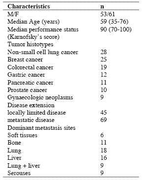
The study included 114 consecutive solid tumor patients with locally limited or metastatic disease, whose clinical characteristics are shown in Table 1. Lung cancer and gastrointestinal tumors were the most frequent neoplasms in our patients. For the immune detections, venous blood samples were collected in the morning after an overnight fast. Operable patients and metastatic patients were investigated before the surgical operation and before the onset of chemotherapy, respectively, in an attempt to exclude the possible influence of the various anticancer therapies on the immune status of patients.

In each sample, we have evaluated total lymphocyte count and the various lymphocyte subpopulations by a flow cytometric assay and monoclonal antibodies, including T helper lymphocytes (CD4), T cytotoxic lymphocytes (CD8), NK cells (CD16CD56), and T regulatory (T-reg) lymphocytes (CD4CD25). Normal values (95% confidence limits) of T-reg observed in our laboratory were below 240/mm3. Moreover, because of its importance in regulating lymphocyte functions and proliferation (Claman, 1998; Sthephens et al, 2004), the circadian rhythm of cortisol was also investigated by collecting blood samples at 8.00 A.M. and at 4.00 P.M., and cortisol serum concentrations were measured in duplicate by using the ECLA method. Data were reported as mean +/- SE, and statistically analyzed by the Student’s t test, the analysis of variance and the chi-square test, as appropriate.

III. Results
As reported in Table 2, an abnormally high number of T-reg was seen in 52/114 (46%) patients. Moreover, the percentage of cases with elevated number of T-reg observed in metastatic patients was significantly higher with respect to that found in non-metastatic patients (44/69 (64%) vs 8/45(18%), p < 0.01). Table 3 shows the mean number of T-reg and the mean percentages of T-reg with respect to both total lymphocytes and T helper (CD4+) lymphocytes observed in cancer patients in relation to their disease extension. The mean number of T-reg observed in metastatic patients was higher with respect to that found in patients with locally limited disease, without, however statistically significant differences. In contrast, the mean percentages of T-reg with respect to that of both lymphocytes and CD4 cells were significantly higher in metastatic patients than in the non-metastatic ones (p< 0.05 and p< 0.001,respectively). Moreover, within the metastatic group, patients with a normal lymphocyte count greater than 1500/mm3 showed a significantly higher mean number of T-reg with respect to the non-metastatic patients, whereas no difference was seen between nonmetastatic patients and metastatic patients with lymphocytopenia, consisting of lymphocyte count lower than 1500/mm3. In contrast, the mean percentages of T-reg with respect to total lymphocytes and CD4+ cells observed in both groups of metastatic patients with normal or low total lymphocyte count were significantly higher than in non-metastatic patients (lymphocytes: p< 0.025, CD4+ cells: p< 0.001).

Table 1. Clinical characteristics of 114 solid tumor patients
090722_clinicalstud1

Table 2. Percentages of abnormally high values of CD4+CD25+ lymphocytes

090722_clinicalstud2

* P < 0.01 vs non-metastatic patients

Table 3. Mean number of CD4+CD25+ lymphocytes and their mean percentages with respect to total lymphocytes and CD4+ lymphocytes in metastatic and non-metastatic patients

090722_clinicalstud3

* p<0.05 vs non-metastatic patients; ** p<0.025 vs non-metastatic patients; *** p<0.01 vs non-metastatic patients; **** p<0.001 vs nonmetastatic patients

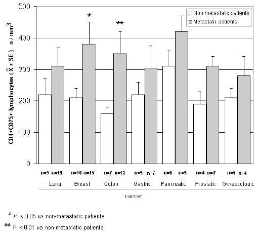
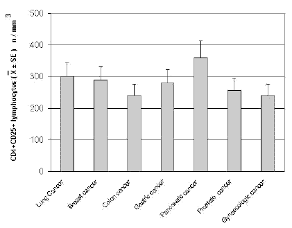
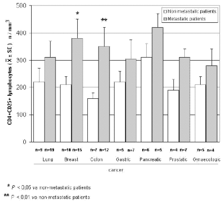
The mean counts of NK and CD8 cells in relation to that of T-reg are reported in Table 4. As shown, no significant difference in the mean number of CD8 lymphocytes was found between patients with normal or abnormally elevated number of T-reg. On the contrary, patients with elevated number of T-reg showed a significantly lower number of NK cells with respect to that found in those with normal T-reg count. Finally, Table 5 shows the circadian rhythm of cortisol in relation to total lymphocytes, CD4+ cells and T-reg mean number. A normal cortisol rhythm, with morning values greater at least than 50% with respect to the values occurring during the afternoon, was found in 85/114 (75%). Total lymphocyte and CD4+ cell mean numbers observed in patients with altered cortisol rhythm were significantly lower than those found in patients with normal cortisol circadianicity (p<0.01), whereas no significant difference was seen in the mean number of T reg. Figure 1 and Figure 2 illustrate T-reg mean numbers in relation to tumor histoptypes in the overall patients and with respect to their disease extension, respectively. No significant difference was seen in relation to tumor histotype. The highest values of T-reg were observed in pancreatic cancer patients, without however significant differences with respect to the overall other histotypes. The metastatic disease was associated with a higher number of T-reg with respect to the non-metastatic group in all tumor histotypes, even though a statistically significant differences occurred for the only breast cancer (p<0.05) and colorectal cancer (p< 0.01).

Table 4. Mean values of NK cells and CD8+ lymphocytes in cancer patients with normal or abnormally high values of CD4+CD25+ lymphocytes

090722_clinicalstud4

* p<0.05 vs normal values of CD4+CD25+ lymphocytes

Table 5. Mean numbers of total lymphocytes, T helper (CD4+) lymphocytes and T regulator lymphocytes (CD4+CD25+) in relation to cortisol circadian secretion in cancer patients
090722_clinicalstud5

* P<0.01 vs patients with altered cortisol rhythm
090722_clinicalstudfig1

Figure 1. CD4+CD25+ lymphocyte mean number in relation to tumor histotype
090722_clinicalstudfig2

Figure 2. CD4+CD25+ lymphocytes in relation to tumor histotype in metastatic and non-metastatic cancer patients

IV. Discussion
According to previous preliminary clinical investigations (Sasada et al, 2003; Cesana et al, 2006), this study confirms in a greater number of cancer patients that the metastatic disease is characterized by the evidence of an abnormally increased percentage of T-reg lymphocytes with respect to both total circulating lymphocytes and CD4+ lymphocytes. This finding does not seem to represent a specific characteristic of some tumor histotypes, then it could constitute a general alteration occurring during the progression of the neoplastic disease, by representing a fundamental immune parameter of cancer-related immunosuppression.

Several immune molecules have appeared to suppress the anticancer immunity, namely IL-6, IL-10, IL-1, TNF-α and TGF-β, but it seems that the common end result of their mechanisms of action may be represented by the stimulation of T-reg generation, with a consequent inhibition of the activation of an effective anticancer immune reaction. On the same way, several immune cells are able to suppress the anticancer immunity, including macrophages, T helper-2 lymphocytes and some myeloidderived suppressor cells, but also in this case they would act in a suppressive way by promoting the generation of Treg.

Then, the detection of T-reg amounts in terms of both absolute number and percentages with respect to total lymphocytes and CD4+ cells could constitute a simple and adequate clinical immune parameter to quantify the whole status of the anticancer immunity in the single cancer patient. Moreover, future clinical studies will be required to establish the possible prognostic significance of changes in T-reg percentage and number in relation to the anticancer efficacy of the various standard antitumor therapies. Moreover, it has to be remarked that T-reg lymphocytes would not represent the only immune cells involved in the suppression of the anticancer immunity. In fact, there is at least another fundamental immunosuppressive system, consisting of the monocytemacrophage cell lineage (Sica and Bronte, 2007). In more detail, it has been observed that the bone marrow may release myeloid precursors provided by suppressive activity on the antitumor immune response and defined as myeloid-derived suppressor cells (MDSC) (Kusmartsev and Gabrilovich, 2005). These cells have appeared to be characterized by the cell surface expression of GR-1, CD11b and CD80 antigens (Anderson et al, 2002; van Ginderachter et al, 2006). The myeloid suppressor cells would promote the generation and activation of T-reg lymphocytes, which at the other side would stimulate MDSC release from the bone marrow and M2 macrophage differentiation (Terabe et al, 2003; Wie et al, 2006).

Moreover, the myeloid suppressive cells would inhibit the anticancer immunity by promoting macrophage differentiation into the M2 sub-type (Ikemoto et al, 2003), which plays a clear inhibitory effect on the anticancer immunity, namely through the release of IL-6 (Ueno et al, 2000), whereas the M1 macrophage sub-type may either stimulate or suppress the antitumor immunity (Mantovani et al, 2004). M1 and M2 macrophage sub-types have appeared to be characterized by a high production of IL-12 or IL-10, respectively (Ueno et al, 2000).

Then, further studies by concomitantly evaluating T reg and MDSC count, will contribute to better define the immune mechanism responsible for the suppression of the anticancer immunity.

References
Anderson CF, Gerber JS, Mosser DM (2002) Modulating macrophage function with IgG immune complexes. J Endotoxin Res 8, 477-81.

Annunziato F, Cosmi L, Liotta F, Lazzeri E, Manetti R, Vanini V, Romagnani P, Maggi E, Romagnani S (2002) Phenotype,localization and mechanism of suppression of CD4+CD25+ human thymocytes. J Exp Med 196, 379-87.

Antony PA, Restito NP (2005) CD4+CD25+ T regulatory cells,immunotherapy of cancer,and interleukin-2. J Immunother 28, 120-8.

Birebent B, Lorho R, Lechartier H, de Guibert S, Alizadeh M,
Vu N, Beauplet A, Robillard N, Semana G (2004) Suppressive properties of human CD4+CD25+regulatory T cells are dependent on CTLA-4 expression. Eur J Immunol 34, 3485-96.

Cesana GC, DeRaffele G, Cohen S, Moroziewicz D, Mitcham J, Stoutenburg J, Cheung K, Hesdorffer C, Kim-Schulze S, Kaufman HL (2006) Characterization of CD4+CD25+ regulatory T cells in patients treated with high-dose interkleukin-2 for metastatic melanoma or renal cell carcinoma. J Clin Oncol 24, 1169-77.

Chen W, Jin W, Hardegen N, Lei KJ, Li L, Marinos N, McGrady G, Wahl SM (2003) Conversion of peripheral CD4+CD25- naïve T cells to CD4+CD25+ regulatory T cells by TGF-β induction of transcription factor Foxp 3. J Exp Med 198, 1875-86.

Claman HN (1998) Corticosteroids and the immune system. Adv Exp Med Biol 245, 203-10.

Dieckmann D, Bruett H, Ploettner H, Lutz MB, Schuler G (2002) Human CD4+CD25+ regulatory contact-dependent T cell induce IL-10 producing,contact-independent type-1-regulatory T cells. J Exp Med 196, 247-53.

Dieckmann D, Plottner H (2001) Ex vivo isolation and characterizationof CD4+CD25+ T cells with regulatory properties from human blood. J Exp Med 193, 1303-10.

Ehrke MJ, Mihich E, Berd D, Mastrangelo MJ (1989) Effects of anticancer drugs on the immune system. Semin Oncol 16, 230-9.

Ghiringhelli F, Larmonier N, Schmitt E, Parcellier A, Cathelin D, Garrido C, Chauffert B, Solary E, Bonnotte B, Martin F (2004) CD4+ CD25+ regulatory T cells suppress tumor immunity but are sensitive to cyclophosphamide which allows Immunotherapy of established tumors to be curative. Eur J Immunol 34, 336-44.

Ikemoto S, Yoshida N, Narita K, Wada S, Kishimoto T, Sugimura K, Nakatani T (2003) Role of tumor-associated macrophages in renal cell carcinoma. Oncol Rep 10, 1843-9.

Jago CB, Yates J, Camara NOS, Lechler RI, Lombardi AG (2004) Differential expression of CTLA-4 among T cell subsets. Clin Exp Immunol 136, 463-71.

Ji HB, Liao G, Faubion WA, Abadía-Molina AC, Cozzo C, Laroux FS, Caton A, Terhorst C (2004) Cutting edge, the natural ligand for glucocorticoid-induced TNF receptorrelated protein abrogates regulatory T cell suppression. J Immunol 172, 5823-7.

Kanamaru F, Youngnak P, Hashiguchi M, Nishioka T, Takahashi T, Sakaguchi S, Ishikawa I, Azuma M (2004) Costimulation via glucocorticoid-induced TNF receptor in both conventional and CD25+ regulatory CD4+ T cells. Immunol 172, 7306-14.

Knutson KL, Disis M, Salazar L (2007) CD4 regulatory T cells in human cancer pathogenesis. Cancer Immunol Immunother 556, 271-85.

Kusmartsev S, Gabrilovich DI (2005) STAT1 signaling regulates tumor-assolciated macrophage-mediate T cell deletion. J Immunol 174, 4880-91.

Lan RY, Ansari AA, Lian ZX, Gershwin ME (2005) Regulatory T cells, development,function,and role in autoimmunity. Autoimmun Rev 4, 351-63.

Leong PP, Mohammad R, Ibrahim N, Ithnin H, Abdullah M, Davis WC, Seow HF (2006) Phenotyping of lymphocytes expressing regulatory and effector markers in infiltrating ductal carcinoma of the breast. Immunol Lett 102, 229-36.

Malek TR, Bayer AL (2004) Tolerance not immunity crucially depends on IL-2. Nat Rev Immunol 4, 665-74.

Mantovani A, Sica A, Sozzani S, Allavena P, Vecchi A, Locati M (2004) The chemokine system in diverse forms of macrophage activation and polarization. Trends Immunol 25, 677-86.

Sasada T, Kimura M, Yoshida Y, Kanai M, Takabayashi A (2003) CD4+CD25+ regulatory T cells in patients with gastrointestinal malignancies, possible involvement of regulatory T cells in disease progression. Cancer 98, 1089-93.

Schwartz RH (2005) Natural regulatory T cells and selftolerance. Nat Immunol 6, 327-30.

Shevach EM (2002) CD4+CD25+ suppressor T cells, more questions than answers. Nat Rev Immunol 2, 389-400.

Sica A, Bronte V (2007) Altered macrophage differentiation and immune dysfunction in tumor development. J Clin Invest 117, 1155-66.

Siddiqui SA, Frigola X, Bonne-Annee S, et al (2007) Tumorinfiltrating Foxp3 CD4+CD25+ T cells predict poor survuival in renal cell carcinoma. Clin Cancer Res 13, 2075-81.

Stephens GL, McHugh RS, Whitters MJ, Young DA, Luxenberg D, Carreno BM, Collins M, Shevach EM (2004) Engagement of glucocorticoid-induced TNFR family-related receptor on effector T cells by its ligand mediates resistance to suppression by CD4+CD25+ T cells. J Immunol 173, 5008-20.

Takahashi T, Tagami T, Yamazaki S, Uede T, Shimizu J, Sakaguchi N, Mak TW, Sakaguchi S (2000) Immunologic and tolerance maintained by CD25+CD4+ regulatory T cells constitutively expressing cytotoxic T lymphocyte-associated antigen. J Exp Med 192, 1285-94.

Terabe M, Matsui S, Park JM, Mamura M, Noben-Trauth N, Donaldson DD, Chen W, Wahl SM, Ledbetter S, Pratt B, Letterio JJ, Paul WE, Berzofsky JA (2003) Transforming growth factor-β production and myeloid cells are an eggector mechanism trhough which CD1d-restricted T cells block cytotoxic T lymphocyte-mediated tumor immunosurveillance, abrogation prevents tumor recurrence. J Exp Med 198, 1741-52.

Thomton AM, Shevach EM (2000) Suppressor effector function of CD4+CD25+ immunoregulatory T cells is antigen nonspecific. J Immunol 164, 183-90.

Ueno T, Toi M, Saji H, Muta M, Bando H, Kuroi K, Koike M, Inadera H, Matsushima K (2000) Significance of macrophage chemo-attractant protein-1 in macrophage recruitment,angiogenesis and survival in human breast cancer. Clin Cancer Res 6, 3282-9.

Van Ginderachter JA, Movahedi K, Hassanzadeh Ghassabeh G, Meerschaut S, Beschin A, Raes G, De Baetselier P (2006) Classical and alternative activation of mononuclear phagocytes, picking the best of both worlds for tumor promotion. Immunobiology 211, 487-501.

Vasu C, Prabhakar BS, Holterman MJ (2004) Targeted CTLA-4 engagement induces CD4+CD25+CTLA-4 high T regulatory cells with target alloantigens specificity. J Immunol 173, 2866-76.

von Boehmer H (2005) Mechanisms of suppression by suppressor T cells. Nat Immunol 6, 338-44.

von Herrath MG, Harrison LC (2003) Regulatory lymphocytes, antigen-induced regulatory T cells in autoimmunity. Nat Rev Immunol 3, 223-32.

Wang XB, Zheng CY, Giscombe R, Lefvert AK (2001) Regulation of surface and intracellularexpression of CTLA-4 on human peripheral T cells. Scand J Immunol 54, 453-8.

Wie S, Kryczeck I, Zou W (2006) Regulatory T-cell compartmentalization and trafficking. Blood 108, 426-31.

Yang R, Cai Z, Zhang Y, Yutzy WH 4th, Roby KF, Roden RB (2006) CD80 in immune suppression by mouse ovarian carcinoma-associated Gr-1+CD11b+ myeloid cells. Cancer Res 66, 6807-15.

Ziegler SF (2006) FOXP3 of mice and men. Annu Rev Immunol 24, 209-26.

Zou W (2006) Regulatory T cells, tumour immunity and immunotherapy. Nat Rev Immunol 6, 295-307.

(download PDF version)



A phase II study of anastrozole plus the pineal anticancer hormone melatonin in the metastatic breast cancer women with poor clinical status

lissoniResearch Article (download PDF version)

Paolo Lissoni1*, Giuseppe Di Fede1, Antonio Battista2, Giusy Messina1, Remo Egardi1, Fernando Brivio3, Franco Rovelli1, Massimo Colciago4, Giuseppe Brera5

1 Institute of Biological Medicine, Milan
2 Azienda Sanitaria locale 2, Avellino;
3 Surgery Division, Bassini Hospital, Cinisello,Milan
4 I.N.R.C.A, Casatenovo, Lecco, Italy
5 Ambrosian University, Milan, Italy

dr. Paolo Lissoni

*Correspondence: Dr. Paolo Lissoni, Divisione di Radioterapia Oncologica, Ospedale S.Gerardo, 20052 Monza, Milano, Italy; Fax: +390392332284, E-mail: p.lissoni@hsgerardo.org
Key words: Anastrozole, breast cancer, melatonin, pineal gland
Abbreviations: melatonin, MLT; estrogen receptor, ER;

Received: 9 March 2009; Revised 1 April 2009;
Accepted: 13 April 2009; electronically published: 28 May 2009

Summary

The recent advances in the psychoneuroendocrinology have suggested the possibility to modulate tumor hormone dependency through a neuroendocrine approach. In particular, it has been proven that the pineal neurohormone melatonin (MLT) may stimulate estrogen receptor (ER) expression in breast cancer cells and inhibit the aromatase activity. On this basis, a study was planned to evacuate the efficacy of a concomitant treatment with the aromatase inhibitor anastrozole plus MLT in metastatic breast cancer. The study included 14 metastatic breast cancer women of poor clinical conditions with ER positive or unknown. Both anastrozole and MLT were given orally at a dose of 1 mg at noon and of 20 mg in the evening, respectively. The clinical response consisted of complete response in 2 and partial response in 6 patients. Then, an objective tumor regression was achieved in 8/14 (57%) patients, with a median duration of 26 months. No neoplastic cachexia occurred on treatment. This preliminary study shows that a neuroendocrine strategy with anastrozole plus the pineal hormone MLT may represent a new effective and well tolerated regimen in the treatment of metastatic breast cancer women, including those with poor clinical status, with therapeutic results apparently superior to those reported in the literature with the only aromatase inhibitor. Then, these results would justify further randomized studies of aromatase inhibitors with or without a concomitant administration of MLT, in an attempt to establish whether the pineal hormone may enhance the efficacy of the aromatase inibibitors in the treatment of human advanced breast cancer.

I. Introduction
Recent experimental studies have demonstrated that the hormone dependency is at least in part under a psychoneuroendocrine regulation (Cos et al, 2008;Grant et al, 2009). In particular, it has been shown that the pineal hormone melatonin (MLT), whose anticancer properties have been well demonstrated (Bartsch et al, 1981; Maestroni, 1993; Reiter et al, 2002), may in vitro stimulate estrogen receptor (ER) expression on breast cancer cell lines (Molis et al, 1995). Therefore, the hormone dependency of breast cancer cells would not depend only on intrinsic characteristics of cancer cells themselves, but also on host neuroendocrine regulation of tumor cell proliferation and differentiation (Bartsch et al, 2000). Moreover, cancer progression has been proven to be associated with pineal alterations, consisting of a progressive decline in MLT nocturnal production. (Maestroni, 1993). Therefore the advanced cancer would require a substitutive endocrine therapy with MLT (Bartsch et al, 1981; Maestroni, 1993). Previous preliminary clinical studies had already suggested that the concomitant administration of the pineal hormone MLT may apparently increase the efficacy of tamoxifen therapy in the treatment of metastatic breast cancer (Lissoni et al, 1995). Moreover, experimental studies have shown that the activity of aromatase enzyme, which is responsible for the peripheral production of estrogens from testosterone (Bagatell et al, 1994), is under a light/dark circadian rhythm (Bhatnagar et al, 1992). Because of the fundamental role of the pineal hormone MLT in the regulation of the daily photoperiod (Bartsch et al, 1981), it is possible to hypothesize that MLT may be involved in the control of the aromatase activity. In fact, recent studies have demonstrated an inhibitory action of MLT on the aromatase activity (Cos et al, 2005). This finding could reserve a prosiming application in the treatment of both early and advanced breast cancer. This statement is justified by the fact that the aromatase inhibitors represent a new class of agents in the endocrine treatment of breast cancer Plourde et al, 1994), with a potential efficacy superior to that achieved by the previous hormonal therapies with anti-estrogens, such as tamoxifene, even though tumor response rate obtained by the aromatase inhibitors are generally not greater than 40%. On this basis, a phase II study was planned in an attempt to evaluate the efficacy of a neuroendocrinotherapeutic regimen consisting of a concomitant administration of the aromatase inhibitor anastrozole and the pineal hormone MLT in metastatic breast cancer women with poor clinical conditions.

II. Materials and methods
The study included 14 consecutive metastatic breast cancer women (median age: 72 years, range 51-82), who were followed at Biological Medicine Institute in Milan, or at Health Local Unit 2 of Avellino, from Feb. 2002 to Sept. 2003. Eligibility criteria were, as follows: histologically proven metastatic breast cancer, measurable lesions, ER positive or unknown, no ability to tolerate chemotherapy because of age, low performance status (PS), important clinical illnesses other than cancer and/or heavy chemotherapeutic pre-treatments, no previous endocrine therapies for the metastatic disease, no double tumor and life expectancy less than 1 year. Previous heavy chemotherapeutic treatment consisting of at least 3 chemotherapeutic lines was made in 11/14 (79 %) patients. Dominant metastasis sites were, as follows: soft tissues:1; bone:1; lung:7 (neoplastic lymphangitis:2); liver:1; lung + liver:1; bone marrow:3. Time-span since first diagnosis of the primary tumor was 44 months (31-66 months). All patients had an acceptable social conditions. The minimum and median follow-up periods were 60 months and 72 months respectively. In all patients, in the case of disease progression, at least to other endocrine therapeutic lines with other aromatase-inhibitors were planned. The experimental protocol, wich was approved by the Health Direction of Biological Medicine Institute of Milan, was explained to each patient and informed consent was obtained. The treatment consisted of anastrozole at a dose of 1 mg/day orally at noon, plus MLT at 20 mg/day orally in the evening, generally half-hour before sleeping, to correct cancer progression-related decline in MLT night secretion (10). Patients were considered to be evaluable when they were treated for at least 3 consecutive months. The clinical response was evaluated according to WHO criteria. Complete response (CR) was the complete disappearance of all neoplastic lesions for at least 1 month. Partial response (PR) was a reduction greater than 50 % of the sum of all neoplastic lesions, for at least 1 month. Stable disesase (SD) was no increase or decrease greater tha 25 % of tumor volume. Progressive Disease (PD) was an increase in tumor volume greater than 25 % or the appearance of new neoplastic lesions. PS was assessed according to Karnofsky’s score, consisting of the evaluation of the quality of life in relation to patient activity and bed-rest period. ER was positive in 10 and unknown in the remaining 4 patients. The median PS was 80% (range 70-100). Data were statistically evaluated by the chi-square test and the Student’s t test, as appropriate.

090722_phase2study

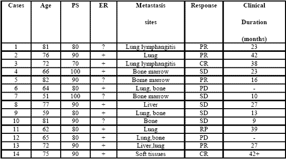
Table 1: Clinical characteristics of metastatic breast cancer women and their clinical response (WHO criteria) to a neuroendocrine regimen consisting of anastrozole plus the pineal hormone melatonin.

III. Results
All patients were fully evaluable for the clinical response. The clinical characteristics of patients and their individual clinical response to the treatment are reported in Table 1. As reported, a complete response (CR) was achieved in 2/14 (14%) (soft tissues:1; lung lymphangitis:1). A partial response (PR) was obtained in other 6/14 (43%) (bone:1; lung:3; liver:1; bone marrow:1). Then, an objective tumor response (CR + PR) was reached in 8/14 (57%) patients. The median duration of response was 26 months (range 9-42 months). A stable disease (SD) was seen in other 4/14 (29%), with a median duration of 25 months (range 10-27). Therefore, a disease-control (DC:CR + PR + SD) was achieved in 12/14 (86%) patients, whereas the remaining 2/14 (14%) patients had a progressive disease (PD). No significant difference in tumor response rate was observed between patients with positive or unknown ER ( 6/10(60%) vs 2/4(50%) ). An overall survival at 1 year and at 3 year was achieved in 11/14 (79 %) and in 5/14 (36 %) patients, respectively. Moreover, 3/14 (21%) patients were still alive at 5 years. The treatment was well tolerated in all patients.

Moreover, most patients experienced a relief of asthenia under the treatment and in no patient the neoplastic cachexia occurred. Finally, an evident increase in PS mean values was achieved under treatment, even though it did not reach the statistical significance (86 ±5 vs 93 ± 4, mean ± SE).

IV. Discussion
The results of this preliminary phase II study, by showing a percentage of 1-year survival greater than 70% in patients with live expectancy less than 1 year, would suggest that a neuroendocrine regimen consisting of the aromatase inhibitor anastrozole plus the pineal neurohormone MLT may represent a new effective therapeutic strategy in the treatment of metastatic breast cancer women, also in patients with poor clinical conditions, who would not be able to tolerate the most aggressive therapies. The concomitant administration of the pineal hormone would seem to enhance the efficacy of the aromatase inhibitor in terms of objective tumor regressions with respect to the results commonly reported in the literature with the only aromatase inhibitor (Plourde et al, 1994), which are generally lower than 40%.

The time to progression would seem to be apparently increased by the concomitant treatment with MLT.This finding is not surprising, since MLT could enhance the therapeutic anticancer acitivity of the aromatase inhibitors by either exerting direct antiproliferative antitumor effects (Bartsch et al, 1981; Maestroni, 1993; Reiter et al, 2002), or further inhibiting the aromatase activity by acting on gene and oncogene expression (Molis et al,1995; Cos et al, 2005). In addiction, MLT appeared to stimulate ER expression of breast cancer lines, by transforming ER negative into ER positive breast cancer, as observed in experimental conditions (Danforth et al, 1983).

Since the prognosis of ER positive breast cancers is clearly better than that of ER negative ones, MLT could per se improve the clinical couse of mammary tumors. Finally, because of its interesting therapeutic efficacy as a supportive care (Reiter et al, 2002), MLT would be responsible for the evident improvement in the relief of asthenia and in preventing the occurrence of the neoplastic cachexia. On the other hand, because of the inhibitory effect of MLT (Grant et al, 2009; Reiter et al, 2002) on cancer cell proliferation, the anticancer activity of this polyendocrine regimen would be due not only to an indirect effect, depending on a diminished estrogen production following aromatase enzyme inhibition, but also on a direct inhibition of cancer cell growth, due to MLT itself. Therefore, the results of this preliminary study may justify further clinical randomized investigations with the only aromatase inhibitor versus the concomitant treatment with MLT, in an attempt to confirm the ability of the pineal hormone to enhance the antitumor properties of the aromatase inhibitors in the treatment of metastatic breast cancer women with poor clinical conditions.

References
Bagatell CJ, Dahl KD, Bremner WJ (1994). The direct pituitary effect of testosterone to inhibit gonadotrophin secretion in men is partially mediated by aromatization to estradiol. J Androl 15:15-21.

Bartsch C, Bartsch H (1981) effect of melatonin on experimental tumors under different photoperiods and times of administration. J Neural Transm 52:269-279.

Bartsch H, Buchberger A, Franz H,Bartsch C,Maidonis I,Mecke D, Bayer E (2000) Effect of melatonin and pineal extracts on human ovarian and mammary tumor cells in a chemosensitivity assay. Life Sci 67:2953-2960.

Bhatnagar AS, Muller P, Schenkel L, Trunet PF, Beh I, Schieweck K. (1992). Inhibition of oestrogen biosynthesis and its consequences on gonadotrophin secretion in the male. J of Steroid Biochemistry and Molecular Biology;41:1021-1027

Cos S, Martinez-Campa C,Mediavilla MD, Sanchez-Barcelo E (2005). Melatonin modulates aromatase activity in MCF-7 human breast cancer cells. J Pineal Res 7:136-142.

Cos S, Gonzales A, Martinez-Campa C, Mediavilla MD, Alonzo-Gonzales C, Sanchez-Barcelo EJ (2008): Melatonin as a selective estrogen enzyme modulator. Curr Cancer Drug Targets. Dec 8(8): 691-702.

Danforth DN, Tamarkin L, Lipmann LE: (1983) Melatonin increase oestrogen receptor binding activity of human breast cancer cells. Nature, 305:323-325.

Grant S.G, Melan MA, Latimer JJ, Witt-Enderby PA (2009) : Melatonin and Breast cancer : cellular mechanism, clinical studies and future perspective. Expert Rev Mol Med 11:e5

Lissoni P, Barni S, Meregalli S, Fossati V, Cazzaniga M, Esposti D, Tancini G (1995) Modulation of cancer endocrine therapy by melatonin:a phase II study of tamoxifen plus melatonin in metastatic breast cancer patients progressing under tamoxifen alone. Br J Cancer 71:854-856.

Maestroni GJM (1993) The immunoneuroendocrine role of melatonin. J Pineal Res 14:1-10.

Molis TM, Spriggs LL,Jupiter Y,Hill SM (1995) Melatonin modulation of estrogen-regulated proteins, growth factors,and protooncogenes in human breast cancer. J Pineal Res 18:93-103.

Plourde PV, Dyroff M, Dukes MD (1994) Arimidex: a potent and selective fourth generation aromatase inhibitor. Breast Cancer Res Treat 30:103-111.

Reiter RJ, Tan DX, Sainz RM, Mayo JC, Lopez-Burillo S. (2002) Melatonin:reducing the toxicity and increasing the efficacy of drugs. Pharm Pharmacol 54:1299-1321

(download PDF version)


La terapia con campi magnetici è tradizionalmente usata per patologie ortopediche; negli ultimi anni alcuni studi hanno dimostrato l’utilità anche in altre patologie ed in particolare quelle di pertinenza neurologica.

Nel nostro Istituto dal 2001 il trattamento con campi elettromagnetici di bassa ed alta frequenza viene utilizzata singolarmente o in associazione con trattamenti farmacologici in varie situazioni. Lo scopo della relazione è di illustrare tre casi clinici in cui la terapia con campi elettromagnetici a frequenza ultrabassa si è dimostrata efficace; la tecnologia utilizzata è la risonanza ciclotronica endogena.

Per tutti i casi lo schema terapeutico prevedeva 2 frazioni settimana della durata di 30 minuti ciascuna per un totale di 12 sedute con una valutazione clinica a metà trattamento.

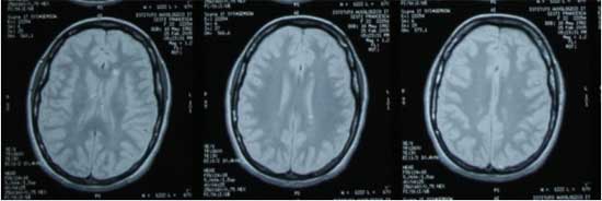
Primo caso: uomo di 41 anni con diagnosi radiologica di sclerosi multipla (luglio 2003-lesioni all’encefalo e midollo spinale); gli esami evidenziarono alti valori di stress ossidativo.

La sintomatologia consisteva in dolori in sede cervicale e lombare e un moderato deficit motorio dell’arto inferiore destro.
Trattamento terapeutico: trattamento con prodotti antiossidanti e terapia con campi elettromagnetici con frequenza variabile da 10 a 30 Hz e intensità variabile tra 10 e 25 microTesla.
Al termine del trattamento si era verificato un significativo miglioramento dei sintomi; la RM di controllo ha evidenziato una riduzione del numero e dimensioni delle lesioni attive. Tale quadro si mantiene anche alla ultima RM(febbraio 2007)

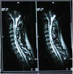
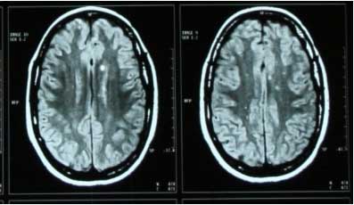
Secondo caso: donna di 23 anni con quadro radiologico (RM 2005)di sclerosi multipla e lesioni encefaliche e midollari con importanti deficit motori neurologici;gli esami evidenziarono alti livelli di stress ossidativo; la paziente era in cura con Interferone da un anno con malattia stabile.
0801_1
0801_2

Trattamento terapeutico:campi elettromagnetici con frequenza variabile tra 10 e 20 Hz e intensità variabile tra 20 e 30 microTesla;come terapia di supporto trattamento energico con prodotti antiossidanti.
L’ultima RM(dicembre 2006) ha mostrato una significativa riduzione delle lesioni ed in particolare modo di quelle midollari.
0801_4
0801_3

Terzo caso: uomo di 70 anni con broncopneumopatia cronica severa( stadio III Gold) in cura da alcuni anni con broncodilatatori e steroidi associata disfunzione sistodiastolica ventricolare sinistra.
I sintomi più evidenti erano la dispnea ed episodi continui di apnea notturna che limitavano molto la qualità di vita.
Dopo cinque sedute con campi elettromagnetici il paziente riferiva un notevole miglioramento della quantità e qualità del sonno e seppure meno evidente anche della dispnea.
La frequenza era di 40-45 Hz con una intensità tra 70 e 90 microTesla.
Alla fine del ciclo di terapia era ben evidente un miglioramento dei parametri respiratori ed anche della qualità di vita. La malattia era radiologicamente stabile.

In tutti e tre i casi riportati l’aggiunta di campi elettromagnetici al trattamento farmacologico ha evidenziato un miglioramento della qualità di vita con quadro radiologico di regressione in 2 casi e stabilità nel terzo.
In considerazione degli aspetti prognostici,psicologici e socioeconomici di queste patologie a nostro avviso è un risultato importante.
Oltretutto il trattamento con campi elettromagnetici è stato ben tollerato e senza effetti collaterali.
Pertanto riteniamo che la terapia con campi elettromagnetici a frequenza ultrabassa possa rappresentare una valida opzione in associazione con altri presidi in una strategia terapeutica integrata; inoltre la possibilità di eseguire eventualmente il trattamento a domicilio risulta particolarmente interessante.

Comunque,in futuro è auspicabile la costituzione di adeguati studi clinici controllati al fine di approfondire in maniera sempre più dettagliata gli aspetti concernenti i parametri tecnici,la sinergia con altre terapie e l’interazione con tessuti biologici.

del Prof. Giuseppe di Fede e Marco G. M. Mancuso

da ELECTROMAGNETIC BIOLOGY AND MEDICINE Volume 26, Numero 4 Anno 2007


Copyright by IMBIO 2017. All rights reserved.