Articoli

Tutto sul nostro settore


Aprile 10, 2015 Newsletter

40 mila nuovi casi di tumore al seno, è il dato agghiacciante che si registra ogni anno in Italia. Questo tipo di cancro è la prima causa di morte per malattia delle donne che vivono nei Paesi industrializzati. È una patologia che fa paura a molte e alle loro famiglie, per questo vi voglio parlare di prevenzione, l’arma più efficace per combattere la malattia. In particolare, vi parlo di un test innovativo per la prevenzione primaria del cancro mammario, in grado di svelare il fattore di rischio e, quindi, di poter adottare tempestivamente delle misure per contrastarlo, l’Estramet.

Come sempre e per quasi ogni aspetto che riguarda la nostra salute, buona parte della prevenzione deve partire dallo stile di vita e dall’alimentazione. Mangiare tanta frutta e verdura e limitare il consumo di cibi grassi e alcool, allontana il rischio di sviluppare il tumore al seno.

Ma ahimè non basta! È stato dimostrato che i fattori di rischio, oltre a uno stile di vita non salutare, sono di tipo genetico/familiare ed endocrini.

I ricercatori hanno scoperto che alla base dello sviluppo della malattia, nelle famiglie dove ha una forte incidenza, c’è una mutazione genetica di due geni identificati come BRCA1(Breast Carcinoma 1) e/o BRCA2 (Breast Carcinoma 2). Questi geni sono alla base dei tumori mammari ereditari.

Non è tutto, il terzo fattore che, secondo medici e ricercatori, provoca l’insorgere del tumore al seno è di tipo ormonale. Cioè, una prolungata esposizione agli estrogeni è determinante per lo sviluppo della malattia. In quest’ottica, le donne con menarca precoce, prima dei 12 anni, e con menopausa tardiva, oltre i 50 anni, corrono un rischio maggiore.

Proprio su quest’ultimo fattore di rischio si è concentrata la medicina negli ultimi tempi. I ricercatori hanno studiato il ruolo degli estrogeni nella formazione del tumore al seno. Le scoperte ottenute sono state in grado di sviluppare un test capace di determinare il fattore di rischio di sviluppare il cancro al seno, l’Estramet.

Non c’è bisogno che vi dica l’importanza di questa scoperta scientifica. Con un semplice esame dell’urina possiamo sapere quanto siamo a rischio e intraprendere delle azioni per modificare il nostro indice di rischio e, quindi, contrastare lo sviluppo della malattia.

Un esempio su come impedire l’insorgere della malattia è semplicemente assumendo una sostanza protettiva naturale, l’indolo3carbinolo, particolarmente presente nelle crucifere: cavolfiori, cavoli e cavoletti di Bruxelles. Questa sostanza da sola è in grado di modificare il metabolismo degli estrogeni.

in collaborazione con Synlab


Luglio 7, 2014 Newsletter

Esiste un legame tra pesticidi e autismo? L’ambiente esterno, secondo i sospetti di numerosi scienziati e ricercatori, influenza lo sviluppo del feto, durante i primi mesi di gestazione, fino a condizionare le caratteristiche di quando sarà adulto.

Nello specifico, i ricercatori dell’UC Davis MIND Institute hanno analizzato il rapporto tra vita rurale e uso di pesticidi nell’agricoltura e autismo. Lo studio ha rivelato che, il rischio di avere un bambino affetto da autismo o da un altro ritardo dello sviluppo aumenta di oltre il 60% per le donne che vivono nelle vicinanze di campi e fattorie in cui si utilizzano pesticidi chimici. Lo studio,  pubblicato su Environmental Health Perspectives, ha mostrato, inoltre, come questo collegamento aumenti quando l’esposizione ai pesticidi avviene tra il secondo e il terzo trimestre della gravidanza.

In diverse zone della California, gli scienziati hanno analizzato l’associazione tra l’esposizione durante la gravidanza a specifiche classi di pestidici, tra cui organofosfati, piretroidi e carbammati, e una diagnosi di ritardo dello sviluppo o autismo nella prole. Lo studio confermerebbe i risultati di ricerche precedenti in merito alla relazione tra la nascita di un bambino autistico e l’esposizione prenatale ad agenti chimici, usati per l’agricoltura in California. Anche se i ricercatori devono ancora scoprire se alcuni sottogruppi sono più vulnerabili all’esposizione a questi composti rispetto ad altri, il messaggio è chiaro: le donne incinte dovrebbero evitare a tutti i costi il contatto con gli agenti chimici usati per l’agricoltura.

La ricerca ha evidenziato che ci sono diverse classi di pesticidi utilizzati più di frequente vicino alle zone in cui risiedono madri i cui bambini sono affetti da autismo o da ritardi. L’esposizione agli insetticidi, secondo i ricercatori, potrebbe essere dannosa durante la gestazione perché il cervello del feto, ancora in via di sviluppo, è più vulnerabile a quello di un adulto. Questi pesticidi contengono neurotossine, e l’esposizione in utero potrebbe disturbare lo sviluppo strutturale dei neuroni, determinando così alterazioni nei meccanismi di eccitazione e inibizione che governano l’umore, l’apprendimento, le interazioni sociali e il comportamento.

Lo studio enfatizza, inoltre, l’importanza dell’alimentazione materna durante la gravidanza, raccomandando soprattutto l’utilizzo di vitamine prenatali per diminuire il rischio di avere bambini affetti da autismo. Una dieta materna corretta, prima del concepimento, aiuta a creare le condizioni favorevoli per il futuro impianto e sviluppo del feto. Un’ alimentazione controllata e bilanciata, consente anche di prevenire malattie, carenze nutrizionali, allergie e intolleranze alimentari da adulto. Un controllo genetico per concepimento, sulla madre, può indirizzare un corretto stile di vita e soprattutto una corretta alimentazione, in modo da non causare o amplificare deficit nutrizionali importanti, per il feto e la madre.

Riferimenti: Environmental Health Perspectives doi: 10.1289/ehp.1307044


Ottobre 5, 2013 RASSEGNA STAMPA

IL LIBRO TRUCCHI E RICETTE PER CHI SOFFRE DI QUESTA INTOLLERANZA POCO NOTA

Nonna blogger insegna a vivere senza nichel – scarichi l’immagine dell’articolo (68 kb)

CASALINGA, madre, nonna. E ora scrittrice. La prima in Italia a pubblicare un libro di ricette per chi soffre di intolleranza al nichel. Tiziana Colombo, 53enne di Cavenago, è una piccola celebrità: da qualche tempo gira l’Italia per raccontare la sua esperienza, per spiegare agli alunni delle scuole alberghiere come preparare piatti gustosi depurandoli dell’odiato metallo.

130411_colomboCONFERENZE a Bari, Roma, Bolzano, Piacenza, Catania, Milano. Dunque conferenziera, appunto scrittrice, in partenza blogger. Perché tutto nasce dal suo blog «nonnapaperina» che fa il pieno di contatti raccontando che è possibile mangiare bene, anzi benissimo, dribblando allergie e intolleranze. Troppo appassionata della buona cucina, Tiziana, per arrendersi. E così comincia a studiare, a confrontarsi con i medici specialisti, ad ascoltare il vissuto di chi è stato costretto a modificare le proprie abitudini. «Ho sofferto per anni di disturbi di cui nessuno aveva capito l’origine: solo nel 2009 ho saputo di essere intollerante al nichel». Invece di scoraggiarsi, inizia la battaglia: «Da allora mi sono impegnata per sensibilizzare la popolazione e le istituzioni. Non c’è abbastanza considerazione, tant’è che l’esame specifico non viene nemmeno passato dalla mutua».

DA INTERNET alla carta il passo è stato breve: nel volume pubblicato da Silvana Editoriale, intitolato «Nichel. L’intolleranza? La cuciniamo», c’è un vasto campionario di ricette, almeno 115, e un lungo elenco di consigli sui metodi di cottura e le pentole da utilizzare, sui detersivi o i cosmetici da evitare. «Un libro così è una novità in Italia perché non esiste una sensibilità diffusa verso questa intolleranza. Ma gli incontri in giro per l’Italia testimoniano che un piccolo interesse sta nascendo. Anche perché l’intolleranza al nichel è sempre più frequente e colpisce soprattutto le donne. E i disturbi sono gravi, dalle crisi d’asma ai mal di testa ricorrenti». Il ricavato servirà a finanziare il sodalizio nato per sensibilizzare le istituzioni: «Sono riuscita a ricostruire la mia quotidianità, ora voglio aiutare altre persone che vivono questa condizione».

Rassegna stampa: La Stampa – 27 maggio 2013 – di MARCO DOZIO


Enhancement of the Efficacy of Chemotherapy with Oxaliplatin Plus 5-Fluorouracil by Pretreatment with IL-2 Subcutaneous Immunotherapy in Metastatic Colorectal Cancer Patients with Lymphocytopenia Prior to Therapy

Research Article (download PDF version)

P. LISSONI1,*, F. BRIVIO2, L. FUMAGALLI2, G. DI FEDE3 and G. BRERA4

1 Division of Radiation Oncology, San Gerardo Hospital, Milan, Italy
2 Division of Surgery, San Gerardo Hospital, Milan, Italy
3 Institute of Biological Medicine, Milan, Italy
4 Ambrosian University, Milan, Italy

*Correspondence: Dr. Paolo Lissoni, Divisione di Radioterapia Oncologica, Ospedale S.Gerardo, 20052 Monza, Milano, Italy; Fax: +390392332284, e-mail: p.lissoni@hsgerardo.org

Abstract
The present study was carried out to evaluate the influence of a short-period IL-2 administration on the efficacy of chemotherapy in metastatic colorectal cancer patients with pretreatment lymphocytopenia, which was defined as a lymphocyte count of less than 1500/mm3.

The study included 144 consecutive metastatic colorectal cancer patients, who underwent chemotherapy with oxaliplatin plus 5-fluorouracil. Lymphocytopenia was seen in 41/144 (28%) patients, who were randomized to receive chemotherapy alone or chemotherapy after a prechemoimmunotherapy with IL-2 (3 MIU twice/day for 3 consecutive days), whereas patients with a normal pretreatment lymphocyte count received only chemotherapy.

A normalization of the lymphocyte number was achieved in 12/19 lymphocytopenic patients pretreated with IL-2. The objective tumor regression rate achieved in patients with a normal lymphocyte count prior to chemotherapy was significantly higher compared to that obtained in lymphocytopenic patients treated with chemotherapy alone (54/103 vs. 3/22, p<0.01), whereas no significant difference occurred between patients with normal lymphocyte count and lymphocytopenic patients pretreated with IL-2 (54/103 vs. 8/19).

This study confirms that pretreatment lymphocytopenia is associated with reduced efficacy of chemotherapy in metastatic colorectal cancer patients. Moreover, it suggests that pretreatment with IL-2 before the onset of chemotherapy may enhance the efficacy of chemotherapy in lymphocytopenic patients. Therefore, the administration of IL-2 before the onset of chemotherapy to improve the immune status of cancer patients may be considered as a new chemoimmunotherapeutic combination, which may be recommended in the treatment of advanced cancer patients, particularly in those with cancer-related immune alterations.

(download PDF version)


Modulation of the anticancer immunity by natural agents: inhibition of T regulatory lymphocyte generation by arabinoxylan in patients with locally limited or metastatic solid tumors

lissoniResearch Article (download PDF version)

Paolo Lissoni1,*, Giusy Messina1, Fernando Brivio2, Luca Fumagalli2, Luigi Vigoré3, Franco Rovelli3, Luisa Maruelli4, Mauro Miceli4, Paolo Marchiori4, Giorgio Porro1, Michael Held5, Giuseppe di Fede6, Toshi Uchiyamada7

1 Division of Radiation Oncology, San Gerardo Hospital, Milan, Italy
2 Division of Surgery, San Gerardo Hospital, Milan, Italy
3 Laboratory of Immunomicrobiology, San Gerardo Hospital, Milan, Italy
4 Natur-Spiritual, Milan, Italy
5 Biological Medicine Center, Rome, Italy
6 Institute of Biological Medicine, Milan, Italy
7 Daiwa Pharmaceuticals, Tokyo, Japan

dr. Paolo Lissoni

*Correspondence: Dr. Paolo Lissoni, Divisione di Radioterapia Oncologica, Ospedale S.Gerardo, 20052 Monza, Milano, Italy; Fax: +390392332284, e-mail: p.lissoni@hsgerardo.org
Key words: Anticancer immunity, arabinoxylan, immunostimulation, T regulatory lymphocytes
Abbreviations: interleukin 10, (IL-10); interleukin 6, (IL-6); interleukin-2, (IL-2); interleukn 12, (IL-12); NK cells, (CD16+CD56+); T cytotoxic lymphocytes, (CD8+); T helper lymphocytes, (TH), (CD4+); T lymphocites, (CD3+); Transforming growth factor beta, (TGF-β) T-regulatory lymphocytes, (T-reg), (CD4+CD25+)

Received: 30 September 2008; Revised: 1 November 2008
Accepted: 17 November 2008; electronically published: December 2008

Summary

In the last years, several immunomodulating antitumor agents have demonstrated in the nature, particularly from Aloe plant and rice bran. However, the major problem concerning the natural antitumor agents is to define their immune mechanisms of action in relation to the more recent advances in tumor immunobiology. At present, the main cause responsible for the lack of an effective antitumor response in advanced cancer patients is belived to be represented by the generation of a subtype of T helper lymphocytes (CD4+) with suppressive activity on anticancer immunity, the so-called T regulatory lymphocytes (T reg), which may be clinically identified as CD4+CD25+ cells. On this basis, a study was planned to evaluate the effect of rice bran extract arabinoxylan on T reg cell count and percentage in solid tumor patients in relation to the various lymphocyte subpopulations. The study included 22 evaluable cancer patients, 16 of whom had an untreatable metastatic solid tumor. Arabinoxylan was given orally at a dose of 2000 mg/day for the first month, followed by a dose of 1000 mg/day for the next month. In each patient we evaluated by monoclonal antibodies the absolute number of lymphocytes, T lymphocytes (CD3+), T helper (TH) lymphocytes (CD4+), T cytotoxic lymphocytes (CD8+), NK cells (CD16+CD56+), T reg lymphocytes (CD4+CD25+) and TH/T reg ratio before and after 2 months of therapy. No substantial change occurred on therapy in the mean number of lymphocytes, CD3+, CD8+ and NK cells. On the other hand, the mean number of TH cells increased, whereas that of T reg cell decreased on treatment, even though none of these differences was statistically significant. On the contrary, TH/T reg mean ratio significantly enhanced after arabinoxylan therapy. In addition to its previously demonstrated stimulatory action on NK function, this study shows that arabinoxylan may inhibit the production of T reg cells, which are responsible for cancer-related immunosuppression, with a following improvement in the anticancer immunity. If further studies will confirm these results, arabinoxylan could be successfully associated with chemotherapy to induce not only a cytotoxic destruction of cancer cells, but also an improvement in the immune status.

I. Introduction
The recent advances in the definition of the mechanisms responsible for tumor progression have suggested the possibility to control cancer growth not only trough chemotherapy-induced cancer cell destruction, but also by stimulating the anticancer immunity. In addiction to the exisence of endogenous antitumor molecules, several agents capable of stimulating the anticancer immunity have alsso isolated from plants. However, the immunomodulatory effects of most natural immunomodulating agents need to be better investigated in an attempt to establish their mechanisms of action in relation to the most recent discoveries concerning the physiopathology of the anticancer immunity. At present, Aloe extracts (Lissoni et al, 1998) and arabinoxylan extract from rice bran (Ghoneum and Jewett, 2000) would represent some of the potential natural agents which could be utilized in the complementary therapy of human neoplasms. Today, it is known that the antitumor immune response is the end-result of several interactions involving cytokines and immune cells, provided by stimulatory or suppressive effects on the anticancer immunity (Atzpodien and Kirchner, 1990; Rosenberg, 1992). Therefore, the lack of an effective anticancer immune response in most cancer patients with advanced disease would simply depend on the prevalence of immunosuppressive mechansisms with respect to the immunostimulatory ones (Atzpodien and Kirchner, 1990). The anticancer immunity is mainly activated by T helper-type 1 lymphocytes by releasing IL-2 (Whittington and Faulds, 1993), and by dentritic cells, which act as antigen-presenting cells producing IL-12 (Banks et al, 1995), T cytotoxic lymphocytes and NK-LAK system, which are involved in the induction of the antigen-dependent and antigen-independent cytotoxicity, respectively (Atzpodien and Kirchner, 1990). Therefore, IL-2 and IL-12 would represent the main anticancer cytokines in humans. On the contrary, the suppression of the anticancer immune response is mediated by several cytokines, namely IL-10 (Moore et al, 1993), IL-6 (Matsuda and Hirano, 1990) and TGF-β (Shevach, 2002). Recently, however, it has been demonstrated that the various endogenous suppressive factors would exert their inhibitory immune effect through a common end-mechanism, consisting of the generation of a subtype of T helper lymphocytes (CD4+cells), provided by a fundamental suppressive activity on the anticancer immunity, the so-called T regulatory lymphocyte (T reg) (Dieckmann et al, 2001), which at present seems to constitute the main mechanism responsible for cancer-related immunosuppressive status. T reg cells may be identified by the simultaneous expression of the alpha-chain of IL-2 receptor (CD25) and CD4 antigen (Dieckmann et al, 2001). Then, T reg cells may be clinically recognized as CD4+CD25+ lymphocytes. Therefore, each eventual natural immunomodulating agent would have to be investigated in relation to its possible effect on T reg generation since, at least from a theoretical point of view, each natural agent capable of counteracting T reg activity could positively influence the prognosis of the neoplastic disease by improving the efficacy of the anticancer immune response. Moreover, our previous preliminary studies have suggested that the percentage of T reg cells with respect to the total number of T helper cells, as expressed as CD4/CD4CD25 ratio, may represent an optimal synthetic immune index to investigate the functional status of the anticancer immunity in the single cancer patient, by representing the synthesis of the actions of the great number of immunostimulating and immunosuppressive factors involved in the modulation of the anticancer immunity (Dieckmann et al, 2001). Within the great number of natural agents derived from plants and potentially usefull to be employed in the complementary therapy of cancer, arabinoxylan would seem to represent one of the potential natural agent, because of its efficacy in improving the clinical status of cancer patients (Ghoneum and Jewett, 2000; Ghoneum and Gollapudi, 2005; Markus et al, 2006; Ghoneum et al, 2007). The immunomodulating properties of this nautral substances extracted from plants have been confirmed by experimental studies, but unfortunately most experiments have been limited to the investigations of they effects on non-specific immune parameters for the anticancer immunity, such as NK cell cytotoxicity. In contrast, since reg cells play a fundamental role in suppressing the generation of the anticancer immunty, each potential antitumor immunomodulatory natural substances, would have to be investigated also in relation to their eventual influence on T reg cell system. On the basis of the recent discoveries in tumor immunobiology (Dieckmann et al, 2001; Shevach, 2002), a study was planned to investigate the possible influence of arabinoxylan on both absolute number of T reg cells and their ratio with respect to the total CD4+ T cells in a group of solid tumor patients, affected by locally limited or metastatic disease.

II. Materials and methods
The study included 24 consecutive patients, 18 of whom had a metastatic solid tumor, which did not respond to the conventional anticancer chemotherapies and for whom no other effective standard treatment was available, while the remaining 6 patients had been surgically treated for a locally limited neoplasm. Patients were followed at Biological Medical Institute of Milan and the protocol was approved by the Director of the Institute. Eligibility criteria were, as follows:histologically proven locally limited or metastatic solid tumor, no double tumor, no chronic therapy with corticosteroids because of their immunosuppressive effects and no concomitant treatment with other immunomodulating agents,such as interferons,interleukins and monoclonal antibodies. At the time of the start of arabinoxylan therapy, patients with untreatable metastatic cancer were under treatment with the only supportive care, consisting of anti-inflammatory agents for pain, anti-dopaminergic drugs for nausea and vomiting and with the pineal hormone melatonin for the therapy of the neoplastic cachexia (Banks et al, 1995). Patients were considered as fully evaluable when they had received arabinoxylan therapy for at least 2 consecutive months. Arabinoxylan was given orally at a dose of 1000 mg twice/day for the first month, followed by a dose of 1000 mg/day for the next month. Arabinoxylan was supplied by DAIWA Pharmaceutical (Tokyo, Japan). It was derived from rice bran treated enzymatically with an extract of the shiitake mushrooms. It is a polysaccharide containing β-1,4-xylopironase hemicellulose, commercially available and known as Biobran. For the immune investigation, venous blood samples were collected in the morning after an overnight fast before the onset of arabinoxylan therapy and after 2 consecutive months of treatment. In each blood sample, we evaluated the absolute number of total lymphocytes, T lymphocytes (CD3+), T helper (TH) lymphocytes (CD4+), T cytotoxic lymphocytes (CD8+), NK cells (CD16+ CD56+ and T regulatory (T reg) lymphocytes (CD4+ CD25+). The different lymphocyte subsets were measured with a flow cytometric assay by using specific monoclonal antibodies supplied by Becton-Dickinson (Milan, Italy). Moreover, because of the importance not only of their absolute number, but also of their percentage with respect to the other lymphocyte subsets, namely to that of CD4+ cells, CD4/CD4CD25 ratio, corresponding to TH/T reg ratio, was also determined before and after therapy. Normal values (95% confidence limits) of T reg number and TH/T reg ratio observed in our laboratory were below 240/mm3 and above 4.0, respectively. Data were reported as mean +/- SE and statistically analyzed by the Student’s t test, the analysis of variance and the chi-square test, as appropriate.

III. Results
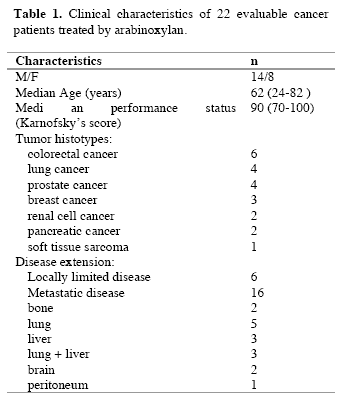
Evaluable patients were 22/24, while the remaining 2 patients, both affected by untreatable disseminated liver metastases due to colorectal cancer, rapidly died for disease progression before concluding the two planned months of arabinoxylan therapy. The clinical characteristics of the evaluable patients are reported in Table 1. Figure 1 illustrates changes in the mean number of total lymphocytes, T lymphocytes, T cytotoxic lymphocytes and NK cells occurring after 2 months of arabinoxylan therapy. No substantial variation was found in the mean number of lymphocytes, T lymphocytes, T cytotoxic lymphocytes and NK cells under arabinoxylan treatment. In contrast, as illustrated in Figure 2, TH and T reg mean numbers increased and decreased, respectively, after arabinoxylan therapy, without, however statistically significant differences with respect to the values seen prior to therapy. On the contrary, a statistically significant increase in TH/T reg mean ratio was achieved after arabinoxylan therapy (p<0.025). The increase in TH/T reg ratio under arabinoxylan therapy was more pronounced in patients with an abnormally low ratio prior to therapy with respect to that occurring in those with normal pre-treatment ratio, however without statistically significant differences ( 2.3 +/- 0.4 vs 1.7 +/- 0.5). In more detail,
090722_linfocitiT_table1
090722_linfocitiT_1

Figure 1. Changes in the number of lymphocytes, Tlymphocytes (CD3), T cytotoxic lymphocytes (CD8) and NK cells (CD16 CD56) after 2 months of arabinoxylan therapy.
090722_linfocitiT_2

Figure 2. Changes in the mean number of T helper (TH) lymphocytes (CD4) and T regulatory lymphocytes (cd4 cd24) and in TH/T reg mean ratio.

before arabinoxylan therapy, an abnormally low TH/T reg ratio was present in 12/22 (55%) evaluable patients. Arabinoxylan treatment induced a normalization of TH/T reg ratio in 5/12 (42%) patients with an abnormally low ratio prior to therapy. The percentage of arabinoxylan-induced TH/T reg normalization obtained in lymphocytopenic patients was not significantly different from that achieved in patients with normal pre-treatment lymphocyte count ( 3/7(43%) vs 2/5(40%) ). No toxicity was observed under arabinoxylan treatment, which was well tolerated in all patients. Asthenia was present in 8/22 (36%) evaluable patients. An evident relief of asthenia, as assessed by a specific patient report, was obtained under arabinoxylan therapy in 5/8 (63%) patients.

IV. Discussion
Previous experimental studies had already demonstrated some immunomodulating properties of arabinoxylan, in particular consisting of stimulation of NK cytotoxic function (Ghoneum, 1998), whereas NK cell number did not seem to be influenced by arabinoxylan administration. However, it has to be remarked that NK cells were belived to be fundamental in the antitumor immunity until some years ago, before the discovery of the essential role played by the antitumor cytokines, such as IL-2 and IL-12 (Whittington and Faulds, 1993) and dendritic cells, because of their function as antigen-presenting cells (Banks et al, 1995). In fact, it has to be considered that the cytotoxic activity of NK cells is effective only against artificial laboratory cancer cell lines, whose biological malignant properties are different from those presented by fresh human tumor cells (Whittington and Faulds, 1993). In addition, NK cells have been proven to be also able to destroy fresh human cancer cells only after the activation of their cytotoxic function by IL-2 (Atzpodien and Kirchner, 1990). From this point of view, arabinoxylan had been already proven to amplify the stimulatory effect of IL-2 on NK-mediated antitumor cytotoxicity (Ghoneum and Jewett, 2000). In contrast, no study has been performed up to now to evaluate the possible influence of arabinoxylan not only on the mechanisms responsible for the generation of an effective anticancer immune response, but also on those involved in the suppression of anticancer immunity. The results of this preliminary study, carried out to evaluate the influence of arabinoxylan on T reg cells, which represent the most important cells involved in the suppression of the antitumor cytotoxic immune response, demonstrates that arabinoxylan may counteract T reg cell generation by reducing their number and percentage with respect to the total amounts of CD4+ cells and circulating lymphocytes. Since NK cell function is inhibited by T reg activation (Shevach, 2002), the previously demonstrated arabinoxylan-induced stimulation of NK cell cytotoxic function might depend at least in part on its capacity of counteracting T reg generation (Dieckmann et al, 2001). Moreover, this study would suggest that the inhibitory action of arabinoxylan on T reg generation is more pronounced in patients with an abnormally high percentage of T reg cells prior to therapy, with a following pre-treatment abnormally low TH/T reg ratio before therapy, whereas its effect was less evident in patients with a pre-treatment value of TH/T reg ratio within the normal range. Therefore, the influence of arabinoxylan on T reg generation would consist of a modulatory action rather than an inhibitory activity. This finding could explain a potential favourable immunomodulatory effect of arabinoxylan also in patients with autoimmune diseases (Ghoneum, 1998), who in contrast to cancer patients would tend to present abnormally low amounts of T reg cells. In any case, the importance of the inhibition of T reg generation in the induction of an effective anticancer immune response has been recently confirmed by the evidence that the block of T reg activity by specific monoclonal antibodies may induce objective tumor regressions in humans (Yang et al, 2007). Obviously, the major problem is the exact identification of he T reg cell population. Even though T reg cells may express other immune markers, namely FOX-p2 cytoplasmatic antigen, most clinicians are in agreement to identify the CD4+CD25+ cells as T reg lymphocytes (12). In any case, further studies, by evaluating other immune markers, will be required to better identify T reg cells population, namely FOX-p3, even though recently some Authors have shown that FOX-p3 expression by T reg cells is associated with a lower suppressive activity (Dieckmann et al, 2001; Shevach, 2002). Moreover, it has to be remarked that several patients included in the present study were concomitantly under palliative therapy with the anti-cachectic pineal hormone melatonin (Brzezinski, 1997), which may also play immunomodulating effects (Maestroni, 1993). Therefore, further randomized studies with arabinoxylan alone versus arabinoxylan plus melatonin will be required to better define the immunomodulating action of arabinoxylan. If further clinical and experimental studies will confirm the inhibitory action of arabinoxylan on T reg cell system, it could be included in cytokine-based immunotherapies to enhance their efficacy by counteracting T reg cell generation.

References
Atzpodien J, Kirchner H (1990) Cancer, cytokines and cytotoxic cells:interleukin-2 in the immunotherapy of human neoplasms. Klin Wochenschr 14, 1-10.
Banks RE, Patel PM, Selby PJ (1995) Interleukin-12:a novel clinical player in cytokine therapy. Br J cancer 71, 655-659.
Brzezinski A (1997) Melatonin in humans. N Engl J Med 336, 185-195.
Dieckmann D, Plottner H, Berchtold S, Berger T, Schuler G (2001) Ex vivo isolation and characterization of CD4+CD25+ T cells with regulatory properties from human blood. J Exp Med 193, 1303-1310.
Ghoneum M (1998) Enhancement of human natural killer cell activity by modified arabinoxylane fro rice bran(MGN-3). Int J Immunother 14, 89-99.
Ghoneum M, Gollapudi S (2005) Synergistic of arabinoxilan rice bran (MGN-3/Biobran in S. Cerevisiae-induced apoptosis of monolayer breast cancer MFC-7 cells. Anticancer Res 25(6B), 4187-96.
Ghoneum M, Brown J, Gollapudi S (2007) Yeast therapy for the treatment of cancer and its enhancement by MGN-3/Biobran, an arabinoxylan rice bran. Cellular Signaling and Apoptosis Research (Ed. Alex R. Demasi) Cap IV: 185-200.
Ghoneum M, Jewett A (2000) Production of tumor necrosis factor-alpha and interferon-gamma from human peripheral blood lymphocytes by MGN-3, a modified arabinoxylan from rice bran, and its synergy with interleukin-2 in vitro. Cancer Detect Prevent 24, 314-324.
Lissoni P, Giani L, Zerbini S, Trabattoni P, Rovelli F (1998) Biotherapy with the pineal immunomodulating hormone melatonin versus melatonin plus Aloe vera in untreatable advanced solid neoplasms. Nat Immun 16, 27-33.
Maestroni JGM (1993) The immunoneuroendocrine role of melatonin. J Pineal Res 14, 1-10.
Markus J, Miller A, Smith M, Orengo I (2006) Metastatic hemangiopericytoma of the skin treated with wide local excision and MGN-3. Dermatol Surg 32, 145-147.
Matsuda T, Hirano T (1990) Interleukin-6 (IL-6). Biotherapy 2, 363-371.
Moore KW, O’Garra A, De Waal-Malefyt R (1993) Interleukin-10. Ann Rev Immunol 11, 165-174.
Rosenberg SA (1992) The immunotherapy and gene therapy of cancer. J Clin Oncol 10, 181-191.
Shevach EM (2002) CD4+CD25+ suppressor T cells:more questions than answers. Nat Rev Immunol 2, 389-400.
Whittington R, Faulds D (1993) Interleukin-2. Drugs 46, 446-514.
Yang JC, Hughes M, Kammula U, Royal R, Sherry RM, Topalian SL, Suri KB, Levy C, Allen T, Mavroukakis, Lowy I, White DE, Rosenberg SA (2007) Ipilimubab (anti-CTLA4 antibody)causes regression of metastatic renal cell cancer associated with enteritis and hypophysitis. J Immunother 30, 825-830.

(download PDF version)


A clinical study of T-regulatory lymphocyte function in cancer patients in relation to tumor histotype, disease extention, lymphocyte subtypes and cortisol secretion

Research Article (download PDF version)

Luigi Vigorè1, Fernando Brivio2, Luca Fumagalli2, Roberto Vezzo1, Giusy Messina6, Franco Rovelli6, Massimo Colciago3, Giovanna D’Amico4, Giuseppe Di Fede5, Paolo Lissoni6

1 Laboratory of Immunomicrobiology, St.Gerardo Hospital, Monza, Milan, Italy
2 Division of Surgery, St.Gerardo Hospital, Monza, Milan, Italy
3 I.N.R.C.A Laboratory of Analyses, Casatenovo, Lecco, Italy
4 Research Center “Fondazione Tettamanti” Clinica Pediatrica Università Milano-Bicocca, Italy
5 Institute of Biological Medicine, Milan, Italy
6 Division of Radiation Oncology, St.Gerardo Hospital, Monza, Milan, Italy

*Correspondence: Dr. Paolo Lissoni, Divisione di Radioterapia Oncologica, Ospedale S.Gerardo, 20052 Monza, Milano, Italy; Fax: +390392332284, E-mail: p.lissoni@hsgerardo.org
Key words: Anticancer immunity, immunosuppression, T regulatory lymphocytes
Abbreviations: cytotoxic T lymphocyte-associated antigen-4, (CTLA-4); glucocorticoid-induced TNF-α receptor, (GITR); myeloidderived suppressor cells, (MDSC); NK cells, (CD16CD56); T cytotoxic lymphocytes, (CD8); T helper lymphocytes, (CD4); Tregulatory lymphocytes, (T-reg)

Received: 24 July 2008; Revised: 11 September 2008
Accepted: 12 September 2008; electronically published: October 2008

Summary

Several clinical investigations showed that the immune status is a prognostic variable in cancer patients, even tough the evaluation of the anticancer immunity is not generally considered in the medical oncology. Several immune parameters, including lymphocyte subsets and cytokine blood concentration, had been proposed to quantify the functional status of the anticancer immunity, but recent discoveries would suggest that the end-result of the various immune interactions is represented by a subtype of CD4 lymphocytes capable of suppressing the antitumor immune reaction, the so called T-regulatory lymphocytes (T-reg). This study was performed to detect T-reg count and percentage in solid tumor patients, in relation to tumor histotype, disease extension, lymphocyte sub-populations and cortisol circadian secretion. The study included 114 consecutive cancer patients affected by the most frequent tumor histotypes, 69 of whom showed a metastatic disease. In each patient we evaluated T-reg cells, identified as CD4+CD25+, in relation to T helper (CD4), T cytotoxic (CD8) and NK (CD16CD56) cells. Abnormally high values of T-reg cells were seen in 52/114 (46%) patients, and the percentage of high values of T-reg was significantly higher in metastatic patients than in non-metastatic ones. In contrast, no significant difference was seen in relation to tumor histotype. Patients with increased T-reg count had a significantly lower NK cell number. Finally no significant difference in T-reg number was seen between patients with altered or normal rithm of cortisol. The study confirmed that, irrespectively of tumor histotype the metastatic disease is associated with a progressive and increased T-reg generation, with a following suppression of anticancer immunity.

I. Introduction
At present, there is no doubt about the existence of a sub-type of T lymphocytes, the so-called T regulatory lymphocytes (T-reg), capable of suppressing the cellular immune responses,including the anticancer immunity (Thomton and Shevach, 2000; Shevach, 2002; von Herrath and Harrison, 2003; Schwartz, 2005; von Boehmer, 2005; Ziegler, 2006; Zou, 2006). However, the exact definition
of T-reg cells in terms of cell surface marker expression still remains controversial, particularly from a clinical point of view. All authors are in agreement to consider Treg lymphocytes as CD4+CD25+ cells, but at present it is still unknown whether the expression of CD4 and CD25 antigens may be sufficient to identify T-reg cells (Thomton and Shevach, 2000; Shevach, 2002; von Herrath and Harrison, 2003; Schwartz, 2005; von Boehmer, 2005; Ziegler, 2006; Zou, 2006), since several authors retain that the intracytoplasmatic expression of the FOX p3 protein is essential for the differentiation into T-reg cells (Ziegler, 2006; Zou, 2006).Recently, however, some preliminary observations would suggest that the cytoplasmatic expression of FOX p3 by CD4+CD25+ cells may be associated at least in some experimental conditions with a diminished, rather than with an enhanced immunosuppressive activity of T-reg cells (Siddiqui et al, 2007). In contrast, all authors agree that the expression of CD152 antigen, also called cytotoxic T lymphocyteassociated antigen-4 (CTLA-4) (Vasu et al, 2004), is fundamental for the immunosuppressive activity of T-reg cells (Takahashi et al, 2000), since the block of its expression by using anti-CTLA-4 monoclonal antibodies may abolish the suppressive activity of T-reg cells, with a following stimulation of the anticancer immunity in cancer patients (Knutson and Disis, 2007) and an enhanced incidence of autoimmune diseases in the healthy subjects (Lan et al, 2005). Therefore, the addition of a third marker, such as CD152 antigen, may allow to define a more homogeneous cell population provided by a regulatory activity with respect to the simple CD4+CD25+ expression (Dieckmann and Plottner, 2001). In fact, the suppressive regulatory action of CD4+CD25+CD152+ has appeared to be clearly higher than that played by the simple CD4+CD25+ T lymphocytes (Leong et al, 2006).This finding is not surprising, since the simple expression of CD25 marker, corresponding to the !-chain of IL-2 receptor, is not an exclusive characteristic of T-reg lymphocytes, but it is a non-specific property of the overall activated T lymphocytes (Thomton and Shevach, 2000; Shevach, 2002; von Herrath and Harrison, 2003; Schwartz, 2005; von Boehmer, 2005; Ziegler, 2006; Zou, 2006). At present, preliminary clinical studies would show that the percent of circulating CD4+CD25+ cells may be about 10% of the all CD4+ lymphocytes, and that of CD4+CD25+CD152+ cells may be about 40% of the total CD4+CD25+ cells, then the expected percent of CD4+CD25+CD152+ in the healthy subjects would be less than 5% of the total circulating CD4+ lymphocytes (Jago et al, 2004). Finally, the expression of glucocorticoidinduced TNF-α receptor (GITR) is also associated with an evident suppressive activity by T-reg lymphocytes (Kanamaru et al, 2004), which in fact are stimulated by
cortisol (Sthephens et al, 2004), that in contrast may inhibit the activity of the most other T lymphocytes, namely that of T helper lymphocytes, with a following diminished production of IL-2 (Claman, 1998). As far as the mechanisms responsible for T-reg-induced suppression of the anticancer immunity are concerned, several experimental observations have shown that T-reg cells may suppress the antitumor immune response through the release of immunosuppressive cytokines, namely IL-10 and TGF-β (Dieckmann et al, 2002), even though other authors would suggest that the suppressive activity of Treg cells on CD4+ and CD8+ lymphocyte activation may be relatively independent from the action of cytokines, by mainly requiring cell surface contact (Birebent et al, 2004). IL-2 has been proven to be essential for T-reg generation and some authors consider IL-2 as the main growth factor of T-reg lymphocytes (Antony and Restito, 2005), but more adequate studies have demonstrated that IL-2 may induce both stimulation and inhibition of T-reg generation and activation (Malek and Bayer, 2004). In fact, IL-2 has appeared to induce and promote T-reg differentiation only in the presence of TGF-β (Chen et al, 2003). Therefore, IL-2 would constitute the main human cytokine in influencing the characteristics of the anticancer immunity, since it may be responsible for both activation and suppression of an effective immune response against cancer cell proliferation and dissemination (Wang et al, 2001), namely depending on the whole status of the cytokine network, in particular on the presence or in the absence of adequate concentrations of TGF-β. In the absence of TGF-β, IL-2 stimulates the anticancer immunity, whereas it counteracts the generation of an effective antitumor immunity in the presence of TGF-β. In other words, IL-2 would physiologically control both tolerance and immunity, depending on the presence of TGF-β and other less known factors (Annunziato et al, 2002). In fact, under cancer immunotherapy with IL-2 the percent of T-reg cells has been shown to decrease in responding patients and to enhance in those with disease progression (Cesana et al, 2006). However, the regulation of T-reg functions does not depend only on immune factors, since it is also under a neuroendocrine control (Ji et al, 2004). In particular, cortisol has appeared to stimulate T-reg cell generation (Ji et al, 2004), with a following enhanced release of IL-10, by representing the main mechanism responsible for cortisol-induced immunosuppression. From a clinical oncological point of view, preliminary observations showed an enhanced percent of circulating CD4+CD25+ lymphocytes in cancer patients, namely in those with advanced disease (Sasada et al, 2003). The present study was performed to better establish which is T-reg behaviour in cancer patients in relation to both tumor histotype and disease extension.

II. Materials and methods
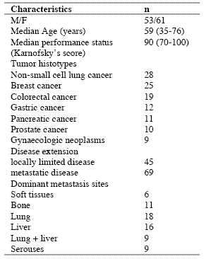
The study included 114 consecutive solid tumor patients with locally limited or metastatic disease, whose clinical characteristics are shown in Table 1. Lung cancer and gastrointestinal tumors were the most frequent neoplasms in our patients. For the immune detections, venous blood samples were collected in the morning after an overnight fast. Operable patients and metastatic patients were investigated before the surgical operation and before the onset of chemotherapy, respectively, in an attempt to exclude the possible influence of the various anticancer therapies on the immune status of patients.

In each sample, we have evaluated total lymphocyte count and the various lymphocyte subpopulations by a flow cytometric assay and monoclonal antibodies, including T helper lymphocytes (CD4), T cytotoxic lymphocytes (CD8), NK cells (CD16CD56), and T regulatory (T-reg) lymphocytes (CD4CD25). Normal values (95% confidence limits) of T-reg observed in our laboratory were below 240/mm3. Moreover, because of its importance in regulating lymphocyte functions and proliferation (Claman, 1998; Sthephens et al, 2004), the circadian rhythm of cortisol was also investigated by collecting blood samples at 8.00 A.M. and at 4.00 P.M., and cortisol serum concentrations were measured in duplicate by using the ECLA method. Data were reported as mean +/- SE, and statistically analyzed by the Student’s t test, the analysis of variance and the chi-square test, as appropriate.

III. Results
As reported in Table 2, an abnormally high number of T-reg was seen in 52/114 (46%) patients. Moreover, the percentage of cases with elevated number of T-reg observed in metastatic patients was significantly higher with respect to that found in non-metastatic patients (44/69 (64%) vs 8/45(18%), p < 0.01). Table 3 shows the mean number of T-reg and the mean percentages of T-reg with respect to both total lymphocytes and T helper (CD4+) lymphocytes observed in cancer patients in relation to their disease extension. The mean number of T-reg observed in metastatic patients was higher with respect to that found in patients with locally limited disease, without, however statistically significant differences. In contrast, the mean percentages of T-reg with respect to that of both lymphocytes and CD4 cells were significantly higher in metastatic patients than in the non-metastatic ones (p< 0.05 and p< 0.001,respectively). Moreover, within the metastatic group, patients with a normal lymphocyte count greater than 1500/mm3 showed a significantly higher mean number of T-reg with respect to the non-metastatic patients, whereas no difference was seen between nonmetastatic patients and metastatic patients with lymphocytopenia, consisting of lymphocyte count lower than 1500/mm3. In contrast, the mean percentages of T-reg with respect to total lymphocytes and CD4+ cells observed in both groups of metastatic patients with normal or low total lymphocyte count were significantly higher than in non-metastatic patients (lymphocytes: p< 0.025, CD4+ cells: p< 0.001).

Table 1. Clinical characteristics of 114 solid tumor patients
090722_clinicalstud1

Table 2. Percentages of abnormally high values of CD4+CD25+ lymphocytes

090722_clinicalstud2

* P < 0.01 vs non-metastatic patients

Table 3. Mean number of CD4+CD25+ lymphocytes and their mean percentages with respect to total lymphocytes and CD4+ lymphocytes in metastatic and non-metastatic patients

090722_clinicalstud3

* p<0.05 vs non-metastatic patients; ** p<0.025 vs non-metastatic patients; *** p<0.01 vs non-metastatic patients; **** p<0.001 vs nonmetastatic patients

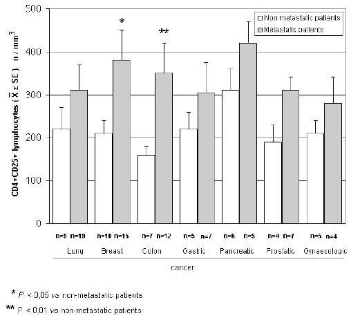
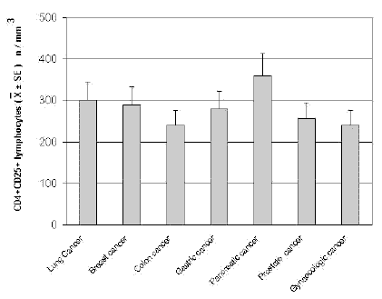
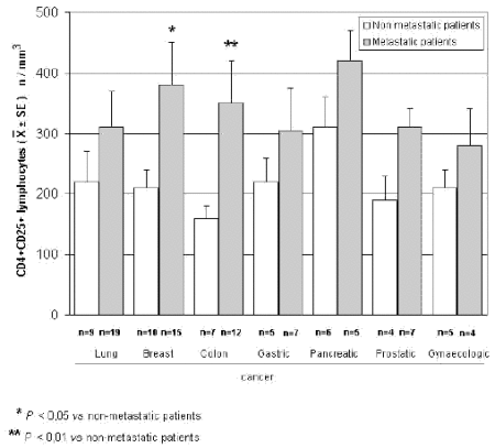
The mean counts of NK and CD8 cells in relation to that of T-reg are reported in Table 4. As shown, no significant difference in the mean number of CD8 lymphocytes was found between patients with normal or abnormally elevated number of T-reg. On the contrary, patients with elevated number of T-reg showed a significantly lower number of NK cells with respect to that found in those with normal T-reg count. Finally, Table 5 shows the circadian rhythm of cortisol in relation to total lymphocytes, CD4+ cells and T-reg mean number. A normal cortisol rhythm, with morning values greater at least than 50% with respect to the values occurring during the afternoon, was found in 85/114 (75%). Total lymphocyte and CD4+ cell mean numbers observed in patients with altered cortisol rhythm were significantly lower than those found in patients with normal cortisol circadianicity (p<0.01), whereas no significant difference was seen in the mean number of T reg. Figure 1 and Figure 2 illustrate T-reg mean numbers in relation to tumor histoptypes in the overall patients and with respect to their disease extension, respectively. No significant difference was seen in relation to tumor histotype. The highest values of T-reg were observed in pancreatic cancer patients, without however significant differences with respect to the overall other histotypes. The metastatic disease was associated with a higher number of T-reg with respect to the non-metastatic group in all tumor histotypes, even though a statistically significant differences occurred for the only breast cancer (p<0.05) and colorectal cancer (p< 0.01).

Table 4. Mean values of NK cells and CD8+ lymphocytes in cancer patients with normal or abnormally high values of CD4+CD25+ lymphocytes

090722_clinicalstud4

* p<0.05 vs normal values of CD4+CD25+ lymphocytes

Table 5. Mean numbers of total lymphocytes, T helper (CD4+) lymphocytes and T regulator lymphocytes (CD4+CD25+) in relation to cortisol circadian secretion in cancer patients
090722_clinicalstud5

* P<0.01 vs patients with altered cortisol rhythm
090722_clinicalstudfig1

Figure 1. CD4+CD25+ lymphocyte mean number in relation to tumor histotype
090722_clinicalstudfig2

Figure 2. CD4+CD25+ lymphocytes in relation to tumor histotype in metastatic and non-metastatic cancer patients

IV. Discussion
According to previous preliminary clinical investigations (Sasada et al, 2003; Cesana et al, 2006), this study confirms in a greater number of cancer patients that the metastatic disease is characterized by the evidence of an abnormally increased percentage of T-reg lymphocytes with respect to both total circulating lymphocytes and CD4+ lymphocytes. This finding does not seem to represent a specific characteristic of some tumor histotypes, then it could constitute a general alteration occurring during the progression of the neoplastic disease, by representing a fundamental immune parameter of cancer-related immunosuppression.

Several immune molecules have appeared to suppress the anticancer immunity, namely IL-6, IL-10, IL-1, TNF-α and TGF-β, but it seems that the common end result of their mechanisms of action may be represented by the stimulation of T-reg generation, with a consequent inhibition of the activation of an effective anticancer immune reaction. On the same way, several immune cells are able to suppress the anticancer immunity, including macrophages, T helper-2 lymphocytes and some myeloidderived suppressor cells, but also in this case they would act in a suppressive way by promoting the generation of Treg.

Then, the detection of T-reg amounts in terms of both absolute number and percentages with respect to total lymphocytes and CD4+ cells could constitute a simple and adequate clinical immune parameter to quantify the whole status of the anticancer immunity in the single cancer patient. Moreover, future clinical studies will be required to establish the possible prognostic significance of changes in T-reg percentage and number in relation to the anticancer efficacy of the various standard antitumor therapies. Moreover, it has to be remarked that T-reg lymphocytes would not represent the only immune cells involved in the suppression of the anticancer immunity. In fact, there is at least another fundamental immunosuppressive system, consisting of the monocytemacrophage cell lineage (Sica and Bronte, 2007). In more detail, it has been observed that the bone marrow may release myeloid precursors provided by suppressive activity on the antitumor immune response and defined as myeloid-derived suppressor cells (MDSC) (Kusmartsev and Gabrilovich, 2005). These cells have appeared to be characterized by the cell surface expression of GR-1, CD11b and CD80 antigens (Anderson et al, 2002; van Ginderachter et al, 2006). The myeloid suppressor cells would promote the generation and activation of T-reg lymphocytes, which at the other side would stimulate MDSC release from the bone marrow and M2 macrophage differentiation (Terabe et al, 2003; Wie et al, 2006).

Moreover, the myeloid suppressive cells would inhibit the anticancer immunity by promoting macrophage differentiation into the M2 sub-type (Ikemoto et al, 2003), which plays a clear inhibitory effect on the anticancer immunity, namely through the release of IL-6 (Ueno et al, 2000), whereas the M1 macrophage sub-type may either stimulate or suppress the antitumor immunity (Mantovani et al, 2004). M1 and M2 macrophage sub-types have appeared to be characterized by a high production of IL-12 or IL-10, respectively (Ueno et al, 2000).

Then, further studies by concomitantly evaluating T reg and MDSC count, will contribute to better define the immune mechanism responsible for the suppression of the anticancer immunity.

References
Anderson CF, Gerber JS, Mosser DM (2002) Modulating macrophage function with IgG immune complexes. J Endotoxin Res 8, 477-81.

Annunziato F, Cosmi L, Liotta F, Lazzeri E, Manetti R, Vanini V, Romagnani P, Maggi E, Romagnani S (2002) Phenotype,localization and mechanism of suppression of CD4+CD25+ human thymocytes. J Exp Med 196, 379-87.

Antony PA, Restito NP (2005) CD4+CD25+ T regulatory cells,immunotherapy of cancer,and interleukin-2. J Immunother 28, 120-8.

Birebent B, Lorho R, Lechartier H, de Guibert S, Alizadeh M,
Vu N, Beauplet A, Robillard N, Semana G (2004) Suppressive properties of human CD4+CD25+regulatory T cells are dependent on CTLA-4 expression. Eur J Immunol 34, 3485-96.

Cesana GC, DeRaffele G, Cohen S, Moroziewicz D, Mitcham J, Stoutenburg J, Cheung K, Hesdorffer C, Kim-Schulze S, Kaufman HL (2006) Characterization of CD4+CD25+ regulatory T cells in patients treated with high-dose interkleukin-2 for metastatic melanoma or renal cell carcinoma. J Clin Oncol 24, 1169-77.

Chen W, Jin W, Hardegen N, Lei KJ, Li L, Marinos N, McGrady G, Wahl SM (2003) Conversion of peripheral CD4+CD25- naïve T cells to CD4+CD25+ regulatory T cells by TGF-β induction of transcription factor Foxp 3. J Exp Med 198, 1875-86.

Claman HN (1998) Corticosteroids and the immune system. Adv Exp Med Biol 245, 203-10.

Dieckmann D, Bruett H, Ploettner H, Lutz MB, Schuler G (2002) Human CD4+CD25+ regulatory contact-dependent T cell induce IL-10 producing,contact-independent type-1-regulatory T cells. J Exp Med 196, 247-53.

Dieckmann D, Plottner H (2001) Ex vivo isolation and characterizationof CD4+CD25+ T cells with regulatory properties from human blood. J Exp Med 193, 1303-10.

Ehrke MJ, Mihich E, Berd D, Mastrangelo MJ (1989) Effects of anticancer drugs on the immune system. Semin Oncol 16, 230-9.

Ghiringhelli F, Larmonier N, Schmitt E, Parcellier A, Cathelin D, Garrido C, Chauffert B, Solary E, Bonnotte B, Martin F (2004) CD4+ CD25+ regulatory T cells suppress tumor immunity but are sensitive to cyclophosphamide which allows Immunotherapy of established tumors to be curative. Eur J Immunol 34, 336-44.

Ikemoto S, Yoshida N, Narita K, Wada S, Kishimoto T, Sugimura K, Nakatani T (2003) Role of tumor-associated macrophages in renal cell carcinoma. Oncol Rep 10, 1843-9.

Jago CB, Yates J, Camara NOS, Lechler RI, Lombardi AG (2004) Differential expression of CTLA-4 among T cell subsets. Clin Exp Immunol 136, 463-71.

Ji HB, Liao G, Faubion WA, Abadía-Molina AC, Cozzo C, Laroux FS, Caton A, Terhorst C (2004) Cutting edge, the natural ligand for glucocorticoid-induced TNF receptorrelated protein abrogates regulatory T cell suppression. J Immunol 172, 5823-7.

Kanamaru F, Youngnak P, Hashiguchi M, Nishioka T, Takahashi T, Sakaguchi S, Ishikawa I, Azuma M (2004) Costimulation via glucocorticoid-induced TNF receptor in both conventional and CD25+ regulatory CD4+ T cells. Immunol 172, 7306-14.

Knutson KL, Disis M, Salazar L (2007) CD4 regulatory T cells in human cancer pathogenesis. Cancer Immunol Immunother 556, 271-85.

Kusmartsev S, Gabrilovich DI (2005) STAT1 signaling regulates tumor-assolciated macrophage-mediate T cell deletion. J Immunol 174, 4880-91.

Lan RY, Ansari AA, Lian ZX, Gershwin ME (2005) Regulatory T cells, development,function,and role in autoimmunity. Autoimmun Rev 4, 351-63.

Leong PP, Mohammad R, Ibrahim N, Ithnin H, Abdullah M, Davis WC, Seow HF (2006) Phenotyping of lymphocytes expressing regulatory and effector markers in infiltrating ductal carcinoma of the breast. Immunol Lett 102, 229-36.

Malek TR, Bayer AL (2004) Tolerance not immunity crucially depends on IL-2. Nat Rev Immunol 4, 665-74.

Mantovani A, Sica A, Sozzani S, Allavena P, Vecchi A, Locati M (2004) The chemokine system in diverse forms of macrophage activation and polarization. Trends Immunol 25, 677-86.

Sasada T, Kimura M, Yoshida Y, Kanai M, Takabayashi A (2003) CD4+CD25+ regulatory T cells in patients with gastrointestinal malignancies, possible involvement of regulatory T cells in disease progression. Cancer 98, 1089-93.

Schwartz RH (2005) Natural regulatory T cells and selftolerance. Nat Immunol 6, 327-30.

Shevach EM (2002) CD4+CD25+ suppressor T cells, more questions than answers. Nat Rev Immunol 2, 389-400.

Sica A, Bronte V (2007) Altered macrophage differentiation and immune dysfunction in tumor development. J Clin Invest 117, 1155-66.

Siddiqui SA, Frigola X, Bonne-Annee S, et al (2007) Tumorinfiltrating Foxp3 CD4+CD25+ T cells predict poor survuival in renal cell carcinoma. Clin Cancer Res 13, 2075-81.

Stephens GL, McHugh RS, Whitters MJ, Young DA, Luxenberg D, Carreno BM, Collins M, Shevach EM (2004) Engagement of glucocorticoid-induced TNFR family-related receptor on effector T cells by its ligand mediates resistance to suppression by CD4+CD25+ T cells. J Immunol 173, 5008-20.

Takahashi T, Tagami T, Yamazaki S, Uede T, Shimizu J, Sakaguchi N, Mak TW, Sakaguchi S (2000) Immunologic and tolerance maintained by CD25+CD4+ regulatory T cells constitutively expressing cytotoxic T lymphocyte-associated antigen. J Exp Med 192, 1285-94.

Terabe M, Matsui S, Park JM, Mamura M, Noben-Trauth N, Donaldson DD, Chen W, Wahl SM, Ledbetter S, Pratt B, Letterio JJ, Paul WE, Berzofsky JA (2003) Transforming growth factor-β production and myeloid cells are an eggector mechanism trhough which CD1d-restricted T cells block cytotoxic T lymphocyte-mediated tumor immunosurveillance, abrogation prevents tumor recurrence. J Exp Med 198, 1741-52.

Thomton AM, Shevach EM (2000) Suppressor effector function of CD4+CD25+ immunoregulatory T cells is antigen nonspecific. J Immunol 164, 183-90.

Ueno T, Toi M, Saji H, Muta M, Bando H, Kuroi K, Koike M, Inadera H, Matsushima K (2000) Significance of macrophage chemo-attractant protein-1 in macrophage recruitment,angiogenesis and survival in human breast cancer. Clin Cancer Res 6, 3282-9.

Van Ginderachter JA, Movahedi K, Hassanzadeh Ghassabeh G, Meerschaut S, Beschin A, Raes G, De Baetselier P (2006) Classical and alternative activation of mononuclear phagocytes, picking the best of both worlds for tumor promotion. Immunobiology 211, 487-501.

Vasu C, Prabhakar BS, Holterman MJ (2004) Targeted CTLA-4 engagement induces CD4+CD25+CTLA-4 high T regulatory cells with target alloantigens specificity. J Immunol 173, 2866-76.

von Boehmer H (2005) Mechanisms of suppression by suppressor T cells. Nat Immunol 6, 338-44.

von Herrath MG, Harrison LC (2003) Regulatory lymphocytes, antigen-induced regulatory T cells in autoimmunity. Nat Rev Immunol 3, 223-32.

Wang XB, Zheng CY, Giscombe R, Lefvert AK (2001) Regulation of surface and intracellularexpression of CTLA-4 on human peripheral T cells. Scand J Immunol 54, 453-8.

Wie S, Kryczeck I, Zou W (2006) Regulatory T-cell compartmentalization and trafficking. Blood 108, 426-31.

Yang R, Cai Z, Zhang Y, Yutzy WH 4th, Roby KF, Roden RB (2006) CD80 in immune suppression by mouse ovarian carcinoma-associated Gr-1+CD11b+ myeloid cells. Cancer Res 66, 6807-15.

Ziegler SF (2006) FOXP3 of mice and men. Annu Rev Immunol 24, 209-26.

Zou W (2006) Regulatory T cells, tumour immunity and immunotherapy. Nat Rev Immunol 6, 295-307.

(download PDF version)



2009: Lissoni P; Fumagalli L; Brivio F; Rovelli F; Messina G; Di Fede G; Colciago M; Brera G
Division of Radiation Oncology, Milan, Italy

Biotherapy with the pineal hormone melatonin plus aloe and myrrh tincture in untreatable metastatic cancer patients as an essence therapy of cancer

Research Article

P. Lissoni1,*, F. Rovelli1, G. Messina2, F. Brivio3, B. Boniardi1, G. Porro1, L.Vigore4, G. Di Fede1, P. Marchiori1, G. Brera5

1 Institute of Biological Medicine, Milan, Italy
2 Psychiatric Division, Policlinico Hospital, Milan
3 Division of Surgery, Bassini Hospital, Cinisello,Milan;
4 Laboratory of Immunomicrobiology,San Gerardo Hospital, Monza, Milan;
5 Ambrosian University, Milan, Italy.

*Correspondence: Dr. Paolo Lissoni, Divisione di Radioterapia Oncologica, Ospedale S. Gerardo, 20052 Monza, Milano, Italia. Fax: +390392332284, e-mail: p.lissoni@hsgerardo.org
Key words: Aloe Vera, Melatonin, Mirrh, and Anticancer Immunity
Abbreviations: Melatonin (MLT), complete response (CR), partial response (PR), stable disease (SD), disease control (DC), progressive disease (PD), T helper lymphocytes (TH, CD4+), T regulatory lymphocytes (T reg, CD4+ CD25+)

Received: 30 July 2009; Revised: 18 October 2009
Accepted: 20 October 2009; electronically published: December 2009

Summary

Background: The recent advances in understanding the immunobiological interactions responsible for cancer progression have allowed us to define the mechanisms of action of some plants, whose antitumor properties were already known by the popular Medicine, in particular Aloe and Myrrha, whose mixture was already therapeutically utilized more than 2000 years ago by the Essence medicine. Moreover, some endogenous natural substances, namely the main hormone produced by the pineal gland melatonin (MLT) may also play anticancer activity. On this basis, a study was performed with a biological regimen consisting of MLT, Aloe and Myrrha in untreatable metastatic cancer patients with life expectancy lower than 1 year. Methods: The study included 35 patients. MLT was given orally at 20 mg/day in the evening and a mixed Aloe and Myrrha tincture was administered at a dose of 5 ml/thrice daily. Results: The clinical response consisted of complete response (CR) in 1, partial response (PR) in 2, stable disease (SD) in 19 patients, whereas the remaining 13 patients had a progressive disease (PD). Thus, a disease control (CR + PR + SD) was achieved in 22/35 (63%)patients. Moreover, a survival longer than 1 year was achieved in 17/35 (49%) patients. Finally, DC was associated with an evident improvement in the immune status, namely consisting of a decrease in the number of T regulatory lymphocytes, which are the main cells responsible for the suppression of the anticancer immunity. Conclusion: This preliminary study shows that a biological anticancer regimen consisting of the pineal hormone MLT in association with Aloe and Myrrha mixture, already known at the times of the Essence medical tradition, may induce a control of the neoplastic disease by stimulating the anticancer immunity, in a relevant percentage metastatic cancer patients, who did not respond to the conventional anticancer treatments and for whom no other standard therapy was available.

References
Davis RH, Parker WL, Sampson RT, Murdoch DP. Isolation of a stimulatory system in an Aloe extract. J Am Pediatr Med Assoc 1991; 81:473-8.

Capasso F, Borrelli F, Capasso R, Di Carlo G, Izzo AA, Pinto L et al. Aloe and its therapeutic use. Phytother Res 1998; 12:124-7.

Vogler BK. Aloe Vera: a systematic review of its clinical effectiveness. B J Gen Pract 1999; 49:823-8.

Claeson P, Zygmunt P, Hogestatt ED. Calcium antagonistic properties of the sesquiterpene T- cadinol. Pharmacol Toxicol 1991; 69:173-7.

Qureshi S, Al-Harbi MM, Ahmed M, Raza M, Giangreco AB, Shah AH. Evaluation of the genotixic, cytotoxic and antitumor properties of Commiphora molmol using normal and Erlich ascites carcinoma cell-bearing Swiss albino mice. Cancer Chemother Pharmacol 1993; 33130-8.

Blazquez C, Casanova ML, Planas A, Del Pulgar TG, Villanueva C, Fernandez-Acenero MJ et al. Inhibition of tumor angiogenesis by cannabinoids. FASEB J 2003; 17:529-31.

Grotenhermen F. Pharmacology of cannabinoids. Neuroendocrinol Lett 2004; 25:14-23.

Aggarwall BB, Kumar A, Bharti AC. Anticancer potential of curcumin. Preclinical and clinical studies. Anticancer Res 2003; 23:363-98.

Lodha R, Bagga A. Tradictional Indian system of Medicine. Ann Acad Med Singapore 2000; 29:37-41.

John’s Gospel 19,38-40.

Iguchi H, Kato KI, Ibayashi H. Age-dependent reduction in serum melatonin concentrations in healthy subjects. J Clin Endocrinol Metab 1982; 55:27-9.

Attanasio A, Borrelli P, Gupta D. Circadian rhythms in serum melatonin from infancy to adolescence. J Clin Endocrinol Metab 1985; 61:388-90.

Jankovic BD. Neuroimmunomodulation. Ann NY Acad Sci 1994; 741:3-38.

Brzezinski A. Melatonin in humans. N Engl J Med 1997; 336:185-95

Bartsch H, Bartsch C. Effects of melatonin on experimental tumors under different photoperiods and times of administration. J neural Transm 1981; 52:269-79

Regelson W, Pierpaoli W. Melatonin: a rediscovered antitumor hormone? Cancer Invest
1987; 5:379-85

Lissoni P. Is there a role for melatonin in supportive care? Supp Care Cancer 2002; 10:110-6.
Sze S, Ng T, Liu W. Antiproliferative effect of pineal indoles on cultured tumor cell lines. J Pineal Res 1993; 14:27-33.

Maestroni GJM. The immunoneuroendocrine role of melatonin. J Pineal Res 1993; 14:1-10.

Lissoni P, Brivio F, Fumagalli L, Messina G,Vigorè L, Parolini D et al. : Neuroimmunomodulation in Medical Oncology: application of Psychoneuroimmunology with subcutaneous low-dose IL-2 plus the pineal hormone melatonin in patients with untreatable metastatic solid tumors. Anticancer Res 2008; 28:1377-82.

Shevach EM. CD4+CD25+ suppressor T cells: more questions than answers. Nat Rev Immunol 2002; 2:389-400.

Rorschach H. Psychodiagnosytics. Ed HA Huber, Bern, Stuttgart, Toronto. Verlag, 1921.

Lissoni P, Messina G, Parolini D, Balestra A, Brivio F, Fumagalli L et al.:A spiritual approach in the treatment of cancer. In Vivo 2008; 22:557-82.


Synchronization of Cortisol Circadian Rhythm by the Pineal Hormone Melatonin in Untreatable Metastatic Solid Tumor Patients and its Possibile Prognostic Significance
on Tumor Progression

Research Article (download PDF version – complete)

FERNANDO BRIVIO1, LUCA FUMAGALLI1, GABRIELE FUMAGALLI2, SIMONETTA PESCIA2, RINALDO BRIVIO2, GIUSEPPE DI FEDE3, FRANCO ROVELLI2 and PAOLO LISSONI3

1 Surgical Division, Bassini Hospital, Cinisello Balsamo, Milan, Italy
2 Department of Clinical Pathology, S.Gerardo Hospital, Monza, Italy
3 Institute of Biological Medicine, Milan, Italy

*Correspondence: Dr. Paolo Lissoni, Divisione di Radioterapia Oncologica, Ospedale S.Gerardo, 20052 Monza, Milano, Italy; Fax: +390392332284, e-mail: p.lissoni@hsgerardo.org

Abstract
The present study was carried out to evaluate the influence of a short-period IL-2 administration on the efficacy of chemotherapy in metastatic colorectal cancer patients with pretreatment lymphocytopenia, which was defined as a lymphocyte count of less than 1500/mm3.

The study included 144 consecutive metastatic colorectal cancer patients, who underwent chemotherapy with oxaliplatin plus 5-fluorouracil. Lymphocytopenia was seen in 41/144 (28%) patients, who were randomized to receive chemotherapy alone or chemotherapy after a prechemoimmunotherapy with IL-2 (3 MIU twice/day for 3 consecutive days), whereas patients with a normal pretreatment lymphocyte count received only chemotherapy.

A normalization of the lymphocyte number was achieved in 12/19 lymphocytopenic patients pretreated with IL-2. The objective tumor regression rate achieved in patients with a normal lymphocyte count prior to chemotherapy was significantly higher compared to that obtained in lymphocytopenic patients treated with chemotherapy alone (54/103 vs. 3/22, p<0.01), whereas no significant difference occurred between patients with normal lymphocyte count and lymphocytopenic patients pretreated with IL-2 (54/103 vs. 8/19).

This study confirms that pretreatment lymphocytopenia is associated with reduced efficacy of chemotherapy in metastatic colorectal cancer patients. Moreover, it suggests that pretreatment with IL-2 before the onset of chemotherapy may enhance the efficacy of chemotherapy in lymphocytopenic patients. Therefore, the administration of IL-2 before the onset of chemotherapy to improve the immune status of cancer patients may be considered as a new chemoimmunotherapeutic combination, which may be recommended in the treatment of advanced cancer patients, particularly in those with cancer-related immune alterations.

http://www.imbio.it/wp-content/uploads/2013/10/100430_synchronization_cortisol_circadian_rhythm.pdf(download PDF version – complete)


Psychoneuroendocrine Modulation of Regulatory T Lymphocyte System: In Vivo and In Vitro Effects of the Pineal Immunomodulating Hormone Melatonin

Research Article (download PDF version – complete)

LUIGI VIGORÉ1, GIUSY MESSINA2, FERNANDO BRIVIO3, LUCA FUMAGALLI3, FRANCO ROVELLI4, GIUSEPPE DI FEDE4 and PAOLO LISSONI4

1 Laboratory of Immunomicrobiology, S. Gerardo Hospital, Monza, Milan, Italy
2 Psychiatric Division, Polyclinic Hospital, Milan, Italy
3 Surgical Division, Bassini Hospital, Cinisello, Milan, Italy
4 Institute of Biological Medicine, Milan, Italy

*Correspondence: Dr. Paolo Lissoni, Divisione di Radioterapia Oncologica, Ospedale S.Gerardo, 20052 Monza, Milano, Italy; Fax: +390392332284, e-mail: p.lissoni@hsgerardo.org

Abstract

Background: At present, it is known that cancer-related immunosuppression would mainly depend on an immunosuppressive action mediated by a subtype of CD4+ lymphocytes, the so-called regulatory T lymphocytes (T-reg), which are identified as CD4+CD25+ cells.
Moreover, it has been shown that anticancer immunity is under psychoneuroendocrine regulation, mainly mediated by the pineal hormone melatonin (MLT).
This study was performed to investigate the in vivo and in vitro effects of MLT on T-reg generation.

Materials and Methods: We evaluated the in vivo effects of MLT (20 mg/daily orally in the evening) in 20 patients with untreatable metastatic solid tumor and the in vitro effects of MLT incubation (at 10 and 100 pg/ml) of pure lymphocyte cultures on T-reg cell count.

Results: MLT induced a statistically significant decline in mean T-reg cell numbers in patients who achieved disease control, whereas no effect was seen in those who had progressed. In contrast, no in vitro effect of MLT incubation was apparent.

Conclusion: This preliminary study would suggest that MLT may exert in vivo an inhibitory action on T-reg cell generation in cancer patients which is associated with a control of the neoplastic progression, whereas no direct effect was seen in vitro on lymphocyte differentiation. This finding would suggest that MLT may counteract T-reg cell generation in vivo by inhibiting macrophage activity which is involved in stimulating T-reg cell production.

(download PDF version – complete)


Settembre 30, 2009 Newsletter

A Londra, alla conferenza della WCRF su alimentazione e cancro, si è discusso di temi fondamentali per l’impostazione individuale e sociale dell’alimentazione mondiale per i prossimi 10 anni. I dati presentati parlano della possibilità di ridurre di oltre 1/3 le morti per cancro, curando l’alimentazione.

In questo convegno a Londra, si è parlato di un tale numero di morti evitabili con sole scelte alimentari, cifre da farci riflettere.

Alcuni ricercatori internazionali della WCRF hanno segnalato il fatto che l’attivazione della infiammazione causata dalla ingestione del cibo e quindi la presenza delle citochine (sostanze prodotte dagli anticorpi che vivono sotto la mucosa intestinale) derivanti dal rapporto tra intestino e cibo, sono in grado di partecipare alla induzione delle forme tumorali (un po’ come dire che le allergie alimentari ritardate, cioè le intolleranze alimentari, potrebbero contribuire alla induzione di forme tumorali).

Significa che c’è un fenomeno infiammatorio alimentare, percepito dai pazienti, che deve essere meglio definito e capito, ma c’è. Sappiamo anche che la diagnostica di una intolleranza alimentare è ancora oggi affidata a test non convenzionali.

Esiste in Italia da alcuni anni l’unico test in grado di diagnosticare le allergie alimentari o intolleranze alimentari, ALCAT® Test, utile strumento diagnostico per il medico che può elaborare delle strategie e soluzioni cliniche.

Negli ultimi tempi è stato chiarito che le allergie alimentari hanno due vie preferenziali, la classica con produzione di IgE e la via alternativa, con sviluppo su anticorpi granulociti che si attivano al contatto di un alimento non tollerato dall’organismo.

Un cenno al problema dell’obesità, che è riconosciuta oggi come il più importante fattore di induzione tumorale esistente.
Una recente scoperta rileva che i Macrofagi, cellule connesse alla allergia, si attivano determinando insulino resistenza e ingrassamento.
Questo fatto ci fa ragionare sul ruolo importante della infiammazione allergica da cibo nell’obesità e quindi su come questa favorisca malattie cardiovascolari e addirittura promuova il cancro.

Quello che l’Istituto di Medicina Biologica sta facendo in questi anni di duro impegno professionale e di ricerca, va al di là del conteggio delle calorie: l’I.M.B.IO., mette in prima fila la ricerca di soluzioni per contrastare la genesi tumorale e l’obesità.


Copyright by IMBIO 2017. All rights reserved.